Received August 30, 2017, Revised September 1, 2017, Accepted for publication September 5, 2017
*These authors equally contributed at this work.
Corresponding author: Yong Hyun Jang, Department of Dermatology, Kyungpook National University Hospital, 130 Dongdeok-ro, Jung-gu, Daegu 41944, Korea. Tel: 82-53-420-5838, Fax: 82-53-426-0770, E-mail:
Do Won Kim, Department of Dermatology, Kyungpook National University Hospital, 130 Dongdeok-ro, Jung-gu, Daegu 41944, Korea. Tel: 82-53- 200-5838, Fax: 82-53-426-0770, E-mail: [email protected]
This is an Open Access article distributed under the terms of the Creative Commons Attribution Non-Commercial License (http://creativecommons.
org/licenses/by-nc/4.0) which permits unrestricted non-commercial use, distribution, and reproduction in any medium, provided the original work is properly cited.
Copyright © The Korean Dermatological Association and The Korean Society for Investigative Dermatology
Ann Dermatol Vol. 30, No. 2, 2018 https://doi.org/10.5021/ad.2018.30.2.158
ORIGINAL ARTICLE
Usefulness of Inflammatory Markers for the Prediction of Postherpetic Neuralgia in Patients with Acute Herpes Zoster
Jun Young Kim*, Gyeong-Hun Park1,*, Min Ji Kim, Hyun Bo Sim, Weon Ju Lee, Seok-Jong Lee, Shin-Woo Kim2, Young Hoon Jeon3, Yong Hyun Jang, Do Won Kim
Department of Dermatology, Kyungpook National University School of Medicine, Daegu, 1Department of Dermatology, Hallym University, Dongtan Sacred Heart Hospital, Hallym University College of Medicine, Hwaseong, Departments of 2Internal Medicine and
3Anesthesiology and Pain Medicine, Kyungpook National University School of Medicine, Daegu, Korea
Background: Increasing evidence suggests a pivotal role for neuronal inflammation in response to replicating varicella zoster virus in the development of postherpetic neuralgia (PHN). Objective: In this study, we investigated the value of serum levels of various inflammatory markers in acute her- pes zoster (HZ) as predictors for the development of PHN.
Methods: A total of 116 patients with acute HZ were enrolled in this study. We measured scores on the pain visual ana- logue scale (VAS) at baseline and at 1, 3, and 6 months after diagnosis of HZ. We defined PHN as pain greater than 1 on the VAS lasting for more than 6 months. Serum samples for laboratory assay, including complete blood count were ob- tained at the initial visit. Correlations between the levels of each inflammatory marker and the development of PHN were evaluated. Results: Levels of erythrocyte sedimentation rate (ESR), C-reactive protein (CRP), lymphocyte count, and albumin showed significant correlations with development
of PHN in univariate analysis. Levels of ESR, CRP, and lym- phocyte count also showed significant correlations in multi- variate analysis. ESR level showed stronger correlations with development of PHN than levels of CRP and lymphocyte count. Conclusion: In this study, we confirmed that elevated ESR was an independent and significant predictor of PHN in patients with acute HZ. To validate these results, further well-designed, randomized clinical trials are needed. (Ann Dermatol 30(2) 158∼163, 2018)
-Keywords-
Herpes zoster, Inflammation, Postherpetic neuralgia
INTRODUCTION
Postherpetic neuralgia (PHN) is characterized by sponta- neous pain, pain provoked by trivial stimuli, and altered sensations that accompany herpes zoster (HZ) and that may continue long after the characteristic rash of HZ has healed1. PHN is neuropathic and results from injury of the peripheral nerves and altered central nervous system sig- nal processing2. These changes may be so complex that no single therapeutic approach will ameliorate all of the abnormalities. As a result, antiviral agents and active inter- ventional pain management during the early period of acute HZ are recommended to prevent the development of PHN3. Various risk factors for development of PHN such as old age, female sex, presence of a prodrome, more severe rash, and more severe acute pain reflect dif- ferent mechanisms leading to the development of PHN4. For the timely identification of patients with HZ who
might benefit from preventive strategies, it is important to identify the factors that can best predict the development of PHN. However, prediction of development of PHN is currently limited to clinical factors. A few reports showed that objective assessment tools such as the varicella zoster virus (VZV) skin-test reaction and infrared thermography are useful as predictors of development of PHN in patients with acute HZ, but more objective markers that can be easily available for early prediction of development of PHN are needed5,6.
Recently, neuronal inflammation secondary to replicating VZV has been identified as a potential mediator in the de- velopment of PHN. Therefore, we investigated whether early serum levels of various inflammatory markers in acute HZ could be useful to predict PHN development.
MATERIALS AND METHODS
This study was undertaken in the Department of Derma- tology at Kyungpook National University Hospital. From June 2013 to March 2015, a total of 116 patients with acute HZ were enrolled. HZ was diagnosed based on clin- ical presentation. Inclusion criteria were patients with lab- oratory results obtained within several hours after diag- nosis of HZ and with a follow-up pain visual analogue scale (VAS) score of at least 1, a month later. Exclusion cri- teria included a confirmed history or suspicion of a con- genital immune disorder; any chronic renal, hepatic, and rheumatologic illness; an acute infection within the pre- vious month; trauma and/or fracture within the previous 6 months; and current use of steroids, including inhalers or nonsteroidal anti-inflammatory drugs. The protocol for this study was approved by the institutional review board of the Kyungpook National University Hospital (KNUH 2016-04-001).
We measured pain VAS scores at baseline and at 1, 3, and 6 months after diagnosis of HZ. Serum samples for labo- ratory assays, including complete blood count (CBC), ery- throcyte sedimentation rate (ESR), C-reactive protein (CRP) level, and albumin level were obtained at the initial visit.
The patients were divided into two groups, based on the presence or absence of pain at each month and the data in each group were statistically compared. We defined PHN as score of greater than 1 on the pain VAS scale having lasted for more than 6 months after onset of acute HZ.
Pain at 6 months was criterion for taking statistics on this study.
We first evaluated correlations between levels of each se- rum inflammatory marker and development of PHN in univariate analysis. Already known predictive factors such as age, sex, and initial pain VAS score were also included Table
1. Demographic, clinical, and laboratory characteristics of study participants by follow-up visit CharacteristicAge (yr)FemalePain VASESR (mm/h) CRP (mg/dl)WBC (/μl)RDW (%)PLT (/μl)Neutrophil (/μl)Lymphocyte (/μl)NLRAlbumin (g/dl) Initial visitAll patients (n=116)59.2±15.674 (63.8)4.6±2.315.6±12.30.6±1.16,486.6 ±2,168.312.9±0.9236.4 K ±64.3 K4,318.5 ±1,615.21,524.7 ±727.43.6±2.74.3±0.4 1 moNo pain (n=43)51.9±15.724 (55.8)3.4±1.913.0±13.30.3±0.66,491.2 ±1,871.312.8±0.9241.8 K ±70.8 K4,264.0 ±1,771.91,577.0 ±747.53.6±3.04.5±0.5 Pain (n=73)62.0±14.350 (68.5)5.3±2.217.2±11.40.7±1.26,484.0 ±2,338.013.0±0.8233.2 K ±60.4 K4,350.5 ±1,527.41,493.8 ±718.73.7±2.54.3±0.4 3 moNo pain (n=58)53.0±14.737 (63.8)3.9±2.113.4±12.50.3±0.66,714.5 ±2,338.612.9±0.9249.2 K ±75.3 K4,442.9 ±1,817.01,634.0 ±819.13.6±2.94.4±0.5 Pain (n=42)63.3±15.028 (66.7)5.0±2.118.8±12.70.7±1.25,799.2 ±1,681.313.1±1.0223.4 K ±49.2 K3,866.7 ±1,141.81,369.3 ±619.43.7±2.84.2±0.4 6 moNo PHN (n=73)54.2±15.647 (64.4)4.0±2.113.2±11.80.3±0.56,451.0 ±2,285.312.9±0.8240.6 K ±71.0 K4,287.1 ±1,704.21,617.7 ±755.13.4±2.74.4±0.5 PHN (n=20)69.2±10.614 (70.0)5.5±2.026.2±12.91.1±1.46,046.0 ±1,765.813.2±1.2232.5 K ±59.3 K3,978.0 ±1,288.41,250.0 ±712.84.4±3.64.2±0.4 Values are presented as mean±standard deviation or number (%). VAS: visual analogue scale, ESR: erythrocyte sedimentation rate, CRP: C-reactive protein, WBC: white blood cell, RDW: red blood cell distribution width, PLT: platelet, NLR: neutrophil lymphocyte ratio, PHN: postherpetic neuralgia.
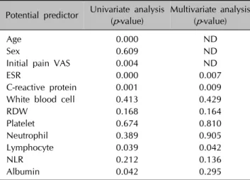
Table 2. Univariate and multivariate analysis of each inflammatory marker and the presence or absence of postherpetic neuralgia at 6 months after diagnosis of herpes zoster
Potential predictor Univariate analysis (p-value)
Multivariate analysis (p-value)
Age 0.000 ND
Sex 0.609 ND
Initial pain VAS 0.004 ND
ESR 0.000 0.007
C-reactive protein 0.001 0.009
White blood cell 0.413 0.429
RDW 0.168 0.164
Platelet 0.674 0.810
Neutrophil 0.389 0.905
Lymphocyte 0.039 0.042
NLR 0.212 0.136
Albumin 0.042 0.295
ND: not done, VAS: visual analogue scale, ESR: erythrocyte sedimentation rate, RDW: red blood cell distribution width, NLR:
neutrophil lymphocyte ratio.
Table 3. Multivariate analyses in each model including each statistically significant inflammatory marker for development of postherpetic neuralgia
Factors of each model
Multiple logistic regression
OR 95% CI p-value
Age 1.12 1.05∼1.19 0.000
Initial pain VAS 1.58 1.15∼2.16 0.004
Age 1.12 1.04∼1.19 0.001
Initial pain VAS 1.61 1.13∼2.30 0.007
ESR 15.72 1.70∼145.16 0.014
Age 1.11 1.04∼1.18 0.001
Initial pain VAS 1.63 1.17∼2.26 0.003
CRP 5.24 1.39∼19.72 0.013
Age 1.11 1.04∼1.18 0.002
Initial pain VAS 1.76 1.24∼2.49 0.001
Lymphocyte 0.17 0.04∼0.71 0.014
OR: odds ratio, CI: confidence interval, VAS: visual analogue scale, ESR: erythrocyte sedimentation rate, CRP: C-reactive protein.
as variables in the analysis. Then, we performed multi- variate logistic regression analysis to identify which in- flammatory markers independently correlated with devel- opment of PHN. To assess the actual influences of each serum inflammatory marker, clinical factors such as age, sex, and initial pain were excluded in multivariate analysis.
Subsequently, cut-off values of the correlated inflammatory markers were established. Models composed of each in- flammatory marker’s cut-off value as well as age and initial pain VAS score were made for confirmation of each mark- er’s influence on the prediction of development of PHN.
The models’ abilities were compared to each other using the area under the receiver operating characteristic (ROC) curves. Differences in area under the curve (AUC) be- tween the reference model derived from only age and ini- tial pain VAS score and a single factor added model were estimated. A p-value <0.05 was defined as statistically significant. All statistical analyses were performed using PASW Statistics ver. 18.0 (IBM Co., Armonk, NY, USA).
RESULTS
The characteristics of the 116 patients, including clinical information and mean initial laboratory results in each group at initial visit and at 1, 3, and 6 months are pre- sented in Table 1. At the initial visit, the mean age of par- ticipants was 59.2 years (standard deviation [SD], 15.6);
there were 42 male patients (36.2%) and 74 female pa- tients (63.8%). Mean initial pain VAS score was 4.60 (SD, 2.30). The pain after diagnosis of HZ persisted for 1 month
in 73 of 116 patients (62.9%), by 3 months in 42 of 100 patients (42.0%), and by 6 months in 20 of 93 patients (21.5%).
In univariate analysis, initial pain VAS score, levels of ESR, CRP, lymphocyte count, and albumin showed signifi- cant correlations with development of PHN at 6 months (p<0.05, Table 2). Other inflammatory markers, including white blood cell (WBC), red blood cell distribution width (RDW), platelet (PLT) count, neutrophil count, and neu- trophil lymphocyte ratio (NLR) did not show significant correlations with development of PHN. There was no sig- nificant correlation between female sex and development of PHN (p>0.05, Table 2). Levels of ESR, CRP, and lym- phocyte count showed significant correlations with devel- opment of PHN in a multivariate analysis conducted ex- cluding clinical factors such as age, sex, and initial pain VAS score (p<0.05, Table 2). Cut-off values of statistically significant inflammatory markers for development of PHN were established and the results were 10.6 (p=0.040) in ESR, 0.34 (p=0.004) in CRP, and 1,297.2 (p=0.032) in lymphocyte count. Multivariate analysis in each model al- so showed significant correlations between each in- flammatory marker’s cut-off value and development of PHN (p<0.05, Table 3). Finally, we evaluated inflam- matory markers that could improve the ability to predict PHN development in combination with age and initial pain VAS score, which are known to be the most im- portant PHN predictors. The AUC increased significantly only when ESR values were combined with age and initial pain VAS scores (p<0.05, Fig. 1).
Fig. 1. Receiver operating characteristic curves of the models adding each inflammatory marker (red) compared with that of a reference model using age and initial pain visual analogue scale (blue, area under curve [AUC]=0.85); (A) erythrocyte sedimentation rate (AUC=0.90, p=0.049), (B) C-reactive protein (AUC=0.89, p=0.221), and (C) lymphocyte count (AUC=0.88, p=0.284).
DISCUSSION
Although PHN is a common neuropathic complication of acute HZ that can reduce quality of life, the exact patho- physiology of PHN is not yet clearly understood7. It is known that pathologic interactions between afferent A be- ta-fibers with impaired function, axon reflexes with im- paired responses, damaged C fibers, and a hyperreflexive spinal cord may play a role in pain associated with HZ as well as PHN8. Additionally, it has been reported that the immune system plays a crucial role in neuropathic pain, which includes interaction between inflammatory im- mune and immune-like glial cells, as well as inflammatory cytokines and chemokines9. This mechanism led to the emergence of the notion of neuropathic pain and sug- gested that the balance between pro- and anti-inflammatory cytokines determines whether neuropathic pain develops or not. However, it is not easy to predict the occurrence of neuropathic pain such as PHN. Several studies have in- dicated the significant contribution of inflammatory cells and their mediators, such as interleukin (IL)-6, to neuro- pathic pain10,11. Although a previous study reported no
significant differences in serum cytokine concentrations between HZ patients with and without PHN, a recent study showed that the levels of IL-6 in patients with PHN were higher than those in patients without PHN12,13. There- fore, it was suggested that patients with nerve injury or an excessive inflammatory response to VZV may have a high- er risk of development of PHN14. However, assessments of proinflammatory cytokines such as IL-6 are difficult to ob- tain in general practice. Therefore, there is a clinical need to identify more easily available inflammatory markers that can predict the development of PHN in patients with acute HZ. During the inflammatory reaction, several proinflammatory cytokines such as IL-1β, IL-6, IL-8, and tumor necrosis factor-α are produced, and these cyto- kines play an important role in the production of acute phase proteins15. That is, acute phase reactants reflect the presence and severity of inflammation. Importantly, CBC and levels of ESR, CRP are widely available, inexpensive, and routinely performed in general practice; moreover, they are accurate and standardized in many settings and can provide objective information to estimate patient prognosis. Therefore, we focused on those inflammatory
markers in this study.
There have been a few reports on ESR and CRP as pre- dictive factors for the development of PHN16. A previous study showed that ESR, CRP, and WBC count are not meaningful predictive factors for the development of PHN, but found that ESR was significantly higher in the 1-month PHN group in univariate analysis7. However, our study showed that levels of ESR, CRP, lymphocyte count, and albumin are significantly correlated with development of PHN at 6 months, and the possibilities of high levels of ESR, CRP, and lymphocyte count as predictors were con- firmed in this study (Table 2). As expected, CBC, including WBC, RDW, PLT count, neutrophil count, and NLR were not predictors of PHN. However, our results differed from that of the previous study in that female sex, one of the al- ready known predictive factors, was not a predictor of PHN in this study (Table 2). When we examined the ac- tual influence of each inflammatory marker in combina- tion with already known clinical factors such as age and initial pain VAS score, a model including ESR showed a better prediction power for development of PHN than models including CRP or lymphocyte count (Fig. 1).
Levels of CRP and lymphocyte count did not show any ad- ditional considerable influence because clinical factors such as age and initial pain VAS score alone had suffi- ciently good predictive power.
PHN has been variably defined as any pain after acute in- fection or any pain at 1 month, 3 months, 4 months, or 6 months after rash onset4,17. In this study, we defined PHN as score greater than 1 on the pain VAS scale, lasting for more than 6 months after onset of acute HZ. This cut-off was chosen because we reasoned that if there was sus- tained pain for a long period of 6 months, it would be suf- ficient to meet any definition of PHN.
In conclusion, we suggest that high levels of ESR in acute HZ may be associated with the development of long-last- ing PHN and that these inflammatory markers in combina- tion with clinical factors such as age and initial pain VAS score will help to predict development of PHN more precisely. Therefore, we need to have more interest in the results of initial routine laboratory tests including these in- flammatory markers as well as already known clinical fac- tors in acute HZ patients. Furthermore, we suggest that an- ti-inflammatory treatment during the early phase of acute HZ is needed to prepare for the development of PHN, es- pecially if the laboratory results show a high ESR. In such cases, patients will require regular long-term follow up for prevention and management of PHN.
This study provides insight into the role of inflammation in the development of PHN. We hope knowledge of these predictive factors may help researchers understand the
pathogenesis of PHN and aid in development and evalua- tion of preventive interventions. A relatively small sample size and data from a single center are limitations of this study. Further investigations employing randomized clin- ical trials with a larger number of patients are required in the future.
ACKNOWLEDGMENT
This research was supported by the Basic Science Research Program through the National Research Foundation of Korea, funded by the Ministry of Education (NRF-2015R1D1A3A01016229).
CONFLICTS OF INTEREST
The authors have nothing to disclose.
REFERENCES
1. Kost RG, Straus SE. Postherpetic neuralgia--pathogenesis, treatment, and prevention. N Engl J Med 1996;335:32-42.
2. Wall PD. Neuropathic pain and injured nerve: central mechanisms. Br Med Bull 1991;47:631-643.
3. Jang YH, Lee JS, Kim SL, Chi SG, Lee WJ, Lee SJ, et al. Do interventional pain management procedures during the acute phase of herpes zoster prevent postherpetic neuralgia in the elderly?: a meta-analysis of randomized controlled trials. Ann Dermatol 2015;27:771-774.
4. Jung BF, Johnson RW, Griffin DR, Dworkin RH. Risk factors for postherpetic neuralgia in patients with herpes zoster.
Neurology 2004;62:1545-1551.
5. Ko EJ, No YA, Park KY, Li K, Seo SJ, Hong CK. The clinical significance of infrared thermography for the prediction of postherpetic neuralgia in acute herpes zoster patients. Skin Res Technol 2016;22:108-114.
6. Imoto K, Okazaki A, Onishi F, Miyazaki Y, Okeda M, Yano S, et al. VZV skin-test reaction, but not antibody, is an important predictive factor for postherpetic neuralgia. J Dermatol Sci 2015;79:235-240.
7. Kim YN, Kim DW, Kim ED. Efficacy of continuous epidural block in acute herpes zoster: Incidence and predictive factors of postherpetic neuralgia, a retrospective single-center study. Medicine (Baltimore) 2016;95:e4577.
8. Baron R. Peripheral neuropathic pain: from mechanisms to symptoms. Clin J Pain 2000;16(2 Suppl):S12-S20.
9. Austin PJ, Moalem-Taylor G. The neuro-immune balance in neuropathic pain: involvement of inflammatory immune cells, immune-like glial cells and cytokines. J Neuroimmunol 2010;229:26-50.
10. Kraychete DC, Sakata RK, Issy AM, Bacellar O, Jesus RS, Carvalho EM. Proinflammatory cytokines in patients with neuropathic pain treated with tramadol. Rev Bras Anestesiol 2009;59:297-303.
11. Wuertz K, Haglund L. Inflammatory mediators in intervertebral
disk degeneration and discogenic pain. Global Spine J 2013;3:175-184.
12. Zak-Prelich M, McKenzie RC, Sysa-Jedrzejowska A, Norval M. Local immune responses and systemic cytokine responses in zoster: relationship to the development of postherpetic neuralgia. Clin Exp Immunol 2003;131:318- 323.
13. Bäckryd E, Ghafouri B, Larsson B, Gerdle B. Plasma pro-inflammatory markers in chronic neuropathic pain: A multivariate, comparative, cross-sectional pilot study. Scand J Pain 2016;10:1-5.
14. Zhu SM, Liu YM, An ED, Chen QL. Influence of systemic immune and cytokine responses during the acute phase of
zoster on the development of postherpetic neuralgia. J Zhejiang Univ Sci B 2009;10:625-630.
15. Gabay C. Interleukin-6 and chronic inflammation. Arthritis Res Ther 2006;8 Suppl 2:S3.
16. Karadag Oncel E, Kara A, Celik M, Karahan S, Cengiz AB, Ceyhan M. Determination and clinical correlation of markers of inflammation in unvaccinated patients with varicella- zoster infection. Eur Rev Med Pharmacol Sci 2013;17:
2032-2039.
17. Desmond RA, Weiss HL, Arani RB, Soong SJ, Wood MJ, Fiddian PA, et al. Clinical applications for change-point analysis of herpes zoster pain. J Pain Symptom Manage 2002;23:510-516.
![Fig. 1. Receiver operating characteristic curves of the models adding each inflammatory marker (red) compared with that of a reference model using age and initial pain visual analogue scale (blue, area under curve [AUC]=0.85); (A) erythrocyte sedimenta](https://thumb-ap.123doks.com/thumbv2/123dokinfo/5167711.105054/4.892.110.804.143.739/receiver-operating-characteristic-inflammatory-compared-reference-erythrocyte-sedimenta.webp)
