105
Occurrence of canine brucellosis in Korea and polymorphism of
Brucella canis isolates by infrequent restriction site-PCR
Dong Hwa Bae, Young Ju Lee*
College of Veterinary Medicine, Kyungpook National University, Daegu 702-701, Korea (Accepted: April 10, 2009)
Abstract : In this study, occurrence of canine brucellosis was surveyed in kennels, indoor dogs and stray dogs in Korea, and infrequent restriction site-polymerase chain reaction (IRS-PCR) was applied to analyze DNA polymorphism of
Brucella canis(
B. canis) isolates. Among a total of 501 dogs tested,
B. canisantibodies by both rapid screening agglutination with 2-mercaptoethanol (2-ME RSAT) and immunochromatographic assay were detected in only 14.1% of kennel dogs. There were no seropositive cases in indoor dogs and stray dogs. DNA polymorphism was observed in 16
B. canisisolates by the IRS-PCR. Sixteen isolates were tested with primers, PsalA, PsalC, PsalG and PsalT, and different primers produced different DNA patterns. In regard to the IRS-PCR pattern of 16 isolates, 9 (56.3%) belonged to the IRS-PCR type I. The remaining 7 were differentiated as type II, III and IV. An application of the primer PsalC provided discrimination between
B. canisisolated in 2005 and others.
Keywords :
Brucella canis, infrequent restriction site-PCR, occurrence
Introduction
Brucellosis is one of the important zoonoses, which affects both animals and humans and leads to serious economic and public health problems. Brucella spp.
that are associated with human brucellosis are Brucella abortus, Brucella melitensis, Brucella suis, and Brucella canis ( B. canis ) . Human B. canis infection is occasionally reported in veterinarians and laboratory workers [2, 15, 24, 31, 34]. Symptoms are usually vague, including prolonged febrile illness with lymph node enlargement.
However, these are infrequently recognized probably due to the lack of serious consideration to the disease.
B. canis cause abortion during the 7 to 9 week period of pregnancy and stillbirths. B. canis has been identified in breeding kennels, and has become a major source of economic loss in large and small dog breeding facilities in Korea [26, 27]. But, clear identification of outbreak sources is rarely achieved because of poor documents sales and breeding practices, absence of regulated testing for B. canis , and a delay in clinical evidence of infection. Moreover, there is no report about its prevalence in indoor dogs
or stray dogs except for those in kennels.
Recently, identification of bacterial strains by DNA fingerprinting facilitates epidemiological studies and disease control. Infrequent restriction site-polymerase chain reaction (IRS-PCR) has been a robust method for the molecular characterization of bacteria [8, 9, 25].
This technique allows for a high level of flexibility and can produce several different genomic fingerprints of varying complexity for each sample analyzed, depending on enzyme combination and primer modification. In this study, occurrence of canine brucellosis was surveyed in indoor dogs, stray dogs and kennels, and IRS-PCR was applied to analyze DNA polymorphism of B. canis isolates.
Materials and Methods
Blood samples
A total of 501 blood samples were collected from three groups: indoor dogs, kennel dogs, and stray dogs.
The indoor dogs consisted of 69 female in a veterinary hospital for therapy of fetation, eutocia, abortion, stillbirth and infertility. A total of 177 kennel dogs
*Corresponding author: Young Ju Lee
College of Veterinary Medicine, Kyungpook National University, Daegu 702-701, Korea
[Tel: +82-53-950-7793, Fax: +82-505-950-5955, E-mail: [email protected]]
consisted of two groups. Group I consisted of 82 dogs in 4 kennels where brucellosis has occurred, and group II consisted of 95 dogs in 6 kennels which had no history of brucellosis. For the stray dogs, 225 dogs were randomly selected during euthanasia at dog shelters.
Isolation of
B. canisTo isolate B. canis, 1 ml of whole blood mixed with 4 ml of leukocyte lysis solution (0.32 M sucrose, 10 mM tris base, 1% triton X-100, 5 mM magnesium chloride in distilled water, pH 7.5) and incubated for 5 min at room temperature [13]. The mixture was centrifuged at 3,000 rpm for 30 min and the supernatant was removed. The cell sediment was spread evenly on 5% sheep blood agar medium (Komed, Korea), and the plates were incubated at 37
oC for 48-72 h. Suspicious colonies sized 1-2 mm in diameter, appearing nonhemolytic and light gray, were confirmed by PCR method [11, 19]. If several colonies were isolated from a plate, only one isolate was randomly chosen and kept frozen at
−70
oC before use.
Rapid screening agglutination with 2-mercaptoet- hanol (2-ME RSAT)
2-ME RSAT antigen using mucoid (M-) variant of B. canis was obtained from National Veterinary Research and Quarantine Service and made as previously described by Carichael and Joubert [5].
Twenty-five
µl of serum was mixed with equal volume of 0.2 M 2-ME solution (Difco, USA) and the mixture stood at least 45 sec. Twenty-five
µl of B. canis antigen was added, the suspension were then mixed and observed for agglutination after 2 min of constant rotation.
Immunochromatographic assay (ICA)
The ICA was done to detect brucella-specific antibodies using a kit by the manufacturer’s indication (Animal Genetic, Korea) [18]. The assay was performed by the addition of one drop of serum directly onto the sample application pad in the sample well of the plastic assay device. Following the addition of 4 drops of diluent, the result is read 10 min later by visual inspection for staining of the antigen and control lines in the test window of the device.
IRS-PCR
A total of 16 B. canis isolates, which consisted of
12 strains (No. 1-12) isolated from 1994 to 2003 and 4 strains (No. 13-16) isolated in the present study, were used for molecular characterization by IRS-PCR.
Genomic DNA of 16 B. canis isolates was extracted by genomic DNA purification kit (Promega, USA) according to the manufacturer’s recommendation. IRS- PCR was performed by the method of Mazurek et al . [25]. In brief, the Pst I adaptor (Pst), which consisted of a Ps1 (5'-GACTCGAC TCGCATGCA-3') and a AH2 (5'-TGCGAGT-3'), was designed to ligate specifically to the cohesive ends of the Pst I restricted fragment of the genomic DNA. The Sal I adaptor (Sal) consisted of a phosphorylated oligonucleotides Sal1 (5'-PO
4-TCGA- TACTGGCAGACTCT-3') and a AX2 (5'-GCCAGTA- 3') and designed to ligate to the cohesive ends of the Sal I restricted fragment of the genomic DNA. Also, the oligonucleotides Ps1 (5'-GACTCGACTCGCATGCA-3') and either PsalA (5'-AGAGTCTGCCAGTATCGAC A- 3'), PsalC (5'-AGAGTCTGCCAGTATCGACC-3'), PsalG (5'-AGAGTCT GCCAGTATCGACG-3'), or PsalT (5'- AGAGTCTGCCAG TATCGACT-3') were used together as primers. Similarity of pattern was calculated by means of computer-based similarity and clusting program (Molecular Analyst Software; Biorad, USA). Dice coefficient was used for similarity calculation and the similarity matrix was expressed graphically by unweighted average linkage (UPGMA).
Results
Among a total of 501 dogs tested, the seropositive rates in indoor dogs, kennel dogs and stray dogs by 2-ME RSAT were 1.5%, 17.5% and 8.2%, respectively.
But, only 14.1% of kennel dogs tested were ICA- positive. The seropositive rates by 2-ME RSAT mismatched those by ICA. Additionally, only 4 kennel dogs were positive for B. canis by culture (Table 1).
When canine brucellosis was diagnosed by both 2- ME RSAT and ICA, it was confirmed in only group I consisted of kennel farms in which brucellosis has occurred. In addition, seropositive rates in female of group I by 2-ME RSAT and ICA were respectively of 35.6% and 33.9% which were higher than those 21.7%
and 21.7% in male (Table 2).
DNA polymorphism was observed in 16 B. canis
isolates by the IRS-PCR. Sixteen isolates were tested
with primers, PsalA, PsalC, PsalG and PsalT, and
different primers produced different DNA patterns.
Especially, primer PsalC and PsalG revealed two and three polymorphic patterns, respectively (Fig. 1).
In regard to the IRS-PCR pattern of 16 isolates, 9 (56.3%) belonged to the IRS-PCR type I. The remaining 7 were differentiated as types II, III and IV.
An application of the primer PsalC provided discrimination between B. canis isolated in 2005 and others (Table 3).
Discussion
Diagnosis of canine brucellosis is based on clinical signs, serological examinations, and isolation of causative bacteria. Serological examinations are relatively easy to perform and provide a practical advantage in determining the prevalence of infection. Classical serological tests, such 2-ME RSAT, tube agglutination Table 1. Occurrence of canine brucellosis in dogs by serological and bacteriological tests
Group Sex No. of tested
samples No. of positive (%)
2-ME RSAT
*ICA
†HC
‡Indoor dogs Female 69 91 (1.5) 90 0
Kennel dogs Female 101 22 (21.8) 20 (19.8) 4 (4.0)
Male 76 99 (11.8) 95 (6.6) 0
Subtotal 177 31 (17.5) 25 (14.1) 4 (2.3)
Stray dogs Female 104 11 (10.6) 90 0
Male 151 10 (6.6) 90 0
Subtotal 255 21 (8.2) 90 0
Total
−501 53 (10.6) 25 (5.0) 4 (0.8)
*
2-ME RSAT, rapid screening agglutination with 2-mercaptoethanol.
†ICA, immunochromatographic assay.
‡HC, hemoculture.
Table 2. Occurrence of canine brucellosis in dogs of kennel farms by serological and bacteriological tests
Groups Farm Sex No. of
tested samples No. of positive samples (%)
2-ME RSAT
‡ICA
§HC
||I
*A Female 20 97 (35.0) 97 (35.0) 4 (20.0)
Male 2 90 90 0
B Female 0 90 90 0
Male 1 91 (100.0) 91 (100.0) 0
C Female 33 13 (39.4) 13 (39.4) 0
Male 19 94 (21.1) 94 (21.1) 0
D Female 6 91 (16.7) 90 0
Male 1 90 90 0
Subtotal Female 59 21 (35.6) 20 (33.9) 4 (6.8)
Male 23 95 (21.7) 95 (21.7) 0
II
†E ND 20 92 (10.0)9 90 (0) 0 (0)
F ND 7 90 (0) 90 (0) 0 (0)
G ND 20 90 (0) 90 (0) 0 (0)
H ND 8 90 (0) 90 (0) 0 (0)
I ND 10 90 (0) 90 (0) 0 (0)
J ND 30 93 (10.0) 90 (0) 0 (0)
Subtotal ND 95 95 (5.3) 90 (0) 0 (0)
Total
− −177 31 (17.5) 25 (14.1) 4 (2.3)
*
Group I consisted of kennel farms in which brucellosis has occurred.
†Group II consisted of kennel farms in which no history
of brucellosis was recognized.
‡2-ME RSAT, rapid screening agglutination with 2-mercaptoethanol.
§ICA, immunochromato-
graphic assay.
||HC, hemoculture.
test, and agar gel immunodiffusion test, are the methods most commonly used to evaluate the status of dogs before breeding or whenever brucellosis is suspected. However, many false-positive results have been found whereas these tests are sensitive [6, 23].
Additionally, blood cultures are essential for diagnosis, especially if serological results are ambiguous. More recently, greater convenience and speed of the test have been achieved by a novel concept of ICA which is a simplified version of ELISA [1, 7, 29].
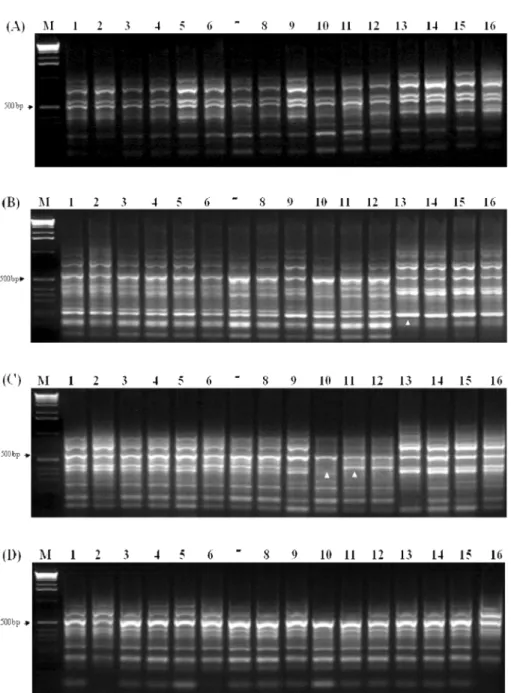
Fig. 1. DNA patterns in
Brucella canisisolates by infrequent restriction site-polymerase chain reaction using primer PsalA (A), PsalC (B), PsalG (C), and PsalT (D). The arrows indicates a different size DNA. Lane 1, JN-GJ2; lane 2, KG-GJ5;
lane 3, KG-YJ29; lane 4, KG-PC56; lane 5, KG-PC87; lane 6, KG-HS2; lane 7, KG-HS11; lane 8, KG-HS20; lane 9,
KG-GJ14; lane 10, KG-GJ56; lane 11, GP-PH38; lane 12, JP-GJ29; lane 13, GP-KW2; lane 14, GP-KW1; lane 15, GP-
KW3; lane 16, GP-KW5; M, 1 kb marker.
In the serological examination for dog populations, B. canis antibodies by both 2-ME RSAT and ICA were detected in only 14.1% of kennel dogs. There were no seropositive cases in indoor dogs and stray dogs. In the previous studies in Korea, the prevalence of canine brucellosis in Seoul was 15.5% [20]. Moon et al . [26]
reported that 33 of 62 dogs in a large kennel in Chonnam area were seropositive and 20 B. canis were isolated. More recently, Park and Oh [27] reported that 82 (42.1%) of 195 kennel dogs were positive for canine brucellosis by both bacteriological and serological tests. In a kennel environment, an aborting female is at high risk for the spread of infection, because vaginal discharge of infective uterine secretions persists for 4~6 weeks following abortion [16]. Likewise, most outbreaks of canine brucellosis in Korea have been reported in kennels, whereas indoor dogs and stray dogs have not been examined for canine brucellosis because of the difficulty to get samples. However, Lovejoy et al . [22] and Fredrickson and Barton [12]
reported that a study in stray dogs in Tennessee demonstrated greater than three-fold rate of infection versus non-stray dogs. Boebel et al . [3], Brown et al . [4], Thierrmann [30] and Wooley et al . [35] also reported an outbreak of brucellosis in stray dogs. In this study we could not confirm the infection in stray
dogs and indoor dogs. However, our study suggested that stray dogs and indoor dogs do not seem to infect the same gender, but these dogs might be infected from kennels. Therefore, continuous survey is needed to monitor the prevalence of canine brucellosis.
In this report only 4 dogs in the kennel group were found to be positive for B. canis isolation. A positive blood culture is the definitive diagnosis for the suspected animal [10, 14, 17, 21]. Dogs were often bacteremia at 2-4 weeks after oral or nasal exposure and became carrier for the next 1-3 years. However, many dog owners have merely using antibiotics for therapy of the dermatosis, respiratory signs, or other diseases without recognizing canine brucellosis. Direct culture is difficult because B. canis is a fastidious organism and therefore, presence of the bacteria is usually overlooked in the clinical samples. Also, the bacteria can not be cultured from clinical samples, if the animal has received previous antibiotic therapy [16].
Brucella is a highly homogenous bacteria with more than 90% DNA homology for all species in DNA- DNA hybridization [32, 33]. Cloeckaert et al. [25]
analyzed the DNA polymorphism of Brucella isolates from marine mammal by the IRS-PCR, and suggested the existence of two new Brucella spp. In this study, 16 B. canis isolates were divided into types I, II, III Table 3. Patterns and types of DNAs determined in 16
Brucella canisisolates by infrequent restriction site-polymerase
chain reaction
No. Strain Year
isolated Geographic
origin PsalA IRS-PCR PsalC
*pattern PsalG PsalT Types
1 JN-GJ2 1994 Gwangju A1 C1 G1 T1 I
2 KG-GJ5 2002 Gwangju A1 C1 G1 T1 I
3 KG-YJ29 2002 Yangju A1 C1 G1 T1 I
4 KG-PC56 2003 Pochun A1 C1 G1 T1 I
5 KG-PC87 2003 Pochun A1 C1 G1 T1 I
6 KG-HS2 2003 Hwasung A1 C1 G1 T1 I
7 KG-HS11 2003 Hwasung A1 C1 G1 T1 I
8 KG-HS20 2003 Hwasung A1 C1 G1 T1 I
9 KG-GJ14 2003 Gwangju A1 C1 G1 T1 I
10 KG-GJ56 2003 Gwangju A1 C1 G2 T1 II
11 GP-PH38 2003 Pohang A1 C1 G3 T1 III
12 JP-GJ29 2003 Gimje A1 C1 G3 T1 III
13 GP-KW2 2005 Gunwi A1 C2 G1 T1 IV
14 GP-KW1 2005 Gunwi A1 C2 G1 T1 IV
15 GP-KW3 2005 Gunwi A1 C2 G1 T1 IV
16 GP-KW5 2005 Gunwi A1 C2 G1 T1 IV
*
IRS-PCR, infrequent restriction site-polymerase chain reaction.
and IV and 4 isolates from 2005 were differentiated to type IV. It was, therefore, suggested that IRS-PCR is possibility as potential tool for molecular epidemiological studies on the B. canis.
References
1. Altuglu I, Zeytino lu A, Bilgic A, Kamcioglu S, Karakartal G, Smits H. Evaluation of Brucella dipstick assay for the diagnosis of acute brucellosis.
Diagn Microbiol Infect Dis 2002, 44 , 241-243.
2. Blankenship RM, Sanford JP.
Brucella canis. A cause of undulant fever. Am J Med 1975, 59 , 424-426.
3. Boebel FW, Ehrenford FA, Brown GM, Angus RD, Thoen CO. Agglutinins to
Brucella canisin stray dogs from certain counties in Illinois and Wisconsin. J Am Vet Med Assoc 1979, 175 , 276-277.
4. Brown J, Blue JL, Wooley RE, Dreesen DW.
Brucella canis
infectivity rates in stray and pet dog populations. Am J Public Health 1976, 66 , 889-891.
5. Carmichael LE, Joubert JC. A rapid slide agglutination test for the serodiagnosis of
Brucellacanis
infection that employs a variant (M-) organism as antigen. Cornell Vet 1987, 77 , 3-12.
6. Carmichael LE, Zoha SJ, Flores-Castro R. Problems in the serodiagnosis of canine brucellosis: dog responses to cell wall and internal antigens of
Brucella canis. Dev Biol Stand 1984, 56 , 371-383.
7. Casao MA, Smits HL, Navarro E, Solera J. Clinical utility of a dipstick assay in patients with brucellosis:
correlation with the period of evolution of the disease.
Clin Microbiol Infect 2003, 9 , 301-305.
8. Choi TY, Kang JO. Application of infrequent- restriction-site amplification for genotyping of Mycobacterium tuberculosis and non-tuberculous mycobacterium. J Korean Med Sci 2002, 17 , 593-598.
9. Cloeckaert A, Grayon M, Grépinet O, Boumedine KS. Classification of Brucella strains isolated from marine mammals by infrequent restriction site-PCR and development of specific PCR identification tests.
Microbes Infect 2003, 5 , 593-602.
10. Currier RW, Raithel WF, Martin RJ, Potter ME.
Canine brucellosis. J Am Vet Med Assoc 1982, 180 , 132-133.
11. Fox KF, Fox A, Nagpal M, Steinberg P, Heroux K.
Identification of Brucella by ribosomal-spacer-region PCR and differentiation of
Brucella canisfrom other
Brucella spp.
pathogenic for humans by carbohydrate profiles. J Clin Microbiol 1998, 36 , 3217-3222.
12. Fredrickson LE, Barton CE. A serologic survey for canine brucellosis in a metropolitan area. J Am Vet Med Assoc 1974, 165 , 987-989.
13. Gaviria-Ruiz MM, Cardona-Castro NM. Evaluation and comparison of different blood culture techniques for bacteriological isolation of Salmonella typhi and
Brucella abortus
. J Clin Microbiol 1995, 33 , 868-871.
14. Greene CE, Carmichael LE. Canine brucellosis. In:
Greene CE (ed.). Infectious Diseases of the Dog and Cat. pp. 369-381, Saunders, Philadelphia, 2006.
15. Hoff GL, Nichols JB. Canine brucellosis in Florida:
serologic survey of pound dogs, animal shelter workers and veterinarians. Am J Epidemiol 1974, 100 , 35-39.
16. Hollett RB. Canine brucellosis: outbreaks and compliance. Theriogenology 2006, 66 , 575-587.
17. Jones RL, Emerson JK. Canine brucellosis in a commercial breeding kennel. J Am Vet Med Assoc 1984, 184 , 834-835.
18. Kim JW, Lee YJ, Han MY, Bae DH, Jung SC, Oh JS, Ha GW, Cho BK. Evaluation of immunochro- matographic assay for serodiagnosis of
Brucella canis. J Vet Med Sci 2007, 69 , 1103-1107.
19. Kim S, Lee DS, Suzuki H, Watarai M. Detection of
Brucella canis
and
Leptospira interrogansin canine semen by multiplex nested PCR. J Vet Med Sci 2006, 68 , 615-618.
20. Lee YS, Mah JS. Serological survey of canine brucellosis in Seoul area. Seoul Univ J Vet Sci 1982, 7 , 101-106
21. Lewis GE. A serological survey of 650 dogs to detect titers for
Brucella canis(
Brucella suis, type 5). J Am Anim Hosp Assoc 1972, 8 , 102-107.
22. Lovejoy GS, Carver HD, Moseley IK, Hicks M.
Serosurvey of dogs for
Brucella canisinfection in Memphis, Tennessee. Am J Public Health 1976, 66 , 175-176.
23. López G, Ayala SM, Escobar GI, Lucero NE. Use of
Brucella canisantigen for detection of ovine serum antibodies against
Brucella ovis. Vet Microbiol 2005, 105 , 181-187.
24. Lucero NE, Escobar GI, Ayala SM, Jacob N.
Diagnosis of human brucellosis caused by
Brucella canis. J Med Microbiol 2005, 54 , 457-461.
25. Mazurek GH, Reddy V, Marston BJ, Haas WH,
Crawford JT. DNA fingerprinting by infrequent-
g
orestriction-site amplification. J Clin Microbiol 1996, 34 , 2386-2390.
26. Moon JS, Oh GS, Park IC, Kang BK, Lee CY, Jung SC, Park YH, Shin SJ. Occurance of canine brucellosis in a large kennel in Chonnam area. Korean J Vet Res 1999, 39 , 1099-1105.
27. Park CK, Oh JY. Bacteriological and serological investigation of
Brucella canisinfection of dogs in Taegu city, Korea. Korean J Vet Res 2001, 41 , 67-71.
28. Piampiano P, McLeary M, Young LW, Janner D.
Brucellosis: unusual presentations in two adolescent boys. Pediatr Radiol 2000, 30 , 355-357.
29. Smits HL, Abdoel TH, Solera J, Clavijo E, Diaz R.
Immunochromatographic
Brucella-specific immunoglobulin M and G lateral flow assays for rapid serodiagnosis of human brucellosis. Clin Diagn Lab Immunol 2003, 10 , 1141-1146.
30. Thiermann AB. Brucellosis in stray dogs in Detroit.
J Am Vet Med Assoc 1980, 177 , 1216-1217.
31. Tosi MF, Nelson TJ.
Brucella canisinfection in a 17- month-old child successfully treated with moxalactam.
J Pediatr 1982, 101 , 725-727.
32. Verger JM, Grimont F, Grimont PAD, Grayon M.
Brucella