486
pISSN 1738-1843 eISSN 2092-8920
© 2013 The Korean Society of Pathologists/The Korean Society for Cytopathology This is an Open Access article distributed under the terms of the Creative Commons Attribution Non-Commercial License (http://creativecommons.org/licenses/
by-nc/3.0) which permits unrestricted non-commercial use, distribution, and reproduction in any medium, provided the original work is properly cited.
Malignant rhabdoid tumors (MRTs) are rare high-grade ma- lignancies in the renal or extrarenal organs. MRTs of the extra- renal organs mostly affect the liver, central nervous system, pel- vis, soft tissue,
1and intra-abdominal cavity.
2Most MRTs occur in infants and young children,
3-8although adult onset MRTs do occur.
9Here, we report a case of primary MRT arising in the liver of a 50-year-old male.
CASE REPORT
A 50-year-old male had admitted for further evaluation of large hepatic mass. The hepatic mass was found accidently in ultrasonography. His major symptom was a 7-kg weight loss in one month. No gastrointestinal symptoms were present. He had no history of hypertension or diabetes. The serologic mark- ers for hepatitis were negative. His mother had died due to cholangiocarcinoma. On physical examination of the patient, a spider angioma was detected. In the laboratory tests, aspartate aminotransferase/alanine aminotransferase/alkaline phosphatase were increased at 148/54/1,215 U/L, alpha-fetoprotein (AFP) 166 ng/mL, and carcinoembryonic antigen (CEA) 8.19 ng/mL, but cancer antigen 19-9 was in the normal range at 26.6 U/mL.
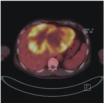
Computerized tomography (CT) showed a very large multinod- ular hypoattenuating mass with rim enhancement in the whole left lobe and anterior segment of the right lobe (Fig. 1). Also the common hepatic lymph nodes were increased in size. There was no abnormality in each right of left kidney. On positron emission tomography-CT, a large hypermetabolic mass was
seen, encompassing the whole left lobe and the anterior segment of the right lobe (Fig. 2).
Pathologic findings
For diagnostic confirmation, CT-guided needle biopsy was performed. The biopsy cores were composed of hypercellular ar- eas alternating with necrosis and hemorrhage. The cellular le- sion showed loosely cohesive tumor cell clusters separated by intermediate fibrous septa. The large round tumor cells showed abundant eosinophilic cytoplasm and eccentric vesicular nuclei with prominent nucleoli (Fig. 3A). Abnormal mitoses were fre- quent. Mucicarmine staining was negative for abundant cyto- plasm. The immunohistochemical stains showed diffuse cyto- plasmic-positive staining for cytokeratin (CK) 19 and vimentin (Fig. 3B, C) and focal positivity for epithelial membrane anti- gen and CK7. The immunohistochemical stains for hepatocyte antigen, alpha-fetoprotein, CK20, CD34, CD117, actin, des- min, S100, and human melanoma black 45 (HMB45) showed no reaction in tumor cells. Also the tumor cells showed partial loss of positivity for integrase interactor-1 (INI-1) (Fig. 3D).
DISCUSSION
MRT is a rare aggressive renal or extrarenal tumor arising in infants and young children. Since Beckwith and Palmer
10first described MRT, a few primary or metastatic liver MRTs have been reported in children.
1,4-8Even after that, primary MRT in the adult liver is very rare. Only one MRT in a young adult has been presented in the liver.
9Generally, primary liver MRTs present as a single voluminous liver mass with no other specific clinical symptoms. On serolog- ic tests, AFP and CEA did not increase in most hepatic MRT patients. Due to voluminous hepatic mass formation and no in- creased tumor markers such as AFP and CEA, primary liver
A Primary Malignant Rhabdoid Tumor in Adult Liver
Yu Na Kang · Sang Pyo Kim · Byoung Kuk Jang
1Departments of Pathology and 1Internal Medicine, Keimyung University School of Medicine, Daegu, Korea The Korean Journal of Pathology 2013; 47: 486-488
http://dx.doi.org/10.4132/KoreanJPathol.2013.47.5.486 ▒ BRIEF CASE REPORT ▒
Corresponding Author Yu Na Kang, M.D.
Department of Pathology, Keimyung University School of Medicine, 1095 Dalgubeol-daero, Dalseo-gu, Daegu 704-701, Korea
Tel: +82-53-580-3814, Fax: +82-53-580-3823, E-mail: [email protected] Received: January 22, 2013 Revised: March 25, 2013
Accepted: March 27, 2013
http://www.koreanjpathol.org http://dx.doi.org/10.4132/KoreanJPathol.2013.47.5.486
A Primary Malignant Rhabdoid Tumor in Adult Liver • 487
Fig. 1. Computed tomography shows a very large hypoattenuating
hepatic mass with rim enhancement in the left lobe of the liver. Fig. 2. Positron emission tomography shows a very large hyper- metabolic hepatic mass.
A B
C D
Fig. 3. Loosely cohesive eosinophilic tumor cells with eccentric nuclei (A) show diffuse positive reaction for cytokeratin 19 (B) and vimentin (C) and loss of reactivity in integrase interactor-1 (D).
http://www.koreanjpathol.org http://dx.doi.org/10.4132/KoreanJPathol.2013.47.5.486 488 • Kang YN, et al.
MRTs should be differentiated from hepatoblastoma, hepatocel- lular carcinoma, and Ewing’s tumor in children, and mass- forming cholangiocarcinoma in adults.
Histologically, loosely cohesive tumor cells had abundant eo- sinophilic cytoplasm and eccentric vesicular nuclei with promi- nent nucleoli. These features are characteristic in renal/extrarenal rhabdoid tumors. Ultrastructurally, intracytoplasmic filaments were detected. Immunohistochemically, almost all MRTs show dual positivity for CK and vimentin but a negative reaction for alpha-fetoprotein, CD34, CD117, actin, desmin, S100, and HMB45. Loss of INI-1 protein nuclear expression in MRTs is known to be related to a mutation of the hSNF5/INI1 gene. We conclude that the characteristic histology showing a rhabdoid feature and immunohistochemistry indicating loss of INI-1 pro- tein nuclear expression are essential for the diagnosis of MRT.
Also, it seems to be significant that this case is the first primary liver MRT arising in an elderly adult among the reports of ex- trarenal hepatic MRTs.
Conflicts of Interest
No potential conflict of interest relevant to this article was reported.
REFERENCES
1. Katzenstein HM, Kletzel M, Reynolds M, Superina R, Gonzalez- Crussi F. Metastatic malignant rhabdoid tumor of the liver treated
with tandem high-dose therapy and autologous peripheral blood stem cell rescue. Med Pediatr Oncol 2003; 40: 199-201.
2. Lee SY, Kim DC, Rha SH, et al. Extrarenal malignant rhabdoid tu- mor: a case report. Korean J Cytopathol 1996; 7: 69-74.
3. Abe T, Oguma E, Nozawa K, et al. Malignant rhabdoid tumor of the liver: a case report with US and CT manifestation. Jpn J Radiol 2009; 27: 462-5.
4. Clairotte A, Ringenbach F, Laithier V, Aubert D, Kantelip B. Malig- nant rhabdoid tumor of the liver with spontaneous rupture: a case report. Ann Pathol 2006; 26: 122-5.
5. Donner LR, Rao A, Truss LM, Dobin SM. Translocation (8;13) (q24.2;
q33) in a malignant rhabdoid tumor of the liver. Cancer Genet Cy- togenet 2000; 116: 153-7.
6. Hunt SJ, Anderson WD. Malignant rhabdoid tumor of the liver: a distinct clinicopathologic entity. Am J Clin Pathol 1990; 94: 645-8.
7. Pogacnik A, Zidar N. Malignant rhabdoid tumor of the liver diag- nosed by fine needle aspiration cytology: a case report. Acta Cytol 1997; 41: 539-43.
8. Ravindra KV, Cullinane C, Lewis IJ, Squire BR, Stringer MD. Long- term survival after spontaneous rupture of a malignant rhabdoid tumor of the liver. J Pediatr Surg 2002; 37: 1488-90.
9. Sibileau E, Moroch J, Teyssedou C, Aubé C. Malignant rhabdoid tu- mors of the liver: an exceptional tumor in adults: a case report and literature review. Eur J Gastroenterol Hepatol 2011; 23: 104-8.
10. Beckwith JB, Palmer NF. Histopathology and prognosis of Wilms tumors: results from the First National Wilms’ Tumor Study. Cancer 1978; 41: 1937-48.