토후박 추출물의 항산화 및 항당뇨 활성
허명록*·JianHe Hu*·왕 란**·김현삼**·김성무**·조동하**,***†
*하남과학기술학원, **강원대학교 BT특성화대학, ***강원대학교 생명공학연구소
Antioxidant and Anti-diabetes Activity of Extracts from Machilus thunbergii S. et Z.
Ming Lu Xu*, Jian He Hu*, Lan Wang**, Hyun Sam Kim**, Cheng Wu Jin** and Dong Ha Cho**,***†
*Henan Institute of Science and Technology, Xinxiang 453-003, PR.China.
**School of Biotechnology, Kangwon National University, Chuncheon 200-701, Korea.
***Institute of Bioscience & Biotechnology, Kangwon National University, Chuncheon 200-701, Korea.
ABSTRACT: Machilus thunbergii was an important medicinal resource and distributed widely in China. In this study, the bioactivities of dichloromethane fraction (DF) and water fraction (AF) from Machilus thunbergii methanol extract were investigated. Total phenolic contents of DF and AF were 57.90 ㎎ Gal/g and 189.92 ㎎ Gal/g, and total flavonoid contents were 17.34 ㎎ Que/g and 58.38 ㎎ Que/g respectively. The EC
50for DPPH radical scavenging activity of DF and AF were 24.37 ㎍ / ㎖ and 2.10 ㎍ / ㎖ . Reducing power and hydroxyl radical scavenging activities of AF were higher than those of DF.
The α -glucosidase inhibitory activity of AF (IC
50= 1.13 ㎍ / ㎖ ) was higher than that of DF (IC
50= 5.34 ㎍ / ㎖ ). The cell via- bility was showed that only the DF had anti-proliferation effect on human cancer cell HT-29. These results suggested that both the DF and AF extract of Machilus thunbergii were potential materials for anti-diabetes and functional food for their radical scavenging activity.
Key Words : Machilus thunbergii, Antioxidant, α-glucosidase, Anti-diabetes
서 언
후박나무 ( Machilus thunbergii ) 는 녹나무과 (Lauracecae) 에 속하며 , 그 수피를 토후박 ( 厚朴 ) 또는 홍남피라고 하는데 , 주 로 남부지역에 자생 또는 재배되고 있다 . 후박나무의 약효로 는 복통 , 천해 , 흉복부팽만의 치료제나 정장 , 이뇨 , 건위 , 소담 등으로 알려져 있으며 , 껍질에서는 미백효과가 있는 machilin A, meso-monomethyl dihydroguaiaretic acid, nectandrin A 와 nectandrin B 같은 lignan 계 화합물 (Li et al. , 2003) 및 sesquiterpene 이 보고되어 있으며 , 잎에서는 quercetin, trifolin
및 quercitrin 등의 flavonoid (Park et al. , 1990) 류가 있다 .
한방에서는 나무껍질을 후박피라 하며 최근에는 약리작용 연 구가 많이 진행되어 수십 가지의 효능이 알려졌다 . 토후박에서 분리된 meso-dihydroguaia-retic acid (MDGA), licarin A 는
glutamate 유도에 의해 신경독성을 억제하는 신경보호활성을 연
구한 결과에서 이 두 물질은 항산화 활성을 통하여 배양한 신 경세포를 뚜렷하게 보호한다고 보고되어 있다 (Ma et al ., 2005) 와 CH
2Cl
2fraction 에서 분리된 (+)-9'-hydroxygalbelgin, isogalcatinB,(7S,8S,8'R)-3',4'-dime-thoxy-3,4-methylenedioxylignan-
7-ol, 1-hydroxy-7-hydroxymethyl-6-methoxyxantho-ne, 5,7- dimethoxy-3',4'- methylenedioxyflavan-3-ol, (+)-(3S,4S,6R)-3,6- dihydroxy-piperitone, protocatechuic acid methyl ester 와
tyrosol 들도 신경보호활성을 연구한 실험에서 rat 의 피질세포 배양을 통하여 신경독성 억제 율을 측정하였을 때 MK-801 과 비교할만한 활성을 나타내었으며 농도 범위는 0.1 µM 에서
10.0 µM 로 나타났다고 보고되어 있으며 (Ma et al ., 2009),
또 Machilin A 는 Melanoma B-16 cell 에서 melanine 의 생성
을 억제한다고 보고되어 있어 미백효과에도 가능성이 있다고 알려져 있다 (Li et al. , 2004).
당뇨병은 고혈당을 특징으로 하는 일련의 대사 질환군으로 ,
만성적인 고혈당은 대혈관 합병증 , 미세혈관합병증 , 당뇨병성 신경병증 및 신장질환과 같은 합병증을 야기한다 (Defronzo, 1981; Steiner et al. , 1984; Young and Stout, 1987). 이에 당뇨병에 대한 치료목표는 지속적으로 이상적인 혈당을 유지 하여 당뇨병성 합병증을 예방하고 지연하는 것이다 . 또한 공 복혈당뿐 아니라 식후혈당을 가능한 정상치에 가깝게 조절하 는 것은 당뇨병의 치료 및 예방에 있어서 매우 중요하다
(Jenkins et al. , 1988; Haller, 1998; Lebovitz, 1998).
†Corresponding author: (Phone) +82-33-250-6475 (E-mail) [email protected]
Received 2009 December 23 / 1st Revised 2010 January 22 / 2nd Revised 2010 February 8 / Accepted 2010 February 11
α -glucosidase 는 식이 중에 함유된 탄수화물의 소화과정 마
지막 단계를 촉매하여 포도당으로 전환시키는 효소로서 , α -
glucosidase 저해제는 탄수화물의 소화와 흡수를 지연시켜 식
후 혈당증가를 완화시킨다 . α -glucosidase 저해제는 고인슐린 혈증이나 저혈당을 유발하지 않고 , 인슐린 분비를 촉진시키며 소장에서 글루카곤 분비를 억제하는 glucagon-like peptide-1 의 분비를 촉진한다 (Mooradian and Thurman, 1999; Baron, 1998). 현재 acarbose 와 voglibose 등의 α -glucosidase 저해제
가 시판되고 있으나 이들 약제를 장기간 복용한 경우 일부환 자에 있어서 복부팽만감 , 구토 , 설사 등 부작용을 나타낼 수 있어 그 사용이 제한될 수 있다 (Hanefeld, 1998). 따라서 이 런 부작용을 줄이고 식후의 혈당강하 효과를 낼 수 있는 대체 식품 및 천연물에 대한 관심이 모아지고 있다 .
따라서 본 연구에서는 항산화효과가 뛰어난 토후박 추출물 이 인류건강과 의약품 , 식품 등 여러 분야의 천연항산화제로
의 가능성과 항암활성의 가능성을 알아 보고 , 항당뇨의 기능
성 식품 , 약품으로서의 활용에 있어 기초자료로 사용하고자 본 실험을 수행하였다 .
재료 및 방법
1. 실험재료 및 추출방법
본 실험에 사용한 토후박나무 껍질은 중국 강소성에서 채집 한 것으로 음건된 2 ㎏을 이용하여 MeOH 로 60 ℃에서 24 시 간씩 세 번 추출하고 농축하여 MeOH 추출물 238.5 g 을 얻었 다 . 상기 추출물을 물로 녹인 후 Hexane, CH
2Cl
2, BuOH 등 용매극성 순으로 세 번씩 분획하고 용매별 분획을 합병하고 농축하여 Hexane (23.7 g), CH
2Cl
2(71.0 g), BuOH (56.2 g)
및 H
2O (96.9 g) 분획을 얻었다 . 상기분획들 중 항당뇨활성이
있는 CH
2Cl
2fraction (DF) 과 H
2O fraction (AF) 을 항산화활
성 실험과 병행하여 본 연구를 진행하였다 . 생리 활성 실험에 서는 모든 샘플과 부동한 농도에서 3 반복으로 실험하였으며 ,
통계 프로그램은 SPSS 11.5 에서 one-way ANOVA 중 Duncan’s multiple range test 를 사용하여 분석하였다 .
2. 총페놀성 및 총플라보노이드 화합물 함량측정
총페놀성 화합물의 함량 측정은 Folin-Denis (Jayaprakasha
et al. , 2007) 법에 의해 정량하였다 . 먼저 1.0 ㎎ / ㎖로 조제한 추출물 1 ㎖에 Folin-reagent 1 ㎖를 가하여 3 분간 정치한 후
10% Na
2CO
31 ㎖을 혼합하고 1 시간 실온에서 방치한 후 725
㎚ 에서 흡광도 (Optizen 2120UV, MECASYS CO. LTD, Daejeon KOREA) 를 측정하였다 . 표준곡선은 gallic acid (Sigma Co., USA) 를 이용하여 작성하였다 . 총 폴리페놀 함량
은 아래의 식에 의해 계산하였다 .
Absorbance = 0.0069 ㎎ gallic acid − 0.042 (R
2= 0.9982)
총플라보노이드 함량은 Davis (Chang et al. , 2002) 변법을
이용하였다 . 1.0 ㎎ / ㎖ 로 조제한 추출물 1 ㎖ 에 diethylene glycol 10 ㎖ 및 1 N NaOH 1 ㎖을 가하고 잘 혼합한 후 30
℃에서 1 시간 반응시킨 후 420 ㎚에서 흡광도를 측정하였다 .
이때 검량곡선은 quercetin (Sigma Co., USA) 을 이용하여 작 성하였으며 . 총플라보이드 함량은 아래의 식에 의해 계산하였다 .
Absorbance = 0.0061 ㎎ quercetin − 0.0281 (R
2= 0.9997)
3. DPPH radical 소거활성 측정
DPPH radical 소거 실험은 광범위하게 쓰이는 간단하고 편
리한 항산화 검색법으로 , 특히 phenol 과 aromatic amine 화합
물의 항산화능 측정에 많이 사용된다 (Blois, 1958).
시료를 10, 50, 100, 500, 1000 ㎍ / ㎖의 농도로 준비한 추 출물 1 ㎖에 0.2 mM DPPH 용액 1 ㎖을 잘 혼합하여 25 분간 실온에서 방치하고 multiplate spectrophotometer (ELx800TM, Biotek, USA) 를 사용하여 515 ㎚에서 흡광도를 측정 (Eom et al. , 2007) 하고 아래와 같이 계산하여 EC
50값으로 나타내었다 . Scavenging effect (%) = [1 − (A
sample– A
blank) / A
control] × 100%
L-Ascorbic acid 를 positive 대조군으로 사용하였다 . EC
50(/)
는 DPPH 라디칼 소거활성이 50% 나타내는 추출물의 농도를
나타내는 것이다 .
4. 환원력의 측정
즉 각각의 농도별로 조제한 시료 0.1 ㎖에 0.2 M 인산 완 충액 (pH 6.8) 0.25 ㎖ 과 1% potassium ferrcyanide [K
3F
3(CN)
6] 0.25 ㎖을 넣은 다음 , 50 ℃에서 20 분간 반응시킨다 . 반
응 후 0.25 ㎖의 10% trichloroacetic acid 를 첨가하고 1000
rpm 10 분간의 원심분리를 통하여 얻어진 상층 액에 0.1% 의
FeCl
30.05 ㎖을 넣어서 발색반응을 유도시킨 다음 , multiplate spectrophotometer (ELx800TM, BioTek, USA) 를 사용하여
750 ㎚에서 흡광도를 측정하였다 (Goh et al. , 2009).
5. •OH 소거능 측정
Hydroxyl radical 소거능 측정은 Fenton 반응으로 생성된
•OH 에 의해 핵산의 구성당인 deoxyriboserk 분해되는 정도를
TBA 발색법을 이용하여 축정한다 . 시험관에 각 추출물 0.2 ㎖ 에 10 mM FeSO
4/EDTA 용액 0.2 ㎖ , 10 mM 2-deoxyribose 0.2 ㎖ , 0.1 M phosphate buffer (pH 7.4) 1 ㎖를 잘 혼합한 후 , 10 mM H
2O
20.2 ㎖를 가하고 37 ℃에서 4 시간 반응시킨
후 2.8% TCA (trichloroacetic acid) 용액 1 ㎖과 0.1% TBA
(Thiobarbituric acid) 를 첨가하여 100 ℃에서 10 분간 가열한 후
급속 냉각시켜 532 ㎚ 에서 UV-visible spectrophotometer
(Optizen 2120UV, Mecasys, Korea) 에서 흡광도를 측정하였다 .
6. 세포독성 및 항암활성 측정
토후박나무껍질 MeOH 추출물의 각 용매분획의 암세포 및
정상세포의 증식 억제효과는 Green et al. (1984) 등의 방법에 따라 MTT assay 를 이용하여 시행하였다 . MTT 분석은 세포 단백질 염색을 이용하여 세포증식이나 독성을 측정하는 방법 으로 10% fetal bovine serum 및 각각의 MDA-MB-231 과
HT-29 를 함유하는 RPMI 164 배지와 293 을 함유하는 DMEM
배지를 5 × 10⁵ cell/ ㎖ 농도로 200 ㎕ 씩 각 well 에 첨가하여 72 시간 동안 배양 (37 ℃ , 5% CO
2) 시킨 후 0.2 M 이하의 DMSO 로 녹인 300 ㎍ / ㎖의 시료를 첨가하여 48 시간 동안 다 시 배양하였다 . 그 후 상등액을 aspirator 로 조심스럽게 제거하 고 새로운 배지와 MTT 용액을 50 ㎖ 씩 첨가해 4 시간 동안 염색시켰다 . 결합되지 않은 MTT 염색액은 네 번 정도 헹구어 ,
건조시킨 후 DMSO 150 ㎕로 염색제를 녹인 후 550 ㎚에서
흡광도 (ELx800 Absorbance Microplate Reader, Biotek, USA) 를 측정하였다 .
7. α-glucosidase 저해활성
토후박의 용매별에 따른 추출물을 10, 50, 100, 200 ㎍ / ㎖ 의 농도로 희석하여 0.75 unit/ ㎖ 의 α -glucosidase 효소액
25 ㎕ 를 50 mM potassium phosphate buffer (pH 6.5) 200
㎕에 넣고 혼합한 후 2 mM p-nitrophenyl- α -D-glucopyrano- side 를 가하여 37 ℃에서 30 분간 반응시켰다 . 0.5 M Na
2CO
3를 가하여 반응을 정지시킨 후 405 ㎚에서 흡광도를 측정하여 효 소 반응으로 생성된 nitrophenyl 의 함량을 정량하여 각 추출물 을 처리하지 않은 대조구와 비교하여 효소활성을 계산하였다
(Kim et al ., 1991).
저해율 (%) = (1 − A / B) × 100. A: 시료 첨가구의 흡광도 , B: 시료 무첨가구의 흡광도 . ( 단 , A, B 모두 대조구의 흡광도
를 제외한 수치임 )
결과 및 고찰
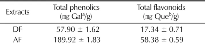
1. 총페놀성 및 총플라보노이드 함량
식물계에 널리 분포되어 있는 페놀성 화합물은 2 차 대사산 물의 하나로 다양한 구조와 분자량을 가진다 . 이들은 phenolic
hydroxyl 기를 가지므로 단백질 및 거대분자들과 쉽게 결합하
여 , 항암 및 항산화 활성과 같은 다양한 생리활성을 나타낸다
(Choi et al ., 2005; Lee and Lee, 1994). 따라서 본 실험에 서는 토후박나무 껍질의 항산화 및 항암활성을 알아보고자 토 후박나무 껍질의 두 가지 fractions 의 총페놀성 및 총플라보노 이드 함량을 측정한 후 그 결과를 Table 1 에 나타내었다 . 토 후박나무 껍질의 DF 와 AF 의 총페놀 함량은 57.90 ㎎ /g 과 189.92 ㎎ /g 으로 나타 내었으며 상기 두 가지 fraction 의 총플
라보노이드 함량은 17.34 ㎎ /g 과 58.38 ㎎ /g 으로 나타났다 .
Park and Lim, (2009) 에 따르면 조릿대 잎 70% 에탄올 조추 출물로부터 얻어진 각 용매별 분획 중 chloroform (155.9 ㎎ / g) 과 ethyl acetate (124.6 ㎎ /g) 및 n-butanol (119.2 ㎎ /g) 분
획은 천연 항산화제로서 특히 , 지질의 과산화를 방지하는 용 도로서의 활용 가치가 있다고 보고하였는데 , 이러한 결과를 토 대로 토후박의 경우 DF 와 AF 가 57.90 ㎎ /g 과 189.92 ㎎ /g 의 총페놀함량을 보이므로 상당한 항산화 활성을 가지고 있음을 확인할 수 있었다 .
2. DPPH radical 소거활성
토후박나무 껍질의 MeOH 추출물의 DF 와 AF 의 DPPH free radical 소거 활성을 측정한 결과는 Table 2 에 나타내었다 .
항산화 활성 중 DPPH radical 에 대한 토후박나무 껍질의 DF
와 AF 두 가지 fractions 의 소거 활성은 EC
5024.37 ±
0.88 ㎍ / ㎖과 2.10 ± 0.11 ㎍ / ㎖로 비교적 높은 항산화 활성을 나타내었다 . 이러한 결과는 토후박나무 껍질 MeOH 추출물이
항산화 활성이 뛰어나 항산화제로의 이용가능성 측면에서도 매우 좋을 것으로 사료된다 . Kwoen et al . (2006) 은 여러 조 건에서 상황버섯을 추출한 후 DPPH radical 을 측정한 후
EC
5017.14 ㎍ / ㎖의 결과 값을 얻어 상황버섯 추출물이 뛰어 난 항산화제임을 제시하였다 . 이와 같은 결과로 미루어 토후 박은 상황버섯보다 더 뛰어난 항산화제로 이용가능성을 확인 할 수 있었다 .
3. 환원력 측정
토후박의 식품 또는 의약품으로의 연구 가치를 측정하는 가
Table 1.Total phenolics and total flavonoid contents of
Machilusthunbergii
extracts.
Extracts Total phenolics
(
㎎Gal
a/g) Total flavonoids (
㎎Que
b/g)
DF 57.90 ± 1.62 17.34 ± 0.71
AF 189.92 ± 1.83 58.38 ± 0.59
aGallic acid (Gal) was used as a standard for measuring of the total phenolic content.
bQuercetin (Que) was used as a standard for measuring of the total flavonoid content.
Note: Values expressed are the mean ± S.D. of three parallel measurements.
Table 2.
DPPH radical scavenging activities of
Machilus thunbergiiextracts.
Sample EC
50(
㎍/
㎖)
DF 24.37 ± 0.88
AF
2.10 ± 0.11
L-Ascorbic acid 12.25 ± 0.39
Note: DF and AF represent CH2Cl2 and H2O fraction from methanol extract of Machilus thunbergii, L-Ascorbic acid is the positive control.장 기초적인 단계로서 , 본 실험에서는 토후박나무 껍질의 메 탄올 추출물로부터 얻은 DF 과 AF 의 환원력을 측정하였다
(Lee et al. , 2004). Fig. 1 에서 나타나는 바와 같이 토후박의 환원력은 100, 500, 1000 ㎍ / ㎖의 농도에서는 DF 는 0.036, 0.260 과 0.476, AF 는 0.357, 1.122 와 1.181 의 흡광도를 보여 농도 의존적으로 환원력을 보임을 확인할 수 있었다 . 그러나 동일 농도에서 항산화제로 널리 쓰이는 BHA (0.129, 0.297, 1.096) 보다 DF 는 약한 환원력을 AF 는 강한 환원력을 나타냄 을 알 수 있었다 .
4. •OH 소거활성 측정
•OH 은 수용액에서 강산 반응성을 나타내어 지질산화를 개 시하고 DNA 에 손상을 주거나 돌연변이를 유발하는 물질로
알려져 있고 생체의 대사과정에서 생성되는 기질의 과산화물 이나 과산화수소가 Fe
2+나 Cu
2+이온의 존재 하에서 생성되며 가장 독성이 강한 자유라디칼로서 소거하는 정도를 측정하였 다 (Bu et al. , 2004). 대조군으로는 항산화제로 알려져 있는
BHT 를 사용하였다 . Fig. 2 에서 보여주는 바와 같이 토후박의
DF 와 AF 분획은 100 ㎍ / ㎖의 농도에서 50% 와 60% 이상의 라디칼 억제 능을 보였으나 , AF 는 오히려 500 과 1000 ㎍ / ㎖ 농도에서는 활성이 큰 변화를 나타내지 않았음을 확인할 수 있었다 .
5. 토후박 추출물의 암세포 증식 억제 효과
항암활성을 측정하는 방법 중 MTT 검색법은 96-well plate
를 사용하며 그 결과를 ELISA reader (Multiwell microplate
reader) 를 이용하여 많은 시료를 간단하게 판독할 수 있어 세
포독성 및 세포증식을 검색하는 법으로서 sulforhodamin B
(SRB) 검색법과 더불어 널리 사용되고 있는 분석 방법이다 . 일
반 세포와 달리 암세포는 대사과정에서 미토콘드리아의 탈수 소 효소 작용에 의해 노란색 수용성 MTT tetraxolium 을 자주 색을 띠는 비수용성의 MTT formazan 으로 환원시킨다 . MTT formazan 의 흡광도는 550 ㎚ 근처 파장에서 최대가 되며 , 대 사적으로 왕성한 세포의 농도를 반영하는 것이다 (Park et al. ,
1987). Fig. 3 과 같이 실험에 사용된 토후박 추출물의 경우
10, 50, 100 ㎍ / ㎖로 조절하였으며 정상세포와 암세포에 성장
에 대한 억제 효과를 검토하였다 . 대조군 5-FU 이 25 ㎍ / ㎖의 농도에서 46.35 ± 0.65% 의 세포 성장 억제력을 나타내었으며 , DF 는 대장암 세포인 HT-29 cell line 에서는 50, 100 ㎍ / ㎖에 서 42.76 ± 1.07% 와 56.35 ± 1.65% 의 억제율을 나타내었으며 ,
유방암 세포인 MDA- MB-231 에서의 샘플은 각각 17.35 ±
1.26% 및 25.48 ± 2.37% 의 성장 억제력을 보였으나 , AF 는 억
제능력을 거의 나타내지 않았다 . 이와 같은 결과는 Kwon et al. (2007) 등 발표한 결과와 비교해 보았을 때 토후박이 뛰어 난 항암 활성도 가지고 있음을 확인할 수 있었다 .
Fig. 1.
Reducing power of
Machilus thunbergiiextracts.
Fig. 2.
Effects of
Machilus thunbergiiextracts on hydroxyl radical (·OH) scavenging assay.
a-b
Values with different letters differ significantly (p < 0.05).
Fig. 3.
Anti-proliferation effect of
Machilus thunbergiiextracts on human cancer cell HT-29.
The final concentration of positive control 5-FU is 25
㎍/
mL.
a-dValues with different uppercase superscripts among the
three concentrations and 5-FU are significantly different
at p < 0.05 by ANOVA with Duncan’s multiple range
test.
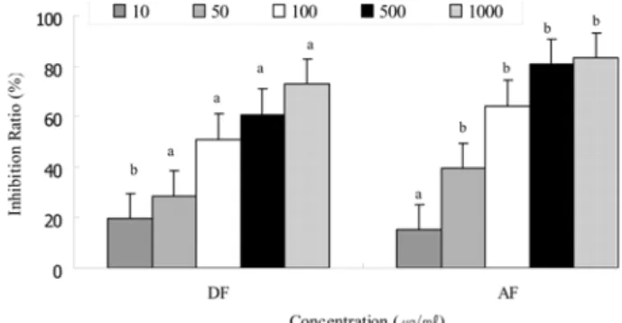
6. α-glucosidase 저해효과
고혈당과 관련된 많은 생화학적 경로들 ( 포도당의 자가산화 , polyol 경로 , 단백질 당화 등 ) 에 의해 자유 라디칼의 생성이 증가됨이 알려졌고 , 그 밖에도 당뇨병에서는 여러 인자들에 의 해 산화 스트레스 및 조직의 산화적 손상이 증가될 수 있다
(Baynes and Thorpe, 1999). 당뇨병에서 혈장 , 적혈구막 , 저밀 도 지단백 등의 지질과산화가 증가됨이 밝혀졌다 (Sato et al. ,
1979). 이런 산화스트레스를 막을 수 있는 기전은 자유 라디칼
의 직접적인 소거 , 지질과산화의 억제 혹은 생체 내 항산화 효 소계 (superoxide dismutase, catalase, glutathione peroxidase)
를 활성화 하는 방법으로 나눌 수 있다 .
당뇨병은 암 및 순환기 질환과 더불어 3 대 질병의 하나로 지 목되고 있다 . α -Glucosidase 는 소장의 brush-border membrane
에 존재하는 소화효소로서 이당류나 다당류 형태의 탄수화물 이 소화 흡수되기 위한 상태인 단당류로 가수분해하는 역할을 한다 . α -Glucosidase 저해제는 탄수화물 식이 후 혈당상승을 억제할 수 있다 .
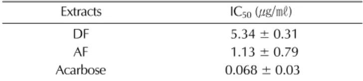
토후박의 DF 와 AF 시료를 농도별로 조제하여 효모 기원의 α -glucosidase 에 대한 저해활성을 검토한 결과 100 ㎍ / ㎖ 농도 에서 DF 와 AF 이 각각 79.97% 와 56.99% 로 비교적 높은 억
제효과를 보였다 . Table 3 에 표시한 바와 같이 양성대조군으
로 사용한 acarbose 의 IC
50은 0.068 ± 0.03 ㎍ / ㎖으로 나타났으
며 , DF 와 AF 의 IC
50은 각각 5.34 ± 0.31 ㎍ / ㎖과 1.13 ± 0.79
㎍ / ㎖으로 나타내었다 . Cho 등 (2007) 은 오미자 추출물에서 α -glucosidase 저해 활성을 측정한 결과 200 ㎍ / ㎖의 농도에서 물 추출물이 97.4%, 60.0%, 에탄올 추출물이 84.5% 의 활성 을 나타낸다고 보고한 바 있다 . 따라서 토후박 추출물이 오미 자의 물 추출물과 에탄올 추출물과 비교하였을 때 더욱 강한 활성을 나타냄은 알 수 있었으며 , 토후박의 두 가지 추출분획
으로 부터 높은 억제 효과를 나타내는 물질 분리 정제를 통해 강력한 α -glucosidase 억제제를 개발 할 수 있다고 판단된다 .
시판되고 있는 acarbose 와 같은 α -glucosidase 저해제는 장 기간 복용할 경우 일부 환자에 있어서 복부팽만감 , 구토 , 설사 등 부작용을 나타낼 수 있어 그 사용이 제한될 수 있다 . 이에 부작용이 적은 천연물로부터 혈당강하제를 찾으려는 연구가 활발히 진행되고 있으며 , 상엽 , 상백피 (Asano et al. , 1994;
Goldman et al. , 1990), 황금 (Nishioka et al. , 1998) 등의 추 출물이 α -glucosidase 저해활성이 높았고 이들 추출물로부터
α -glucosidase 저해제를 분리하기도 하였다 .
본 연구에서도 천연물인 토후박 추출물로부터 α -glucosidase
의 저해활성을 확인함으로써 혈당강하제로서의 토후박 추출물 의 기능성을 검정하였다 . 하지만 , 본 연구에서는 토후박 추출 물이 α -glucosidase 에 대한 높은 저해활성을 나타냄을 밝혔을 뿐이기에 , 앞으로 토후박의 추출물 중 어떤 물질에 의해 탄수 화물 소화효소의 저해활성이 높아졌는지에 대한 추후 연구가 필요하다 .
LITERATURE CITED
Asano N, Tomioka E, Kizu H and Matsui K.
(1994). Sugars nitrogen in the ring isolated from the leaves of Morus bomhycis. Carbohydrate Research. 253:235-245.Baron AD.
(1998). Postprandial hyperglycemia and α-glucosidase inhibitors. Diabetes Research and Clinical Practice. 40:S54-S55.Baynes JW and Thorpe SR.
(1999). Role of oxidative stress in diabetic complications: a new perspective on an old paradigm.Diabetes. 48:1-9.
Blois MS.
(1958). Antioxidant determination by the use of a stable free radical. Nature. 181:1199-1200.Bu HJ, Lee HJ, Yoo ES, Jung DS, Riu KZ and Lee S.
(2004).Antioxidant effects and inhibitory effet on NO synthesis by etracts of Canavalia lineate. Korean Journal of Phramcognosy.
35:338-345.
Chang CC, Yang MH, Wen HM and Chern JC.
(2002).Estimation of total flavonoid content in propolis by two complementary colorimetric methods. Journal of Food and Drug Analysis. 10:178-182.
Choi SY, Lin SH, Ha TY, Kim SR, Kang KS and Hwang IK.
(2005). Evaluation of the estrogenic and antioxidant activity of some edible and medicinal plant. Korean Journal of Food Science and Technology. 37:549-556.
Cho YJ, Ju IS, Kim BC, Lee WS, Kim MJ, Lee BG, An BJ, Kim JH and Kwon OJ.
(2007). Biological activity of Omija (Schizandra chinensis Baillon) extracts. Journal of the Korean Society for Applied Biological Chemistry. 50:198-203.Defronzo RA.
(1981). The effect of insulin on renal sodium metabolism. Diabetologia. 21:165-171.Eom SH, Park HJ, Jin CW, Park SM, Kim MJ, Yu CY and Cho DH.
(2007). Changes of antioxidant activity in Juglans mandshurica Maxim. leaves by far infrared ray inradiation.Korean Journal of Medicinal Crop Science. 15:266-270.
Goh EJ, Seong ES, Lee JG, Na JK, Lim JD, Kim MJ, Kim NY, Lee GH, Seo JS, Cheoi DS, Chung IM and Yu CY.
(2009). Antioxidant activities according to peelling and cultivated years of Astragalus membranaceus roots. Korean Journal of Medicinal Crop Science. 17:233-237.
Goldman A, Milat ML and Ducrot PH.
(1990). Tropane derivatives from Calystegia sepium. Phytochemistry. 29:2125- 2128.Green LM, Reade JL and Ware CF.
(1984). Rapid colometric assay for cell viability: Application to the quantitation of cytotoxic and growth inhibitory lymphokines. Journal ofTable 3. α
-glucosidase inhibitory activity of
Machilus thunbergiiextracts.
Extracts IC
50(
㎍/
㎖)
DF 5.34 ± 0.31
AF 1.13 ± 0.79
Acarbose 0.068 ± 0.03
Immunological Methods. 70:257-268.
Haller H.
(1998). The clinical importance of postprandial glucose.Diabetes Research and Clinical Practice. 40:S43-S49.
Hanefeld M.
(1998). The role of acarbose in the treatment of non- insulin- dependent diabetes mellitus. Journal of Diabetes and its Complications. 12:228-237.Jayaprakasha GK, Negi PS, Jena BS and Rao LJM.
(2007).Antioxidant and antimutagenic activities of Cinnamomum zeylanicum fruit extracts. Journal of Food Composition and Analysis. 20:330-336.
Jenkins DJ, Wolever TM and Jenkins AL.
(1988). Starchy foods and glycemic index. Diabetes Care. 11:149-159.Kim BN, Park HK, Kwon TB and Maeng YS.
(1991). Analysis of rutin contents in buckwheat noodles. Journal of Korean Food Scienceand Nutrition. 7:61-66.Koivisto VA.
(1993). Insulin therapy in type II diabetes. Diabetes Care. 16: 29-39.Kwoen DJ, Youn SJ, Cho JG, Choi UK and Kang SC.
(2006).Antioxidant activities and biological properties of Phellinus linteus extract according to different extraction methods. Journal of the Korean Society for Applied Biological Chemistry. 49:91-
Kwon OW, Kim CH, Kim HS, Kwon MC, Ahn JH, Lee HJ,
96.Kang HY and Lee HY.
(2007). Comparison of immuno modulatary and anticancer activities according to the parts of the Styrax japonica Sieb. et Zucc. Korean Journal of Medicinal Crop Science. 15:170-176.Lebovitz HE.
(1998). Postprandial hyperglycemic state: importance and consequences. Diabetes Research and Clinical Practice.40:S27-S28.
Lee JH and Lee SR.
(1994). Analysis of phenolic substances content in Korean plants foods. Korean Journal of Food Science and Technology. 26:310-316.Lee YR, Kang MY, Koh HJ, Chin JH and Nam SH
. (2004).Screeing of physiological functionality of germainatedgiant embryonic rices. Journal of the Korean Society for Applied Biological Chemistry. 47:216-221.
Li gao, Lee CS, Woo MH, Lee SH, Chang HW and Son JK.
(
2004). Lignans from the bark of Machilus thunbergii and Their DNA topoisomerase I and II inhibition and cytotoxicity.Biological & Pharmaceutical Bulletin. 27:1147-1150.
Li gao, Ju HK, Chang HW, Jahng YD, Lee SH and Son JK.
(
2003). Melanin biosynthesis inhibitors from the bark of Machilus thunbergii. Biological & Pharmaceutical Bulletin.26:1039-1041.
Ma CJ, Kim SR, Kim JW and Kim YC.
(2005). Meso- dihydroguaiaretic acid and licarin A of Machilus thunbergii protect against glutamate-induced toxicity in primary cultures of a rat cortical cells. British Journal of Pharmacology. 146:752-Ma CJ, Kim YC and Sung SH.
759. (2009). Compounds with neuroprotective activity from the medicinal plant Machilus thunbergii. Journal of Enzyme Inhibition and Medicinal Chemistry. 24:1117-1121.Mooradian AD and Thurman JE.
(1999). Drug therapy of postprandial hyperglycemia. Drugs. 57:19-29.Nishioka T, Kawabata J and Aoyama Y.
(1998). Baicalein, an alpha-glucosidase inhibitor from Scutellaria baicalensis. Journal of Natural Products. 11:1413-1415.Park JC, Kim BW and Young HS.
(1990). Further study on the flavonoids from the leaves of Machilus thunbergii in Korea.Korean Journal Pharmacognosy. 21:197.
Park JG, Kramer BS, Steinber SM, Carmichael J, Collins JM, Minna JD and Gazar AF.
(1987). Chemosensitivity testing of human colorectal carcinoma cell lines using a teterazolium- based colorimetric assay. Cancer Research. 47:5875-5897.Park YO and Lim HS.
(2009). Antioxidant activities of bamboo (Sasa Borealis) leaf extract according to extraction solvent.Journal of the Korean Society of Food Science and Nutrition.
38:1640-1648.
Sato Y, Hotta N, Sakamoto N, Matsuoka S, Ohishi N and Yagi K.
(1979). Lipid peroxide level in plasma of diabetic patients.Biochemical Medicine. 21:104-107.
Steiner G, Haynes F and Yoshino G.
(1984). Hyperinsulinemia and in vivo very-low-density lipoprotein triglyceride kinetics.American Journal of Physiology. 246:187-192.