This is an open-access article distributed under the terms of the Creative Commons Attribution Non-Commercial License (http://creativecommons.org/licenses/
by-nc/3.0) which permits unrestricted non-commercial use, distribution, and reproduction in any medium, provided the original work is properly cited.
Laparoscopic Resection of Gastric Submucosal Tumors:
Outcomes of 141 Consecutive Cases in a Single Center
Keesang Yoo, M.D., Hoon Hur, M.D., Cheul Su Byun, M.D., Yi Xian, M.D., Sang-Uk Han, M.D., Yong Kwan Cho, M.D.
Department of Surgery, Ajou University School of Medicine, Suwon, Korea
Purpose: The treatment of choice for gastric submucosal tumors (SMT) is surgical resection. Recent advanced techni- ques has facilitated more extensive application of laparoscopic surgery to most types of resectable gastric SMTs. The aim of this study was to verify the efficacy of laparoscopic resection for treatment of gastric SMT through analysis of outcomes obtained at a single center.
Methods: A total of 141 patients who underwent laparo- scopic resection for treatment of gastric SMT were enrolled between April 2003 and June 2011. Analysis of the demo- graphics, tumor characteristics, and surgical or oncological outcomes of these patients was performed.
Results: Gastrointestinal stromal tumors (GIST) were the most common pathologic findings (90 cases), and the upper third of the stomach was the most common location (70 cas- es). Wedge resections were performed in 128 patients and
major gastrectomies were performed in 13 patients. The mean surgical time was 102 minutes, which was reduced to a stable 70 minutes after the 30th case. The surgical time for tumors located on the posterior or lesser portion of the upper third of the stomach was longer than that for other lesions. Twelve postoperative complications, including two cases of intra-ab- dominal bleeding, one case of marginal ulcer bleeding, and one case of leakage occurred. However, there was no occur- rence of complications after the 70th case. During the follow-up period, two patients suffered recurrent GIST.
Conclusion: Laparoscopic surgery for treatment of gastric SMT is safe and feasible, particularly as the surgeon develops greater skill with increased experience. Laparoscopic resection is useful for treatment of any type of gastric SMT.
Key words: Submucosal tumor, Laparoscopy, Gastric neoplasm
Received September 13, 2012, Revised September 28, 2012, Accepted October 5, 2012
※ Corresponding author:Hoon Hur
Department of Surgery, Ajou University School of Medicine, San 5, Woncheon-dong, Yeongtong-gu, Suwon 442-721, Korea Tel:+82-31-219-5200, Fax:+82-31-219-5755
E-mail:[email protected]
The major concept of this manuscript was presented at the Korean Gastric Cancer Congress 2011, Pusan, Korea as an oral presentation.
http://dx.doi.org/10.7602/jmis.2012.15.4.106
INTRODUCTION
A submucosal tumor (SMT) in the gastrointestinal tract describes an epithelial or non-epithelial mesenchymal tumor, accounting for 1% or less of all gastrointestinal tumors.1 The stomach is the most common site for SMTs. The rate of detection of asymptomatic gastric SMTs has increased in Asian countries including Japan and Korea because of the increasing use of screening gastrofiberscopy for detection of gastric cancer.2
The first option for management of a gastric SMT is surgical resection, due to the limited preoperative pathologic diagnosis and the high possibility of malignancy.3 Although the patho- logic result is malignant gastrointestinal stromal tumor (GIST), lymphadenectomy is not usually required because of the low
frequency of lymph node involvement.4 Therefore, wedge re- section of the stomach is possible and often results in complete resection of the gastric SMT without subtotal or total gastrectomy.
This is fortunate as the latter procedures have a significant im- pact on the patient’s postoperative quality of life.5
In recent years, laparoscopic resection of gastric SMTs has been regarded as the most appropriate curative approach. Previously, several reports had suggested limiting the indications for the lapa- roscopic approach to gastric SMTs of 2 cm in diameter, or smaller.6,7 Since several comparative studies have described the efficacy of laparoscopic surgery compared to open surgery for gastric SMTs,4,8,9 the laparoscopic approach has been applied to most types of resectable gastric SMTs.10,11 However, in these reports, the number of enrolled patients was too small to be conclusive. In addition, they did not consider the features of SMTs requiring subtotal or total gastrectomy. Therefore, a clin- ical study involving a large series of patients undergoing lapa- roscopic surgery for gastric SMT can be of significant value and aid in the determination of the safety and effectiveness of the procedure across a broad array of SMTs.
Here, we analyzed prospectively collected data concerning laparoscopic resections of gastric SMTs within a single center.
We aimed to evaluate the feasibility of this procedure through the analysis of surgical and oncologic outcome.
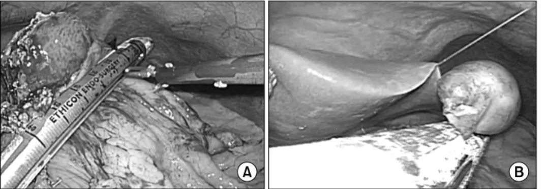
Fig. 1. (A) Laparoscopic resection for endophytic submucosal resection using eversion tech- nique. (B) Laparoscopic re- section for exophytic sub- mucosal tumor using endo- scopic linear staplers.
MATERIALS AND METHODS
Between April 2003 and June 2011, 141 consecutive patients with gastric SMTs underwent laparoscopic surgery in our institution. Indications for surgical treatment of gastric SMTs included, a tumor with a diameter greater than 2 cm; a fine nee- dle aspiration-confirmed gastrointestinal stromal tumor (GIST) of any size; and a clinical GIST suggested by computed tomog- raphy (CT) or endoscopic ultrasonography. Also we resected the mass which cause symptoms or patients want resection un- less the tumor is smaller than 2 cm.
Initially, we applied laparoscopic surgery to gastric SMT of below 5 cm size. However, indication for laparoscopic surgery was extended to 8cm size after surgeon performed the 70th lapa- roscopic surgery for gastric SMT.
Data concerning patient clinicopathologic features, surgical procedures, pathology reports, and follow-up results were retro- spectively collected from the medical records of all patients. In addition, data regarding surgical parameters, including oper- ation times and postoperative complications, were reviewed.
1) Surgical procedures
Two surgeons who had a combined experience of more than 100 laparoscopic surgeries for gastric tumors, including ad- enocarcinomas, performed all of the surgical procedures.
Laparoscopic surgery was performed under general endo- tracheal anesthesia. A 10-mm trocar for the laparoscope was inserted into the infra-umbilical area, and a 12 mm trocar for a flexible linear stapler was inserted, along with 2 or 3 addi- tional trocars, into the upper abdomen after CO2 pneumo- peritoneum was established. Intra-abdominal pressure was maintained at 10∼12 mmHg.
Each abdominal cavity was fully explored and the location of the exophytic tumors was easily identified. For exophytic tu- mors located in posterior wall or lesser curvature of the stom-
ach, the lesser or greater omental fat was dissected to expose the mass. Small endophytic tumors (<2 cm in size) were lo- cated using intraoperative sonography or gastrofiberscopy. To avoid gastric deformity after resection, six endophytic tumors of intermediate size (2∼5 cm) which was not detected at the serosa surface of the stomach were resected by eversion, as suggested by Hyung et al.12 For resection of tumors located in the posterior wall of the stomach, anterior gastrotomy was per- formed (Fig. 1A). After tumors were identified, a flexible linear stapler was inserted through the gastrostomy site and the sta- pling was performed. Most tumors were resected by wedge re- section using 2 or 3 staplers (Fig. 1B). Several intracorporeal sutures were made to reinforce the resection lines to reduce the risk of postoperative bleeding. Laparoscopic gastrectomy was performed for several SMTs located at the gastroesophageal junction or the prepyloric area which can cause narrowing or obstruction of stomach after resection. Anastomoses were per- formed extracorporeally through a 4∼5 cm incision into the epigastric area. Most of the tumors were removed through the trocar site, using a laparoscopic bag. For large tumors (>5 cm), the trocar site was extended 3∼4 cm to extract the tumor from the abdominal cavity.
During operation, tumors were carefully manipulated in order to prevent the tumor rupture. To extract from abdominal cavity, resected specimen was inserted into the laparoscopic bag, and the pull it out without contact of tumor with wound.
2) Perioperative management and outcome measurement
All patients who underwent gastric wedge resection were managed with common clinical pathway. They started sips of water on the second postoperative day, and began on a soft diet on the fourth postoperative day, if they were available. In case of patients who underwent gastrectomy, sips of water was sup- plied on the third postoperative day, and a soft diet on the fifths postoperative day. All patients were discharged once they ex- hibited at least 2 days without specific complaints and had nor-
Table 1. Clinicopathologic and surgical results of 141 enrolled patients
Clinical variables N Percentage
Age (years old)
<65
≥65 Gender
Male Female
Body mass index (kg/m2)
<25
≥25 Location
Upper Middle Lower Circular location
Lesser/Posterior Greater/Anterior Type
Endophytic Mixed Exophytic Size
<2 cm
≥2 cm, <5 cm
≥5 cm Surgical procedure
Wedge resection Distal gastrectomy Proximal gastrectomy Total gastrectomy
111 30 63 78 88 53 70 41 30 72 69 61 35 45 11 89 41 129 9 1 2
78.7 21.3 55.3 44.7 62.4 37.6 49.6 29.1 21.3 51.1 48.9 43.3 24.8 31.9 7.8 63.1 29.1 90.8 7.1 0.7 1.4
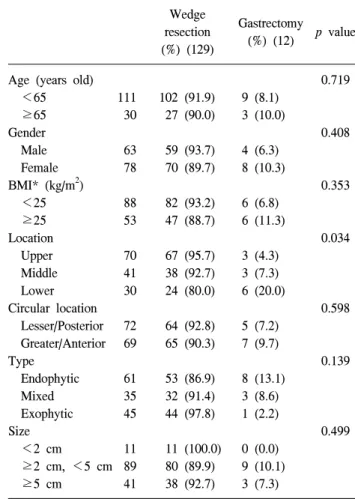
Table 2. Comparison of gastrectomy according to the clinicopathologic features
Wedge resection (%) (129)
Gastrectomy (%) (12) p value
Age (years old)
<65
≥65 Gender Male Female BMI* (kg/m2)
<25
≥25 Location
Upper Middle Lower Circular location
Lesser/Posterior Greater/Anterior Type
Endophytic Mixed Exophytic Size
<2 cm
≥2 cm, <5 cm
≥5 cm
111 30 63 78 88 53 70 41 30 72 69 61 35 45 11 89 41
102 (91.9) 27 (90.0) 59 (93.7) 70 (89.7) 82 (93.2) 47 (88.7) 67 (95.7) 38 (92.7) 24 (80.0) 64 (92.8) 65 (90.3) 53 (86.9) 32 (91.4) 44 (97.8) 11 (100.0) 80 (89.9) 38 (92.7)
9 (8.1) 3 (10.0) 4 (6.3) 8 (10.3) 6 (6.8) 6 (11.3) 3 (4.3) 3 (7.3) 6 (20.0) 5 (7.2) 7 (9.7) 8 (13.1) 3 (8.6) 1 (2.2) 0 (0.0) 9 (10.1) 3 (7.3)
0.719
0.408
0.353
0.034
0.598
0.139
0.499
*Body mass index.
mal clinical status and physical examination after starting soft diet. We decided that postoperative morbidity was occurred when the patient required additional management due to un- usual events.
3) Patient follow-up
Only patients diagnosed with GIST were recommended for follow-up every 6 months to 1 year. Abdominal CT and gastro- fiberscope examinations were performed to evaluate tumor recurrence. Recurrence was diagnosed by further imaging stud- ies or explorative laparotomy.
4) Statistical analysis
Statistical analyses were performed using the Statistical Package for the Social Sciences version 15.0 (SPSS, IBM Corporation, Armonk, NY, USA). Differences in the numbers of major gastrectomies and complication rates were evaluated relative to clinicosurgical features using the chi-squared test.
Operation time, according to the tumor location, was compared using a one-way analysis of variance (ANOVA) test, and a post-hoc test using the Duncan method was performed to make specific comparisons between locations. A p value of less than 0.05 was considered to be statistically significant.
RESULTS
Laparoscopic surgeries for SMT resections were performed on 141 patients without open conversion. Mean age and body mass index of the patients was 53.9±12.4 years old and 24.5±4.3 kg/m2 (mean±S.D.), respectively. Other clinicopatho- logic factors are listed in Table 1. The most common location of the tumors was the upper third of the stomach (70 cases) followed by the middle (41 cases) and lower thirds (30 cases).
More than 90% of masses were larger than 2 cm in diameter, and 41 patients (29.1%) had tumors larger than 5 cm in diameter. Maximum size of tumors was 9 cm and it was re-
Fig. 2. Change in operation time, according to the surgeon’s experi- ence in laparoscopic surgery for gastric submucosal tumors.
Arrow shows the occurrence of postoperative complications.
Fig. 3. Operation time according to tumor locations. There was no significant difference in operation time among the locations, by one-way ANOVA test. However, operation times for gas- tric submucosal tumor located in the posterior wall or lesser curvature of upper portion of the stomach was significantly longer than it was for those in the anterior wall or greater curvature of the stomach. *Means p<0.05 in post-hoc test (Duncan method). PW = posterior wall; LC = lesser curvature;
AW = anterior wall; GC = greater curvature.
Table 3. Clinical features of patients who were undergone laparoscopic gastrectmy for gastric submucosal tumor
Gender/Age Location Type Size (cm) Pathology
1 2 3 4 5 6 7 8 9 10 11 12
Distal gastrectomy Distal gastrectomy Distal gastrectomy Distal gastrectomy Distal gastrectomy Distal gastrectomy Distal gastrectomy Distal gastrectomy Distal gastrectomy Proximal gastrectomy Total gastrectomy Total gastrectomy
F/39 F/75 F/45 F/66 F/32 F/54 M/69 F/42 M/55 M/45 F/45 M/27
Lower Lower Lower Lower Lower Middle Middle Middle Lower Upper Upper Upper
Lesser Greater Posterior Anterior/Greater Posterior Lesser Lesser Greater Greater Greater Lesser/Posterior Lesser/Posterior
Endophytic Mixed Endophytic Endophytic Endophytic Endophytic Endophytic Exophytic Mixed Endophytic Endophytic Mixed
3.7 6 2 2.8 5.5 4.8 3 4.5 2.2 7 4 3.7
Leiomyoma Schwannoma Polyp GIST
Ectopic pancreas GIST
Plasmacytoma Schwannoma Polyp Leiomyoma GIST Leiomyoma GIST = gastrointestinal stromal tumor.
sected by wedge resection. Endophytic masses were found in 61 cases and exophytic masses in 45 cases. GIST was the most common pathologic finding (63.8%); other diagnoses included leiomyoma (14.2%), schwannoma (11%), ectopic pancreas (7%), and polyps (7%).
Among the 141 patients, 129 underwent laparoscopic wedge resections and 12 underwent laparoscopic major gastrectomy.
Nine SMTs, located in the prepyloric antrum, were treated by distal gastrectomy, and 3 gastroesophageal junction tumors were treated by either proximal gastrectomy or total gastrectomy. The characteristics of the tumors resected by wedge resection were compared to those resected by major gastrectomy, by age, sex, body mass index, and tumor size; statistically significant corre- lations were not observed, but gastric SMTs located in the low-
er third of the stomach were more likely to be removed by ma- jor gastrectomy (p=0.034) (Table 2). Tumor rupture or spillage did not occur during any of the procedures. The pathological and surgical features associated with each type of surgery are listed in Table 3.
The mean operation time was 102 minutes, and it was gradu- ally reduced to about 70 minutes, after 30 cases, where it stabilized. However, the surgical time did vary on a case-by-case basis (Fig. 2). A one-way ANOVA test indicated that the oper-
Table 4. Characteristics of patients who had complications No Gender/
Age Resection Location Approach Type Size Complication Treatment
1 2 3 4 5 6 7 8 9 10 11 12
F/45 M/52 M/68 F/40 F/39 M/45 M/65 F/71 F/50 M/67 M/71 M/44
Distal gastrectomy Wedge resection Wedge resection Wedge resection Distal gastrectomy Proximal gastrectomy Wedge resection Wedge resection Wedge resection Wedge resection Distal gastrectomy Wedge resection
Lower Middle Upper Upper Lower Upper Upper Upper Upper Upper Middle Middle
Posterior Lesser Greater Greater Lesser Greater Lesser/Posterior Anterior Lesser Greater Greater Lesser
Endophytic Endophytic Endophytic Endophytic Endophytic Endophytic Exophytic Endophytic Exophytic Exophytic Endophytic Mixed
1.8 4.3 3 3 3.5 3 5 2.2 3.2 1.7 2 1.5
Ulcer bleeding Tonsilitis Bleeding Bleeding Gastric stasis Gastric stasis Leakage Effusion Effusion
Wound hematoma Intraabdominal abscess Wound hematoma
Reoperation
Reoperation
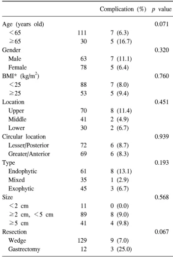
Table 5. Comparison of risk of postoperative complications according to the clinicopathologic features
Complication (%) p value Age (years old)
<65 ≥65 Gender Male Female BMI* (kg/m2) <25 ≥25 Location Upper Middle Lower Circular location Lesser/Posterior Greater/Anterior Type
Endophytic Mixed Exophytic Size <2 cm ≥2 cm, <5 cm ≥5 cm Resection Wedge Gastrectomy
111 30 63 78 88 53 70 41 30 72 69 61 35 45 11 89 41 129 12
7 (6.3) 5 (16.7) 7 (11.1) 5 (6.4) 7 (8.0) 5 (9.4) 8 (11.4) 2 (4.9) 2 (6.7) 6 (8.7) 6 (8.3) 8 (13.1) 1 (2.9) 3 (6.7) 0 (0.0) 8 (9.0) 4 (9.8) 9 (7.0) 3 (25.0)
0.071
0.320
0.760
0.451
0.939
0.193
0.568
0.067
*body mass index.
ation times did not differ significantly relative to the tumor lo- cation (p=0.116). The surgical time for resection of gastric SMTs located in the posterior wall of the lesser curvature of the stomach were relatively longer than they were for tumors of the anterior wall or greater curvature of the stomach (Fig.
3).
The mean time to gas out after surgery was 3.3±2.3 days, and the mean length of hospital stay was 7.1±3.4 days. The discharge of forty seven patients (33.3%) who should be man- aged for postoperative complications and want it to be post- poned was behind the planned schedule.
Operative morbidity was 8.5% (12/141), including 2 cases of intra-abdominal bleeding, 1 case of marginal ulcer bleeding, and 1 case of leakage of resection line. Re-operation was neces- sary only in the 2 cases of bleeding and leakage. There were no mortality cases (Table 4). None among age gender, BMI, location, growth type, size, and type of resection were found to have significant influence on post-gastrectomy complications.
(Table 5), and there were no complications observed after the 70th patient was operated on.
Of the 90 patients diagnosed with GIST, we selected 52 pa- tients who were followed up more than 12 months. At a mean follow-up of 31 months (range, 12.3 to 56.5 months, median:
17.6 months), recurrence was observed in 2 (1.4%) patients, and no local recurrence was observed. Both of these patients had high-risk GIST; 1 had a tumor that was 5.4 cm in diameter and more than 10 mitoses per 50 high-powered fields were ob- served; the other case involved a tumor of 7.5 cm in diameter and more than 15 mitoses per 50 high-powered fields. One case was detected liver metastasis at postoperative 16 months, and the other case peritoneal seeding at postoperative 18 months.
DISCUSSION
To date, many researchers have reported the efficacy of laparo- scopic surgery compared to open surgery for gastric SMTs.4,8,9,13 Laparoscopic surgery for gastric SMTs has been shown to yield pa- tient benefits, including enhancing their postoperative recovery.14,15 This study reports the evaluation of the largest number of pa- tients who underwent laparoscopic surgery for gastric SMTs at a single center. Most of tumors were resected by laparoscopic wedge resection. However, tumors located at the prepyloric junction showed the highest proportion of distal gastrectomies compared to tumors at other locations. The surgical time was variable in spite of sufficient surgeon’s experience, but there were no complications after 70 cases. Results of the long-term follow-up showed that laparoscopic surgery is feasible for gas- tric SMTs.
Although laparoscopic resection has become the treatment of choice for gastric SMTs, not all SMTs are resected using this technique. Some authors have limited the indication of laparo- scopic surgery to only those patients who have a gastric SMT that is less than 5 cm in diameter.13,15,16 This tumor size limi- tation might reflect the technical difficulty of laparoscopic min- imal resection. In the current series, laparoscopic surgery was applied to all tumors below 5 cm in size, but the limitation was increased to 8 cm after the 70th surgery in our institution.
As a result, 41 cases (29.0%) involved tumors ≥5 cm in diameter. All of these laparoscopic surgeries were successfully performed, without open conversion, presumably because of proficiency of the surgeon in the laparoscopic technique. It is possible for surgeons to perform total or subtotal laparoscopic gastrectomy for SMTs that are impossible to resect by wedge resection. Therefore, the indication for laparoscopic surgery of gastric SMTs below 8 cm might have no limitations with re- spect to tumor location.
Although the surgical procedures can vary according to tu- mor size and location, most tumors can be resected by wedge resection. It is important to resect gastric SMTs by wedge re- section, if possible. Major gastrectomy and reconstruction leads to a defect in gastrointestinal function, such as dumping syn- drome and stasis, and can negatively affect the patient’s quality of life. Because resection of gastric SMTs does not require lymph node dissection, it is possible to resect the tumor while preserving most of the stomach. However, stapler resection of endophytic SMTs can lead to stomach deformities because an excessive amount of normal stomach around the tumor needs to be removed due to the extra-luminal approach. Through the
use of the eversion method suggested by Hyung et al.,12 most endophytic SMTs located even in the lower and upper portions of the stomach can be treated by wedge resection. However, the rate of gastrectomy for SMTs in the lower portion of the stomach increased to 20% in this study.
Previous reports have not documented the development of strictures after wedge resection of the stomach near the pylorus, but several authors have reported strictures after endosopic resection.17,18 Such strictures can occur after surgical resection of SMTs near the pyloric ring. Therefore, distal gastrectomy was employed if the resection margin was too close to the pylo- rus, in order to avoid the possible formation of a postoperative stricture around the pyloric ring.
Several reports have described the tail-road approach for re- section of SMTs near the esophagogastric (EG) junction.19,20 Proximal or total gastrectomies of these SMTs are associated with increased postoperative complications, including reflux esophagitis. Most surgeons consider that minimal resection, avoiding major gastrectomy, is required for the removal of SMTs near the EG junction. Several approaches to such tumors can be applied to allow minimal resection. For endophytic le- sions in the posterior stomach wall, transgastric resection is the treatment of choice. In this method, the surgeon can visualize the exact location of the tumor and confirm its margins.21,22 Exophytic SMTs located on the lesser curvature of the stomach or on its posterior wall can be removed completely after dis- section of the gastrocolic or gastrohepatic ligaments to identify the tumor margin. For this reason, the surgical time for SMTs in this region were longer than for those conducted on tumors located in other regions of the stomach, in the present case series. However, most SMTs in this region were resected by wedge resection, except for 3 cases of large tumors where ma- lignancy was expected in the preoperative study. In these in- stances, major gastrectomy was inevitable. One of these cases was later diagnosed as having a 3.7 cm leiomyoma. If an exact diagnosis, by fine needle biopsy, had been available earlier, a minimal resection, like enucleation, could have been performed.
Therefore, thorough preoperative investigations are required to obtain a correct diagnosis and for the selection of appropriate techniques.
Present study showed fair results with regard to the safety of the procedure. There were no mortalities, and no complica- tions were observed after the 70th case. Laparoscopic surgery for gastric cancer has become popular in Korea, because of the high proportion of early gastric cancers. First laparoscopic sur- gery for gastric SMTs was started after they performed the first laparoscopic gastrectomy for early gastric cancers. Now, they
performed over 100 laparoscopic surgeries for gastric cancers a year. Because the laparoscopic gastrectomy for gastric cancer requires the complicated procedure, laparoscopic resection for SMTs is assumed to have become a safe method. The variation of operative time should be considered according to complexity of procedures for tumors that are located in the lesser curvature and posterior wall of the stomach or closed to esophagogastric junction or pylorus.
Two of the 52 patients diagnosed with GIST demonstrated a recurrence of their cancer within 12 months of surgery. In both these cases, the tumors were greater than 5 cm in diameter. Although the number of cases is insufficient to be conclusive, large tumors (≥5 cm) appeared to be more likely to recur than did the smaller ones (<5 cm). Previous clinical studies on patients who underwent open laparotomy for GIST also indicated that tumor sizes greater than 5 cm in diameter were prognostic of poor survival.23-25 We assumed that the lapa- roscopic surgery for tumors with greater than 5cm did not af- fect on the recurrence of them. However, prospective clinical study will be required for confirmative results about it.
CONCLUSION
In conclusion, we confirmed that laparoscopic surgery for gastric SMTs is technically safe and oncologically feasible.
Laparoscopic resection could be a useful and promising proce- dure for any kind of gastric SMT if size is below 8 cm.
However, wedge resection was considered according to the lo- cation and characteristics of tumor.
ACKNOWLEDGEMENTS
This work was supported by a grant from the Korea Healthcare technology R&D project, Ministry of Health, Welfare, & Family Affairs, Republic of Korea (1020410).
REFERENCES
1) Kim CJ, Day S, Yeh KA. Gastrointestinal stromal tumors:
analysis of clinical and pathologic factors. Am Surg 2001;67:
135-137.
2) Iwahashi M, Takifuji K, Ojima T, et al. Surgical management of small gastrointestinal stromal tumors of the stomach. World J Surg 2006;30:28-35.
3) Berindoague R, Targarona EM, Feliu X, et al. Laparoscopic resection of clinically suspected gastric stromal tumors. Surg Innov 2006;13:231-237.
4) Matthews BD, Walsh RM, Kercher KW, et al. Laparoscopic
vs open resection of gastric stromal tumors. Surg Endosc 2002;16:803-807.
5) Roukos DH. Current advances and changes in treatment strategy may improve survival and quality of life in patients with potentially curable gastric cancer. Ann Surg Oncol 1999;6:46-56.
6) Heinrich MC, Corless CL. Gastric GI stromal tumors (GISTs):
the role of surgery in the era of targeted therapy. J Surg Oncol 2005;90:195-207.
7) Demetri GD, Benjamin RS, Blanke CD, et al. NCCN Task Force report: management of patients with gastrointestinal stromal tumor (GIST)--update of the NCCN clinical practice guidelines. J Natl Compr Canc Netw 5 Suppl 2007;2:S1-29.
8) Karakousis GC, Singer S, Zheng J, et al. Laparoscopic versus open gastric resections for primary gastrointestinal stromal tumors (GISTs): a size-matched comparison. Ann Surg Oncol 2011;18:1599-1605.
9) Nishimura J, Nakajima K, Omori T, et al. Surgical strategy for gastric gastrointestinal stromal tumors: laparoscopic vs.
open resection. Surg Endosc 2007;21:875-878.
10) Novitsky YW, Kercher KW, Sing RF, et al. Long-term outcomes of laparoscopic resection of gastric gastrointestinal stromal tumors. Ann Surg 2006;243:738-745.
11) De Vogelaere K, Van Loo I, Peters O, et al. Laparoscopic resection of gastric gastrointestinal stromal tumors (GIST) is safe and effective, irrespective of tumor size. Surg Endosc 2012;26:2339-2345.
12) Hyung WJ, Lim JS, Cheong JH, et al. Laparoscopic resection of a huge intraluminal gastric submucosal tumor located in the anterior wall: eversion method. J Surg Oncol 2005;89:95-98.
13) Ishikawa K, Inomata M, Etoh T, et al. Long-term outcome of laparoscopic wedge resection for gastric submucosal tumor compared with open wedge resection. Surg Laparosc Endosc Percutan Tech 2006;16:82-85.
14) Cheng HL, Lee WJ, Lai IR, et al. Laparoscopic wedge resection of benign gastric tumor. Hepatogastroenterology 1999;46:2100-2104.
15) Shimizu S, Noshiro H, Nagai E, et al. Laparoscopic wedge resection of gastric submucosal tumors. Dig Surg 2002;19:
169-173.
16) Otani Y, Furukawa T, Yoshida M, et al. Operative indications for relatively small (2-5 cm) gastrointestinal stromal tumor of the stomach based on analysis of 60 operated cases. Surgery 2006;139:484-492.
17) Iizuka H, Kakizaki S, Sohara N, et al. Stricture after endoscopic submucosal dissection for early gastric cancers and adenomas. Dig Endosc 2010;22:282-288.
18) Tsunada S, Ogata S, Mannen K, et al. Case series of endoscopic balloon dilation to treat a stricture caused by circumferential resection of the gastric antrum by endoscopic submucosal dissection. Gastrointest Endosc 2008;67:979-983.
19) Hwang SH, Park do J, Kim YH, et al. Laparoscopic surgery
for submucosal tumors located at the esophagogastric junction and the prepylorus. Surg Endosc 2009;23:1980-1987.
20) Song KY, Kim SN, Park CH. Tailored-approach of laparoscopic wedge resection for treatment of submucosal tumor near the esophagogastric junction. Surg Endoc 2007;21:2272-2276.
21) Ibrahim IM, Silvestri F, Zingler B. Laparoscopic resection of posterior gastric leiomyoma. Surg Endosco 1997;11:277-279.
22) Watson DI, Game PA, Devitt PG. Laparoscopic resection of benign tumors of the posterior gastric wall. Surg Endosc 1996;
10:540-541.
23) DeMatteo RP, Lewis JJ, Leung D, et al. Two hundred gastrointestinal stromal tumors: recurrence patterns and prognostic factors for survival. Ann Surg 2000;231:51-58.
24) Ng EH, Pollock RE, Romsdahl MM. Prognostic implications of patterns of failure for gastrointestinal leiomyosarcomas.
Cancer 1992;69:1334-1341.
25) Pierie JP, Choudry U, Muzikansky A, et al. The effect of surgery and grade on outcome of gastrointestinal stromal tumors. Arch Surg 2001;136:383-389.