인산화단백질의 억제를 통한 진세노사이드 Rg6의 혈소판 기능 및 혈전 형성 조절
권 혁 우 극동대학교 임상병리학과
Ginsenoside Rg6 Modulates Platelet Function and Thrombus Formation via Inhibition of Phosphoproteins
Hyuk-Woo Kwon
Department of Biomedical Laboratory Science, Far East University
ABSTRACT The root of Panax ginseng is used in traditional medicine in Eastern Asia and various studies have reported that P. ginseng has inhibitory effects on cardiovascular disease including stroke and myocardial infarction.
Each factor causing cardiovascular disease is known to have various processes. Among these processes, platelets are the most important. Therefore, inhibiting the activity of platelets is an essential factor for the prevention of platelet-medi- ated cardiovascular diseases. This study examined the inhibitory mechanism of ginsenoside Rg6 (G-Rg6) on human platelet aggregation and thrombus formation. Collagen-induced human platelet aggregation was inhibited dose-depend- ently by G-Rg6, and it suppressed the collagen-induced elevation of [Ca2+]i mobilization. In addition, G-Rg6 suppressed collagen-induced fibrinogen binding to αIIb/β3 and clot retraction. Thus, G-Rg6 has an inhibitory effect on human platelet activation and thrombus formation, highlighting its potential as a natural substance for preventing thrombosis and cardiovascular diseases.
Key words: ginsenoside Rg6, [Ca2+]i mobilization, fibrinogen binding, clot retraction
Received 25 November 2019; Accepted 6 January 2020 Corresponding author: Hyuk-Woo Kwon, Department of Biomed- ical Laboratory Science, Far East University, Chungbuk 27601, Korea
E-mail: [email protected], Phone: +82-43-880-3081 Author information: Hyuk-Woo Kwon (Professor)
서 론
지혈을 위한 혈소판 응집반응은 필수적인 과정임과 동시 에 고지혈증과 함께 혈전증, 동맥경화증, 뇌졸중, 심근경색, 죽상동맥경화증에 노출된 고위험군 환자에게는 질환을 촉 진할 수 있는 요인이 될 수도 있다. 그러므로 혈소판 응집을 저해시킬 수 있는 다양한 약물의 탐색은 심혈관계 질환의 예방을 위한 중요한 요소라 할 수 있다(Schwartz 등, 1990;
Jackson, 2011). 혈관의 손상 부위에 동원된 혈소판은 다양 한 생체 agonists에 의해 활성화되며, 활성화된 phospholi- pase C에 의해서 혈소판 막의 phosphatidylinositol 4,5- bisphosphate는 inositol-1,4,5-triphosphate(IP3)와 di- acylglycerol로 가수분해하고, 생성된 IP3는 혈소판의 세포 질 내부의 Ca2+ 농도를 증가시킨다. 혈소판 내 증가한 Ca2+
은 혈소판의 shape change와 granule release를 일으키며 (Nishikawa 등, 1980; Berridge와 Irvine, 1984; Payras-
tre 등, 2000), inside-out signaling pathway를 통하여 혈소 판 막의 integrin(αIIb/β3)을 활성화한다(Phillips 등, 2001;
Estevez 등, 2015). 최종적으로 활성화된 혈소판의 αIIb/β3 는 인접한 혈소판과 fibrin을 매개로 가교를 형성하여 혈전 마개를 생성하여 지혈작용을 수행하며, 지혈마개 형성 후 진행되는 clot retraction 과정은 손상된 혈관의 복구 및 치 유 과정에서 가장 중요한 단계이다(Ruggeri와 Mendolic- chio, 2007; Jennings, 2009). Clot retraction 작용은 활성 화된 혈소판과 내인계 및 외인계 응고인자의 활성에 의해 생성된 fibrin의 작용에 의해서 일어나는 반응으로 지혈마개 를 더욱 견고하게 해주는 작용을 한다(Hantgan 등, 1985).
혈소판의 정상적인 순환에서 내피세포는 prostaglandin I2와 nitric oxide를 분비하여 혈소판 내부의 cyclic adeno- sine monophosphate(cAMP)와 cyclic guanosine mono- phosphate(cGMP)를 각각 생성시키고 cAMP-dependent protein kinase A와 cGMP-dependent protein kinase G 를 활성화해 혈소판의 인산화 단백질인 vasodilator stimu- lated phosphoprotein(VASP)을 인산화시켜 혈소판을 rest- ing 상태로 유지해준다. VASP는 혈소판의 αIIb/β3의 활성 을 조절하는 인자로 잘 알려져 있기 때문에 VASP의 인산화 는 혈소판 αIIb/β3의 활성을 평가하기 위한 지표로서 유용하 다(Laurent 등, 1999; Sudo 등, 2003).
인삼(Panax ginseng Meyer)은 동양의 전통 의학에서 다 양하게 사용되어 왔으며, 인삼의 saponin인 ginsenoside들 의 생리활성은 현재까지도 활발하게 연구되고 있다(Lee와 Kim, 2014). 인삼의 다양한 활성 중 심혈관질환에 대한 연구 또한 진행되고 있으며, 그중 혈소판에 대한 연구는 지속해서 수행되고 있다(Irfan 등, 2019). 인삼은 가공방법에 따라 백 삼, 태극삼, 홍삼 등으로 구분되며, 처리방법에 따라 ginse- noside들의 구성과 생리활성 또한 변화된다(Kim, 2007).
현재까지 ginsenoside Ro, Rg3, F4와 유도체인 Rp1, Rp3, Rp4 그리고 nonsaponin fraction인 gintonin(Irfan 등, 2019;
Shin 등, 2019) 등에서 혈소판의 활성을 억제한다는 연구가 보고되었다. 하지만 고려홍삼의 minor fraction인 ginse- noside Rg6(G-Rg6)가 인체혈소판에 미치는 영향은 아직 보고되지 않았다. 따라서 본 연구에서는 G-Rg6가 인체혈소 판 응집반응과 칼슘 분비작용, 그리고 αIIb/β3의 활성에 미 치는 영향과 관련 신호전달 분자의 인산화를 어떻게 조절하 는지 규명하고자 하였다.
재료 및 방법
재료
G-Rg6는 엠보연구소(Daejeon, Korea)에서 구입하였 다. Collagen은 Chrono-Log(Havertown, PA, USA)에서, lactate dehydrogenase(LDH) cytotoxicity assay kit 은 Cayman Chemical(Ann Arbor, MI, USA)에서 구입하였 다. cAMP activator인 pCPT-cAMP와 그 밖의 시약들은 Sigma-Aldrich(St. Louis, MO, USA)에서 구입하였고, Fi- brinogen Alexa Fluor 488-conjugate는 Invitrogen Mo- lecular Probes(Eugene, OR, USA)에서 구입하였다. West- ern blotting용 antibody들과 lysis buffer는 Cell Signa- ling(Beverly, MA, USA)에서 구입하였고, Polyvinylidene difluoride(PVDF) membrane과 Enhanced chemilumine- scence solution(ECL)은 GE Healthcare(Buckingham- shire, UK)에서 구입하였다.
세척 혈소판 준비
Acid-citrate-dextrose solution(0.8% citric acid, 2.2%
sodium citrate, 2.45% glucose)으로 항응고 처리된 hu- man platelet-rich plasma(PRP)를 한국적십자 혈액원 (Suwon, Korea)으로부터 제공받았다. 미량의 적혈구를 제 거하기 위해 PRP를 125×g에서 10분간 원심분리 한 후 1,300×g에서 10분간 원심분리 하여 platelet pellets을 얻 었다. 이것을 washing buffer(138 mM NaCl, 2.7 mM KCl, 12 mM NaHCO3, 0.36 mM NaH2PO4, 5.5 mM glucose, 1 mM EDTA, pH 6.5)로 두 번 세척하고, 세척된 혈소판을 suspension buffer(138 mM NaCl, 2.7 mM KCl, 12 mM NaHCO3, 0.36 mM NaH2PO4, 0.49 mM MgCl2, 5.5 mM glucose, 0.25% gelatin, pH 6.9)로 재구성하여 최종 108
/mL 농도가 되게 하였다. 위에 있는 모든 과정은 낮은 온도 에서 일어날 수 있는 혈소판 응집을 피하기 위하여 25°C에 서 수행하였다. 이 실험은 The Korea National Institute for Bioethics Policy Public Institutional Review Board (Seoul, Korea)의 승인을 받아 수행되었다(P01-201812- 31-007).
혈소판 응집반응 측정
세척 혈소판(2.5×10⁸/mL)에 여러 농도의 G-Rg6(30~
120 μM)를 첨가하여 37°C에서 3분간 전처리한 후 2.5 μg/
mL collagen으로 응집을 유도하고 5분간 측정하였다. 응집 은 1,000 rpm stirring speed에서 aggregometer(Chrono- Log)로 측정하였고, 응집능은 빛 투과도의 증가한 정도로 산출하였다. Suspension buffer를 투과도 0%의 기준값으 로 사용하였고, G-Rg6는 dimethyl sulfoxide(DMSO)에 녹 여 0.1%의 최종농도로 사용하였다.
세포독성 평가
세척 혈소판(2.5×10⁸/mL)에 여러 농도의 G-Rg6(30~
120 μM)를 첨가하여 37°C에서 5분간 전처리한 후 12,000
×g로 15분간 원심분리 하여 세포 debris를 제거한 상층을 LDH cytotoxicity assay kit(Cayman Chemical)으로 측정 하였다. 0.1% Triton X-100으로 혈소판을 완전히 용해한 값은 양성대조군으로서 100%로 기준을 정하고 G-Rg6의 값을 %로 제시하였다.
세포 내 Ca2+ 동원 측정
세척 혈소판(2.5×10⁸/mL)에 5 μM의 Fura 2-AM을 처 리하고 37°C에서 60분간 전처리하였다. 그 후 1,300×g에 서 10분간 원심분리 하고 suspending buffer에 다시 부유 하여 Ca2+ mobilization 측정용 혈소판을 제조하였다. 세척 혈소판에 여러 농도의 G-Rg6(30~120 μM)를 첨가하여 37
°C에서 3분간 전처리한 후 2.5 μg/mL collagen으로 응집을 유도하고 5분간 반응시켰다. 형광파장은 excitation 340 nm, emission 510 nm에서 분석되었으며, Grynkiewicz 등 (1985)의 방법을 사용하여 spectrofluorometer(SFM-25;
BioTeek Instruments, Milan, Italy)로 측정하였다.
Fibrinogen binding 활성 측정
세척 혈소판(2.5×10⁸/mL)에 여러 농도의 G-Rg6(30~
120 μM)를 첨가하여 37°C에서 3분간 전처리한 후 2.5 μg/
mL collagen으로 응집을 유도하고 5분간 응집반응을 수행 하였다. 그 후 250 μL ice-cold PBS(pH 7.4)와 10 μL의 fibrinogen(alexa Fluor 488-conjugated)을 더한 후 4°C 에서 60분간 전처리하였다. 이후 0.5% paraformaldehyde 로 고정하고 flow cytometry(BD Biosciences, San Diego, CA, USA)를 사용하여 분석하였다.
A B
C
Fig. 1. Effects of G-Rg6 on collagen-induced platelet aggre- gation and cytotoxicity. (A) Effect of G-Rg6 on collagen-in- duced human platelet aggregation. (B) Half-maximal inhibi- tory concentration (IC50) value of G-Rg6 in collagen-induced human platelet aggregation. (C) Effect of G-Rg6 on cytotox- icity. Platelet aggregation and cytotoxicity were carried out as described in ‘Materials and Methods’ section. The data are expressed as the mean±standard deviation (n=4). *P<0.05,
**P<0.01 versus the collagen-stimulated human platelets. NS, not significant.
Western blot을 이용한 IP3RI와 VASP의 인산화 측정 세척 혈소판(2.5×10⁸/mL)에 여러 농도의 G-Rg6(30~
120 μM)를 첨가하여 37°C에서 3분간 전처리한 후 2.5 μg/
mL collagen으로 응집을 유도하고 5분간 반응시켰다. 그 후 동량의 lysis buffer를 첨가함으로써 반응을 정지시켰다.
혈소판 lysate는 BCA protein assay kit(Pierce Biotech- nology, IL, USA)을 사용하여 단백질을 정량하였고 동량의 단백질(15 μg)을 분석에 사용하였다. 전기영동은 8% SDS- PAGE를 사용하였고 PVDF membrane에 단백질을 trans- fer 하였으며, transfer 한 membrane은 ECL 시약으로 발 색하였다.
Clot retraction 측정
인체 PRP(500 μL)를 polyethylene tube로 옮긴 후 여러 농도의 G-Rg6(60, 120 μM)를 첨가하여 37°C에서 10분간 전처리한 후 0.05 U/mL thrombin으로 자극하여 20분간 반 응시켰다. 결과는 digital camera를 사용하여 Image J Soft- ware(NIH, Bethesda, MD, USA)로 분석하였다. Y27632 는 양성대조군으로 사용되었다.
통계분석
측정된 모든 실험 결과들은 mean±SD로 처리하여 anal- ysis of variance(ANOVA)로 분석하였다. 그룹 간의 평균 에 유의적인 차이가 있을 경우 Newman-Keuls method로 비교하여 각 그룹 간에 표기하였다. P<0.05일 때 유의적인 의미가 있는 것으로 판단하였다.
결과 및 고찰
G-Rg6가 collagen 유도 혈소판 응집과 세포독성에 미치는 효과
G-Rg6는 collagen, arachidonic acid, U46619로 유도 한 SD rat에서 응집억제 효과를 보이는 것으로 보고되었다 (Lee 등, 2010). 하지만 매우 제한적인 연구 결과로 platelet aggregation에 대한 결과 한 가지만 보고되었으며, 인체 혈 소판에 미치는 효과 및 정확한 억제 기전을 밝히는 연구는 수행되지 않았다. 따라서 본 연구에서는 G-Rg6의 항혈소판 효과와 그 억제 기전을 명확히 규명하고자 하였다. 혈소판에 collagen을 첨가하여 응집을 유도하였을 때 응집률이 95.5
±1.7%로 나타났고, G-Rg6(30~120 μM)를 첨가하였을 때 농도 의존적으로 강하게 응집이 억제되는 결과를 확인하였 다(Fig. 1A). 이때 G-Rg6의 IC50 값은 55.6 μM을 나타내었 다(Fig. 1B). 천연물의 세포독성을 평가하기 위하여 LDH leakage를 수행하였다. 인체 혈소판에 G-Rg6(30~120 μM) 를 처리하여 LDH leakage를 분석한 결과 G-Rg6는 세포독 성 결과에서 유의성을 나타내지 않았다(Fig. 1C). 이 결과는 G-Rg6가 세포독성 없이 강력한 항혈소판 효과를 가지고 있는 물질임을 제시한다.
G-Rg6가 세포 내 Ca2+ 동원과 IP3RI의 인산화에 미치는 효과
세포 내 Ca2+의 분비([Ca2+]i)는 혈소판 활성에 필수적이 기 때문에 G-Rg6가 세포 내 Ca2+ 동원에 미치는 영향을 확인하였다. Collagen의 자극에 의해 활성화된 phospholi-
A B
C
Fig. 2. Effects of G-Rg6 on [Ca2+]i mobilization and IP3RI (Ser1756) phosphorylation. (A) Effect of G-Rg6 on collagen-induced [Ca2+]i
mobilization. (B) Effect of G-Rg6 on collagen-induced IP3RI (Ser1756) phosphorylation. (C) Effect of G-Rg6 with pCPT-cAMP on collagen-induced IP3RI (Ser1756) phosphorylation. [Ca2+]i mobi- lization and western blot were performed as described in ‘Materials and Methods’ section. The data are expressed as the mean±stand- ard deviation (n=4). *P<0.05, **P<0.01 versus the collagen-stimu- lated human platelets.
pase C는 혈소판 막의 phosphatidylinositol 4,5-bisphos- phate를 IP3와 diacylglycerol로 가수분해하고, 생성된 IP3 는 혈소판의 세포질 내부의 endoplasmic reticulum(ER)의 membrane에 존재하는 IP3 receptor type I(IP3RI)에 결합 하여 ER에 저장된 Ca2+을 세포질로 분비한다. IP3RI는 cAMP-dependent protein kinase A의 기질로 cAMP의 증 가에 의해 인산화되면 IP3와의 결합이 억제되어 세포질로의 Ca2+ 동원이 저해된다. Fig. 2A에서 보이는 바와 같이 [Ca2+]i 의 수준은 intact cell에서 101.2±0.6 nM이었던 [Ca2+]i이 collagen에 의해 590.6±18.3 nM로 강하게 증가하였고, G-Rg6(30~120 μM)를 처리하였을 때 농도 의존적인 억제 활성을 보였다. G-Rg6 120 μM에서의 억제율은 75.8%였 다. pCPT-cAMP는 positive control로 사용되었다. 세포 내 Ca2+의 분비는 ER membrane에 존재하는 IP3RI의 인산 화에 의해 억제되는 것으로 알려져 있다(Varga-Szabo 등, 2009). IP3RI는 cAMP-dependent protein kinase A의 기 질로 cAMP의 증가에 의해 인산화되면 IP3와의 결합이 억제 되어 세포질로의 Ca2+ 동원이 저해된다. Fig. 2B에서 볼 수 있듯이 G-Rg6는 IP3RI의 Ser1756 위치를 농도 의존적으로 인산화시켰고, 그 결과로 [Ca2+]i의 수준이 억제된 것이 확 인되었다. Fig. 2C의 pCPT-cAMP는 cAMP activator로 IP3RI의 Ser1756 위치를 인산화시키는 positive control로 사용되었다.
G-Rg6가 fibrinogen binding 활성에 미치는 효과 αIIb/β3는 혈소판 내부의 신호전달 기전을 통해 발현되는 혈소판 막 integrin으로 인접한 혈소판과 혈중 단백질인 fi- brinogen을 매개로 결합하게 해주는 binding molecule이 다. G-Rg6의 세포 내 Ca2+ 동원 억제 활성은 integrin의 활성에 영향을 줄 것으로 생각하여 fibrinogen binding 활성 을 분석하였다. Collagen의 자극은 αIIb/β3와 fibrinogen의 접합을 85.1±1.0%로 증가시켰다(Fig. 3A-b), 하지만 G- Rg6(30~120 μM)를 처리하였을 때는 농도 의존적인 억제 활성을 보였다(Fig. 3A-c~f, Fig. 3B). G-Rg6 120 μM에서 의 억제율은 78.6%였다. GR144053는 αIIb/β3의 inhibitor 로 사용되었다(Fig. 3A-g, Fig. 3B).
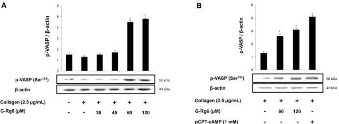
G-Rg6가 VASP 인산화에 미치는 효과
혈소판 integrin, αIIb/β3를 조절하는 인자로는 VASP가 잘 알려져 있다(Laurent 등, 1999; Sudo 등, 2003). VASP 는 인체 혈소판에서 Ser157과 Ser239, 두 군데 인산화 위치를 가지는 것으로 알려져 있으며, Ser157은 cAMP의 증가로 인 한 protein kinase A의 활성에 의해 인산화되고, Ser239는 cGMP 의존성 protein kinase G에 의해서 인산화되는 것으 로 보고되었다(Laurent 등, 1999; Sudo 등, 2003). Colla- gen으로 자극한 인체 혈소판은 fibrinogen과의 결합을 강하 게 촉진하였고(Fig. 3A-b), G-Rg6를 처리했을 때는 농도
A
B
Fig. 3. Effects of G-Rg6 on fibrinogen binding to αIIb/β3. (A) The flow cytometry histograms on fibrinogen binding. (B) Effects of G-Rg6 on collagen-induced fibrinogen binding. Fibrinogen binding was performed as described in ‘Materials and Methods’
section. The data are expressed as the mean±standard deviation (n=4). *P<0.05, **P<0.01 versus the collagen-stimulated human platelets.
A B
Fig. 4. Effects of G-Rg6 on VASP (Ser157) phosphorylation. (A) Effects of G-Rg6 on collagen-induced VASP (Ser157) phosphorylation.
(B) Effects of G-Rg6 with pCPT-cAMP on collagen-induced VASP (Ser157) phosphorylation. Western blot was determined as described in ‘Materials and Methods’ section. The data are expressed as the mean±standard deviation (n=3). *P<0.05 versus the collagen-stimu- lated human platelets.
의존적인 감소 양상을 나타냈다(Fig. 3B). 이 결과는 G-Rg6 로 인한 inside-out signaling의 저하로 인한 αIIb/β3의 비 활성화로 인한 것으로 사료되어 αIIb/β3의 활성화에 밀접한 관련이 있는 VASP의 인산화를 분석하였다. Collagen으로 자극한 인체 혈소판에 G-Rg6(30~120 μM)를 처리하여 VASP의 인산화를 분석한 결과 VASP157에서 농도 의존적인
인산화의 증가 양상을 보였다(Fig. 4A). 하지만 VASP239에 서는 인산화의 증가를 확인하지 못하였다. Fig. 4B의 pCPT- cAMP는 cAMP activator로 VASP157 위치를 인산화시키는 positive control로 사용되었다. 이 결과를 통해 G-Rg6는 cAMP 의존성 활성을 통해 αIIb/β3의 활성을 억제하는 것으 로 사료된다.
A
B
Fig. 5. Effects of G-Rg6 on clot retraction. (A) Photographs of fibrin clot. (B) Effects of G-Rg6 on thrombin-retracted fibrin clot. Quantification of fibrin clot retraction was performed as de- scribe in ‘Materials and Methods’. The data are expressed as the mean±standard deviation (n=4). *P<0.05, **P<0.01 versus the thrombin-stimulated human platelet.
G-Rg6가 혈소판 매개 clot retraction에 미치는 효과 지혈 부위의 자극을 받은 혈소판은 αIIb/β3를 매개로 인 접한 혈소판들과 그물 구조를 형성하게 되어 지혈 마개를 형성하게 되고 이후 수축작용이 발생하여 그물 구조는 더욱 견고해지고 지혈 부위를 단단하게 막아준다. 이전에 결과들 에서 G-Rg6는 세포 내 Ca2+ 동원과 αIIb/β3의 활성을 억제 하였기 때문에 platelet-platelet interaction의 작용을 억 제할 것이라 예상하였고, thrombin을 사용한 clot 형성과정 에서 얼마만큼의 영향을 미치는지 확인하기 위하여 clot retraction test를 수행하였다. 그 결과 G-Rg6는 농도 의존 적으로 clot retraction 반응을 억제하는 효과를 나타내었다 (Fig. 5).
0.05 U/mL thrombin을 PRP에 추가하였을 때 두 번째 tube에서 fibrin을 매개로한 platelet-platelet interaction 을 관찰할 수 있었으며, fibrin과 혈소판의 응집 및 수축작용 에 의한 결과로 혈소판이 부유되어 불투명했던 PRP가 투명 해지는 것을 확인할 수 있다(Fig. 5A). G-Rg6를 60 μM과 120 μM 처리하였을 때는 각각 fibrin-platelet의 수축작용 을 저해하는 것을 확인할 수 있었으며, G-Rg6의 농도가 증 가함에 따라 tube 내부의 clot의 범위가 더 넓게 형성되며 농도 의존적인 지연 효과를 보였다.
결론적으로 G-Rg6는 cAMP-dependent protein kinase
A의 기질인 IP3RI와 VASP를 인산화하였고, 인산화를 통하 여 세포 내 Ca2+ 동원과 αIIb/β3와 fibrinogen과의 결합을 억제하여 clot retraction을 억제하는 효과를 나타냈다. 따 라서 G-Rg6는 혈소판 응집으로 인한 심혈관계 질환에 있어 서 치료 및 예방 약물로 유용한 가치가 있다고 여겨진다.
요 약
G-Rg6는 인삼의 성분으로서 혈소판 억제 기작에 대한 연구 는 아직 미흡하다. 본 연구에서는 G-Rg6가 platelet ag- gregation과 thrombus formation에 미치는 영향을 평가하 였고, 이와 관련된 신호전달 분자인 IP3RI와 VASP의 인산 화를 어떻게 조절하는지 규명하고자 하였다. 그 결과 G-Rg6 는 collagen이 유도한 platelet aggregation을 강력하게 억 제하였고, IP3RI와 VASP의 인산화 및 세포 내 Ca2+ 동원과 fibrinogen binding의 활성을 감소시키며 clot retraction 반응을 농도 의존적으로 억제하였다. 따라서 G-Rg6는 인체 혈소판에서 platelet aggregation과 thrombus formation 에 억제 효과를 나타내는 물질이므로 치료 및 예방약물로서 잠재적 가치가 있다고 여겨진다.
감사의 글
본 연구는 2019년도 극동대학교 교내연구비 지원(FEU 2019S03)에 의해 수행된 것으로 이에 감사드립니다.
REFERENCES
Berridge MJ, Irvine RF. Inositol trisphosphate, a novel second messenger in cellular signal transduction. Nature. 1984. 312:
315-321.
Estevez B, Shen B, Du X. Targeting integrin and integrin signal- ing in treating thrombosis. Arterioscler Thromb Vasc Biol.
2015. 35:24-29.
Grynkiewicz G, Poenie M, Tsien RY. A new generation of Ca2+
indicators with greatly improved fluorescence properties. J Biol Chem. 1985. 260:3440-3450.
Hantgan RR, Taylor RG, Lewis JC. Platelets interact with fibrin only after activation. Blood. 1985. 65:1299-1311.
Irfan M, Kim M, Rhee MH. Anti-platelet role of Korean ginseng and ginsenosides in cardiovascular diseases. J Ginseng Res.
2019. 44:24-32.
Jackson SP. Arterial thrombosis-insidious, unpredictable and deadly. Nat Med. 2011. 17:1423-1436.
Jennings LK. Role of platelets in atherothrombosis. Am J Car- diol. 2009. 103:4A-10A.
Kim DH. Metabolism of ginseng constituents by intestinal mi- croflora and its relation to biological effects. J Ginseng Res.
2007. 1:77-88.
Laurent V, Loisel TP, Harbeck B, Wehman A, Gröbe L, Jock- usch BM, et al. Role of proteins of the Ena/VASP family in actin-based motility of Listeria monocytogenes. J Cell Biol.
1999. 144:1245-1258.
Lee CH, Kim JH. A review on the medicinal potentials of gin- seng and ginsenosides on cardiovascular diseases. J Ginseng
Res. 2014. 38:161-166.
Lee JG, Lee YY, Wu B, Kim SY, Lee YJ, Yun-Choi HS, et al. Inhibitory activity of ginsenosides isolated from processed ginseng on platelet aggregation. Pharmazie. 2010. 65:520-522.
Nishikawa M, Tanaka T, Hidaka H. Ca2+-calmodulin-dependent phosphorylation and platelet secretion. Nature. 1980. 287:863- 865.
Payrastre B, Missy K, Trumel C, Bodin S, Plantavid M, Chap H. The integrin alpha IIb/beta 3 in human platelet signal transduction. Biochem Pharmacol. 2000. 60:1069-1074.
Phillips DR, Nannizzi-Alaimo L, Prasad KS. Beta3 tyrosine phos- phorylation in αIIbβ3 (platelet membrane GP IIb/IIIa) out- side-in integrin signaling. Thromb Haemost. 2001. 86:246-258.
Ruggeri ZM, Mendolicchio GL. Adhesion mechanisms in plate- let function. Circ Res. 2007. 100:1673-1685.
Schwartz SM, Heimark RL, Majesky MW. Developmental mech- anisms underlying pathology of arteries. Physiol Rev. 1990.
70:1177-1209.
Shin JH, Kwon HW, Lee DH. Ginsenoside F4 inhibits platelet aggregation and thrombus formation by dephosphorylation of IP3RI and VASP. J Appl Biol Chem. 2019. 62:93-100.
Sudo T, Ito H, Kimura Y. Phosphorylation of the vasodilator- stimulated phosphoprotein (VASP) by the anti-platelet drug, cilostazol, in platelets. Platelets. 2003. 14:381-390.
Varga-Szabo D, Braun A, Nieswandt B. Calcium signaling in platelets. J Thromb Haemost. 2009. 7:1057-1066.

![Fig. 2. Effects of G-Rg6 on [Ca 2+ ] i mobilization and IP 3 RI (Ser 1756 ) phosphorylation](https://thumb-ap.123doks.com/thumbv2/123dokinfo/5194742.603530/4.892.97.739.666.1157/fig-effects-rg-ca-mobilization-ip-ser-phosphorylation.webp)