Korean J. Environ. Biol. 36(3) : 277~284(2018) https://doi.org/10.11626/KJEB.2018.36.3.277
INTRODUCTION
Green algae (Chlorophyta) have a greater diversity of cel- lular organization, morphological structure and reproductive process than any other algae (Bold and Wynne 1978). Green algae including photosynthetic pigment, pyrenoid that stores photosynthetic products, and tissue of chlorophyll that close- ly related to higher plant (Happy-Wood 1988). The green algae are distinguished by their photosynthetic pigments, carbohydrate reserve, chloroplast structure and flagella (Sze 2003).
Chlorophyta can be classified into 12 classes including Chlorophyceae and Trebouxiophyceae, and there are about 6,500 species reported worldwide. There are about 3,700 and 8,500 species reported in classes Chlorophyceae and Trebouxiophyceae, respectively (Guiry and Guiry 2018).
Domestically, there are 484 and 112 taxa of Chlorophyceae and Trebouxiophyceae reported, respectively (Lee and Kim 2015).
Trebouxiophyceae was first classified by Friedl (1995).
He had used molecular analysis to classification of Chloro- phytes that overlapped morphologically. As a result, some of the coccoid green algae were forming clade, named Tre- bouxiophyceae. These algae live usually terrestrial, often symbiotic in lichens, rarely in fresh water (Friedl 1995).
From this study, the newly recorded species of chloro- phytes in Korea were collected and identified from various freshwater lakes, soil, rocks, and land plants in order to ex- pand the recorded species of the Korean flora.
MATERIALS AND METHODS
These samples were collected 8 stations in Korea, from February 2017 to August 2017 (Table 1). Planktonic algae were collected using phytoplankton net that mesh size 25 μm and diameter 30 cm. The periphytic algae was collected by scrubbing off aquatic plants, submerged land plants and rocks (Sournia 1978). The collected specimens were separat- ed using a Pasteur pipette under an inverted microscope in solid media. The unialgal specimens were cultured in Bold’s
* Corresponding author: Ok Min Lee, Tel. 031-249-9643, Fax. 031-241-0860, E-mail. [email protected]
ⓒ2018. Korean Society of Environmental Biology.
Eight Taxa of Newly Recorded Species of Chlorophytes (Chlorophyceae and Trebouxiophyceae, Chlorophyta) in Korea
Mi Ran Kim, Jee Hwan Kim
1, Do Hyun Kim and Ok Min Lee*
Department of Life Science, College of Natural Science, Kyonggi University, Suwon 16227, Republic of Korea
1
Bioresources Culture Collection Division, Nakdonggang National Institute of Biological Resources, Sangju 37242, Republic of Korea
Abstract - In 2017, the freshwater algae were collected from reservoirs, small ponds, soil, and rocks in Korea. Eight taxa of Chlorophyta (Chlorophyceae and Trebouxiophyceae) have been newly reported in Korea. The unrecorded indigenous species were Chlorolobion braunii, Coelastrum pseudomicroporum, Coelastrum reticulatum var. cubanum, Monoraphidium nanum, Tetrachlorella incerta, Ecdysichlamys obliqua, Gloeotila scopulina, and Stichococcus jenerensis.
Keywords : Chlorophyceae, Chlorophyta, newly recorded species, Trebouxiophyceae
<Original article>
Mi Ran Kim, Jee Hwan Kim, Do Hyun Kim and Ok Min Lee 278
basal media under the following conditions: a temperature of 25℃, light/dark cycle of 16:8, and 40 μmol m
-2s
-1light (Stein 1973; Bold and Wynne 1978). Each sample was ex- amined using an ×400-1000 magnification under a Zeiss Microscope (Axio Imager A2; Carl Zeiss, Germany) and was photographed using an AxioCam HRC camera (Carl Zeiss, Germany). The taxonomic classification system was based on AlgaeBase (Guiry and Guiry 2018) and Komárek and Fott (1983). The taxa were identified based on informa- tion taken from West and West (1908), Prescott (1962) and Hirose et al. (1977).
RESULTS AND DISCUSSION
The eight newly added Korean species were Chlorolobi- on braunii, Coelastrum pseudomicroporum, Coelastrum re- ticulatum var. cubanum, Monoraphidium nanum, Tetrachlo- rella incerta, Ecdysichlamys obliqua, Gloeotila scopulina, and Stichococcus jenerensis.
We described the morphological characteristics of the newly recorded species and provided their microscopic photographs (Figs. 1-8).
Class Chlorophyceae Wille 1884 Order Sphaeropleales Luerssen 1877
Family Selenastraceae Blackman & Tansley 1903 Genus Chlorolobion Korsikov 1953
Chlorolobion braunii (Nag.) Kom. 1979 (Fig. 1) Cells are spindle shaped, and they are straight and slight- ly asymmetrical. The young cells are a bit short and point- ed. In the adulthood, they are rounded with blunted tips.
Chloroplast has one pyrenoid and it is present in the edges besides the incision area which covers the entire cell wall.
The length of the cell is 7-12 μm, and the width of cell is 3.2-4 μm.
Ecology: This species appears freshwater or associate with submerged surfaces in various aquatic habitat (John et al.
2011). We collected this species in planktonic samples from eutrophic reservoirs.
Distribution: Europe: Britain (John and Tsarenko 2002) Asia: Taiwan (Shao 2018).
Site of collection: Cheongpyeongho, Gyeonggi-do.
Date of collection: July 14, 2017.
Specimen Locality: ACKU2017NR01
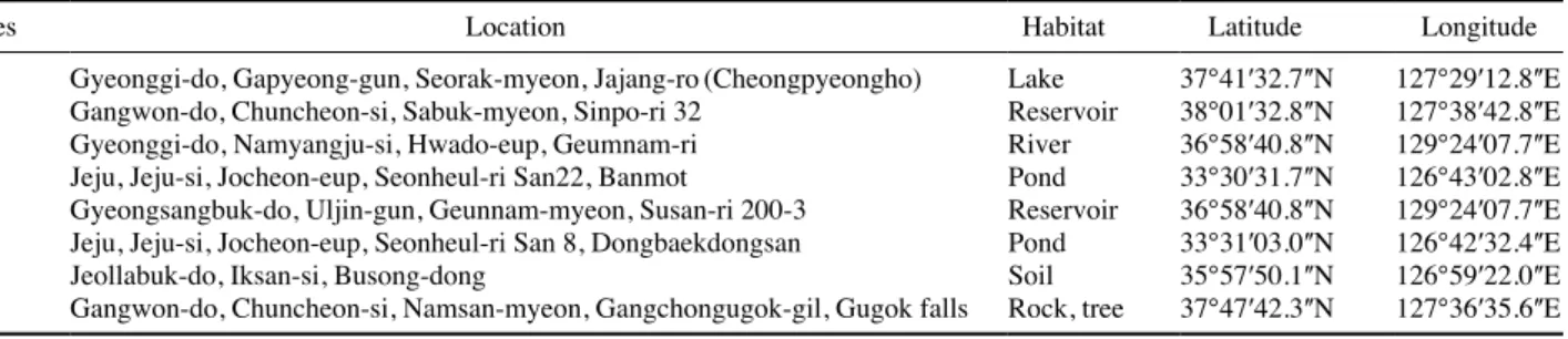
Table 1. The locational information about eight sites from where the phytoplankton was collected in 2017Sites Location Habitat Latitude Longitude 1 Gyeonggi-do, Gapyeong-gun, Seorak-myeon, Jajang-ro(Cheongpyeongho) Lake 37°41ʹ32.7ʺN 127°29ʹ12.8ʺE 2 Gangwon-do, Chuncheon-si, Sabuk-myeon, Sinpo-ri 32 Reservoir 38°01ʹ32.8ʺN 127°38ʹ42.8ʺE
3 Gyeonggi-do, Namyangju-si, Hwado-eup, Geumnam-ri River 36°58ʹ40.8ʺN 129°24ʹ07.7ʺE
4 Jeju, Jeju-si, Jocheon-eup, Seonheul-ri San22, Banmot Pond 33°30ʹ31.7ʺN 126°43ʹ02.8ʺE 5 Gyeongsangbuk-do, Uljin-gun, Geunnam-myeon, Susan-ri 200-3 Reservoir 36°58ʹ40.8ʺN 129°24ʹ07.7ʺE 6 Jeju, Jeju-si, Jocheon-eup, Seonheul-ri San 8, Dongbaekdongsan Pond 33°31ʹ03.0ʺN 126°42ʹ32.4ʺE
7 Jeollabuk-do, Iksan-si, Busong-dong Soil 35°57ʹ50.1ʺN 126°59ʹ22.0ʺE
8 Gangwon-do, Chuncheon-si, Namsan-myeon, Gangchongugok-gil, Gugok falls Rock, tree 37°47ʹ42.3ʺN 127°36ʹ35.6ʺE
A B
C D
Fig. 1. Chlorolobion braunii(Nag.) Kom. 1979. Scale bar: 10μm.
A-D: Spindle-shaped cells, A and C: One pyrenoid.
Class Chlorophyceae Wille 1884 Order Sphaeropleales Luerssen 1877 Family Scenedesmaceae Oltmanns 1904 Genus Coelastrum Nägeli 1849
Coelastrum pseudomicroporum Kors. 1953 (Fig. 2) Coenobia are arranged spherically with 8-32 cells and each cell neighbors with 4-6 connected cells. Cells are egg- shaped with thickened poles. Each cell is connected by means of short connecting projections. The length of cell is 5.2-6.4 μm, and the width of cell is 5.2-7.0 μm. The diame- ter of colony is 35 μm.
Ecology: This is reported on a few occasions from rivers (John et al. 2011). We collected this species in planktonic samples from eutrophic reservoirs.
Distribution: Europe: Bulgaria (Hegewald et al. 2010).
South America: Brazil (Ramos et al. 2015). Asia: China (Cao et al. 2005)
Site of collection: Sinpo-ri reservoir, Gangwon-do Date of collection: August 22, 2017.
Specimen Locality: ACKU2017NR04
Class Chlorophyceae Wille 1884 Order Sphaeropleales Luerssen 1877 Family Scenedesmaceae Oltmanns 1904 Genus Coelastrum Nägeli 1849
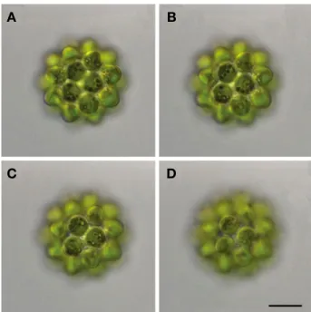
Coelastrum reticulatum var. cubanum Komárek 1975 (Fig. 3)
Coenobia are spherical or oval shaped, with 4-32 cells.
Cells are spherical or slightly ellipsoidal from the side, slightly flattened, rounded in apex. Cells are spherical or slightly flattened from the side, with rounded apices. Neigh- boring cells have grown together with 1-(2) narrow, subapi- cal junction shoots, each forming 5-6 extensions. The gap between cells is triangular to irregularly roundish. A walled chloroplast has with a pyrenoid. The length of cell is 8.3 μm, and the width of cell is 8.4 μm. The diameter of colony is 22 μm.
Ecology: This species lives in planktonic at various water biotopes (Komárek and Fott 1983). We collected this spe- cies in planktonic samples from eutrophic reservoirs.
Distribution: Asia: China (Liu and Hu 2012).
A B
C D
Fig. 2. Coelastrum pseudomicroporum Kors 1953. Scale bar: 10 μm. A and B: Egg-shaped cells with thickened poles, C and D: Short connecting projections.
A B
C D
Fig. 3. Coelastrum reticulatum var. cubanum Komárek 1975. Scale bar: 10μm. A-C: Short cylindrical extension, D: Subapical junction shoots.
Mi Ran Kim, Jee Hwan Kim, Do Hyun Kim and Ok Min Lee 280
Site of collection: Geumnam-ri, Garamsusangnejeo, Gyeo- nggi-do.
Date of collection: August 22, 2017.
Specimen Locality: ACKU2017NR05 Class Chlorophyceae Wille 1884 Order Sphaeropleales Luerssen 1877
Family Selenastraceae Blackman & Tansley 1903 Genus Monoraphidium Komárková-Legernová 1969 Monoraphidium nanum (Ettl) Hindák 1980 (Fig. 4) The cells are small, short kidney-shaped with broadly rounded cell tips, and have no mucus. The chloroplast is pa- rietal, trough-shaped, without a pyrenoid. The length of cell is 2-5 (5.5) μm and the width is 1.4-2 (2.5) μm.
Ecology: This is a freshwater and terrestrial species. They live in soil (especially forest soil) and small water biotope (Komárek and Fott 1983). We collected this species in plank- tonic samples from eutrophic reservoirs.
Distribution: South America: Brazil (Menezes 2010; Ra- mos et al. 2012).
Site of collection: Banmot, Jeju-do.
Date of collection: May 20, 2017.
Specimen Locality: ACKU2017NR10 Class Trebouxiophyceae Friedl 1995 Order Chlorellales Bold & M.J. Wynne 1985 Family Oocystaceae Bohlin 1901
Genus Tetrachlorella Korsikov 1939 Tetrachlorella incerta Hindák 1977 (Fig. 5)
Coenobia are 2 to 4-celled with ellipsoidal to oval shape and rounded at both ends. Two cells are often parallel or at most slightly displaced. Gelatinous envelopes are very fine but sometimes entirely absent. Cell wall is smooth without incrustations. Chloroplast is channel-shaped and pyrenoid is often ambiguous. Asexual reproduction is done by 2 or 4 autospores being released due to fracture of the mother cell wall. The length of cell is 4.5-8 μm and the width is 2-4 μm.
Ecology: This is a planktonic species in a wide range of water (John et al. 2011). We collected this species in plank- tonic samples from eutrophic reservoirs.
A B
C D
Fig. 4. Monoraphidium nanum(Ettl) Hindák 1980. scale bar: 5μm.
A-D: Small, short kidney-shaped cells with rounded tips.
A B
C D
Fig. 5. Tetrachlorella incerta Hindák 1977. Scale bar: 10μm.
A-D: Typically, 4-cells with ellipsoidal shape and two fre- quently parallel cells.
Distribution: Europe: Netherlands (Veen et al. 2015), Slo- vakia (Hindák and Hindáková 2016). Asia: Russia (Medve- deva and Nikulina 2014).
Site of collection: Susan-ri, Gyeongsangbuk-do.
Date of collection: May 31, 2017.
Specimen Locality: ACKU2017IR07 Class Trebouxiophyceae Friedl 1995 Order Chlorellales Bold & M.J. Wynne 1985 Family Oocystaceae Bohlin 1901
Genus Ecdysichlamys G.S. West 1912
Ecdysichlamys obliqua G.S. West 1912 (Fig. 6)
Cells are made of single cell, groups, or small colony.
The mother cell wall is attached to the cell. Cells are shaped like wide spindle, elliptic to ovate, and asymmetrical with a more curved side at the poles with papillary thickening.
Chloroplast is cell wall-shaped with sometime wavy margin and has a distinct pyrenoid. Cell wall is thick, smooth, col- orless, and indistinctly layered. The length of cell is 9-14 μm, and the width of cell is 5-10 μm.
Ecology: This is a freshwater species. They found surface
of soil or periphytic in spring (Komárek and Fott 1983). We collected this species in planktonic samples from eutrophic reservoirs.
Distribution: Europe: Spain (Uher et al. 2005). Caribbean Islands: Cuba (Comas González 2008, 2009).
Site of collection: Dongbaekdongsan, Jeju-do.
Date of collection: May 20, 2017.
Specimen Locality: ACKU2017NR02 Class Trebouxiophyceae Friedl 1995 Order Chlorellales Bold & M.J. Wynne 1985 Family Chlorellaceae Brunnthaler 1913 Genus Gloeotila Kützing 1843
Gloeotila scopulina (Hazen) Heering 1914 (Fig. 7) Filaments are long and bright green. Cells are cylindri- cal shape and they are not constricted at the diaphragm.
Chromatophore is narrow, pale green, without a distinct pyrenoid. This species is distinguished usually by its longer cells, and by its manner of growth in long dense masses of straight filaments. It does not maintain the filamentous state when brought into the laboratory and subsequently breaks A B
C D
Fig. 6. Ecdysichlamys obliqua G.S. West 1912. Scale bar: 10μm.
A-C: The poles with papillary thickening, D: Autospore.
A B
C D
Fig. 7. Gloeotila scopulina(Hazen) Heering 1914. Scale bar: 10 μm. A and B: Cylindrically-shaped cells, C and D: Straight filament.
Mi Ran Kim, Jee Hwan Kim, Do Hyun Kim and Ok Min Lee 282
down into the coccoid state. The cell width is 3-3.5 μm in diameter and its length is 1-10 times as long.
Ecology: This species is planktonic or associated with submerged surfaces in various aquatic habitat (John et al.
2011). We collected this species surface soil crust around moss.
Distribution: Europe: Slovakia (Hindák and Hindáková 2016). Asia: China (Hu and Wei 2006).
Site of collection: Busong-dong, Jeollabuk-do.
Date of collection: April 8, 2017.
Specimen Locality: ACKU2017IA01 Class Trebouxiophyceae Friedl 1995 Order Prasiolales Schaffner 1922
Family Prasiolaceae F.F. Blackman & A.G. Tansley 1902 Genus Stichococcus Nägeli 1849
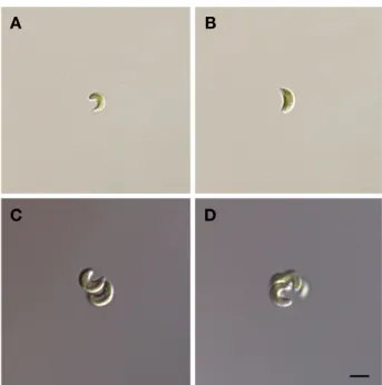
Stichococcus jenerensis Neustupa, Eliás & Sejnohová 2007 (Fig. 8)
This species is composed of easily fragmenting unbranched filaments so that the cells are usually solitary or in two-celled filaments. The cells are cylindrical shape and the cell wall is
thin, without any thickenings. The cells possess a single pari- etal chloroplast with a single starch enveloped pyrenoid. The length of cell is 8-11 μm, and the width of cell is 2.7-3.1 μm.
Ecology: This is a terrestrial species. They live in surface of soil crust on a tree base in a secondary lowland rainforest (Neustupa et al. 2007). We collected this species from sur- face soil around water fall.
Distribution: Asia: Malaysia (Nestupa et al. 2007).
Site of collection: Gugok falls, Gangwon-do.
Date of collection: February 27, 2017.
Specimen Locality: ACKU2017NR03
ACKNOWLEDGEMENTS
This study was supported by a grant from the Natio nal Institute of Biological Resources (NIBR 201701107, NIBR 201801205) and the Nakdonggang National Institute of Bi- ological Resources (NNIBR2017287, The project on collec- tion of freshwater algal strains (Year-1), 2017) funded by the Ministry of Environment (MOE) of the Republic of Korea.
REFERENCES
Bold HC and MJ Wynne. 1978. Introduction to the Algae - Structure and Reproduction. Prentice Hall. Englewood Cliffs. New Jersey.
Cao X, A Strojsová, P Znachor and E Zapomelová. 2005. De- tection of extracellular posphatases in natural spring phyto- plankton of a shallow eutrophic lake(Donghu, China). Eur.
J. Phycol. 40:251-258.
Comas González A. 2008. Algunas características de la Flora de algas y cianoprocariotas de agua dulce de Cuba. AL- GAS. Boletín de la Sociedad Española de Ficología 39:21- 29.
Comas González A. 2009. Catálogo de las algas y cianopro- cariotas dulciacuícolas de Cuba. Editorial Universo Sur, Universidad de Cienfuegos.
Ettl H. 1977. Taxonomische Bemerkungen zu den Xanthophy- ceen. Nova Hedwigia 28:555-568.
Friedl T. 1995. Inferring taxonomic positions and testing genus level assignments in coccoid green lichen algae: a phylo- genetic analysis of 18S ribosomal RNA sequences from Dictyochloropsis reticulata and from members of the ge-
A B
C D
Fig. 8. Stichococcus jenerensis Neustupa, Eliás and Sejnohová 2007. Scale bar; A-C: 10μm, D: 5μm. A-C: Pyrenoid, D:
Neustupa et al. 2007.
nus Myrmecia(Chlorophyta, Trebouxiophyceae cl. nov.). J.
Phycol. 31:632-639.
Guiry MD and GM Guiry. 2018. AlgaeBase. World-wide elec- tronic publication, National University of Ireland, Galway, Available from: http://www.algaebase.org.
Happy-Wood CM. 1988. Ecology of freshwater planktonic green algae. pp. 175-226. In Growth and Reproduction Strategies of Freshwater Phytoplankton(Sandgen CD ed.). Cambridge University Press. Cambridge.
Hazen TE. 1902. The Ulothricaceae and Chaetophoraceae of the United States. Memoirs of the Torrey Botanical Club 11:135-250.
Heering W. 1914. Chlorophyceae III. Ulothrichales, Microspo- rales, Oedogoniales. pp. 1-250. In Süsswasserflora Deutsch- lands, Österreich und der Schweiz.(Pascher A ed.). Gustav Fischer. Jena.
Hegewald E, M Wolf, A Keller, T Friedl and L Krienitz. 2010.
ITS 2 sequence-structure phylogeny in the Scenedesma- ceae with special reference to Coelastrum(Chlorophyta, Chlorophyceae). including the new genera Comasiella and Pectinodesmus. Phycologia 49:325-335.
Hindák F. 1977. Studies on the Chlorococcal algae(Chlorophy- ceae). I. Biologicke Práce 23:1-192.
Hindak F. 1980. Studies on the Chlorococcal algae(Chloro- phyceae) II. Publishing House of the Slovak Academy of Sciences. Bratislava.
Hindák F and A Hindáková. 2016. Algae. In Zoznam nižších a vyšších rastlín Slovenska(on-line list).
Hirose H, M Akiyama, M Hirano, K Imahori, T Ioriya, H Ka- saki, H Kobayasi, S Kumano, E Takahashi, K Tsumura and T Yamagishi. 1977. Illustrations of the Japanese Fresh-wa- ter Algae. Uchidarokakuhe. Tokyo.
Hu H and Y Wei. 2006. The freshwater algae of China. Sys- tematics, taxonomy and ecology. China. www.sciencep.
John DM, BA Whitton and AJ Brook. 2011. The Freshwater com.
Algal Flora of The British Isles. An Identification Guide to Freshwater and Terrestrial Algae. Second edition. Cam- bridge: Cambridge University Press.
John DM and PM Tsarenko. 2002. Order Chlorococcales. pp.
329-409. In The Freshwater Algal Flora of the British Isles (John DM et al. eds.). Cambridge University Press. Cam- bridge.
Komárek J. 1975. New coenobial Chlorococcales algae of Cuba. Preslia(Prague) 47:275-279.
Komárek J. 1979. Änderungen in der Taxonomie der Chl- orokokkalalgen. Archiv für Hydrobiologie 24:239-263.
Komárek J and B Fott. 1983. Das Phytoplankton des Süsswas- sers. Schweiz Verg. Stuttgart.
Komárková-Legnerová J. 1969. The Systematics and Ontogen- esis of the genera Ankistrodesmus Corda and Monoraphid- ium gen. nov. pp. 75-144. In Studies in Phycology(Fott B et al. eds.). Schweiz Verg. Stuttgart.
Korshikov AA. 1939 Contribution to the algal flora of the Gorky District Phytoplankton of the Oka River in August 1932.
Zapiski Gorkovskogo Universiteta 9:101-128.
Korshikov AA. 1953. The Freshwater Algae of the Ukrainian SSR. V. Sub-Class Protococcineae. Vacuolales and Proto- coccales. Kyjv Akad. NAUK URSR.
Kützing FT. 1843. Phycologia generalis oder Anatomie, Phys- iologie und Systemkunde der Tange. F.A. Brockhaus.
Leipzig.
Lee OK and JH Kim. 2015. National List of Species of Korea (Green algae). Jeonghaeng Publ. Co. Seoul.
Liu G and Z Hu. 2012. Flora Algarum Sinicarum Aquae dulcis Tomus XV Chlorophyta Chlorooccales(II) Tetrasporales Dichotomosiphonales Cladophorales. Science Press. Bei- jing.
Medvedeva LA and TV Nikulina. 2014. Catalogue of Fresh- water Algae of the Southern part of the Russian Far East.
Dalnauka, Vladivostok.
Menezes M. 2010. Chlorophyceae. pp. 335-352. In Catálogo de Plantas e Fungos do Brasil(Forzza RC et al. eds.). Insti- tuto de Pesquisas Jardim Botânico do Rio de Janeiro.
Nägeli C. 1849. Gattungen einzelliger Algen, physiologisch und systematisch bearbeitet. Neue Denkschriften der Allg.
Schweizerischen Gesellschaft für die Gesammten Natur- wissenschaften 10:1-139.
Neustupa J, M Eliás and L Sejnohová. 2007. A taxonomic study of two Stichococcus species(Trebouxiophyceae, Chloro- phyta) with a starch-enveloped pyrenoid. Nova Hedwigia 84:51-63.
Prescott GW. 1962. Algae of the Western Great Lakes area.
W.M.C. Brown Co. Inc. Iowa.
Ramos GJP, CEM Bicudo, A Góes-Neto and CW Moura. 2012.
Monoraphidium and Ankistrodesmus(Chlorophyceae, Chlo- rophyta) from Pantanal dos Marimbus. Chapada Diamantina 39:421-434.
Ramos GJP, CEM Bicudo and CW Moura. 2015. Scenedes- maceae(Chlorophyta, Chlorophyceae) de duas áreas do Pantanal dos Marimbus(Baiano e Remanso), Chapada Dia- mantina, Estado da Bahia, Brasil. Hoehnea 42:549-566.
Shao KT. 2018. TaiBNET(Catalogue of Life in Taiwan). http://
taibnet.sinica.edu.tw.
Sournia A. 1978. Phytoplankton Manual. UNESCO, Paris.
Stein JR. 1973. Handbook of Phycological Methods. Culture Methods and Growth Measurements. Cambridge Universi- ty Press. USA.
Mi Ran Kim, Jee Hwan Kim, Do Hyun Kim and Ok Min Lee 284
Sze P. 2003. A Biology of the Algae 2nd. Ed. WCB. Published.
Uher B, M Aboal and L Kovacik. 2005. Epilithic and chas-IA.
moendolithic phycoflora of monuments and buildings in South-Eastern Spain. Cryptogamie. Algologie 26:275-308.
Veen A, CHJ Hof, FAC Kouwets and T Berkhout. 2015. Rijk- swaterstaat Waterdienst. Informatiehuis Water. Taxa Water- management the Netherlands(TWN).
West GS. 1912. Algological notes. V-IX. J. Bot. London 50:79- West W and GS West. 1908. A Monograph of the British De-89.
smidiaceae. Vol. 3. Ray Society. London.
Received: 20 August 2018 Revised: 6 September 2018 Revision accepted: 6 September 2018