53
Field efficacy of a combined vaccine supplemented with recombinant Pasteurella multocida toxin subunits
against atrophic rhinitis
Mi Lan Kang
1, Seung Won Shin
1, Nabin Rayamahji
1, Yeon Soo Seo
1, Su In Lee
1, Won Hyung Lee
2, Han Sang Yoo
1,*
1College of Veterinary Medicine, BK21 for Veterinary Science and KRF Zoonotic Disease Priority Research Institute, Seoul National University, Seoul 151-742, Korea
2XP BioInc, Anseong 456-914, Korea (Accepted: February 20, 2008)
Abstract : We have investigated efficiency of a recombinant subunit Pasteurella multocida toxin (PMT) that was mixed with a vaccine consisted of inactivated whole cells of Bordetella bronchiseptica , P.
multocida (types A and D). For verification of the efficacy of the vaccine, all experimental pigs (suckling piglets, sow and gilts) in the three farms were vaccinated. Antibody titers against B. bronchiseptica and P. multocida type A of the vaccinated pigs by microplate agglutination were significantly higher than those of the control pigs ( p < 0.05). Similar patterns were observed in the analysis of anti- PMT neutralizing antibody by serum neutralizing method using Vero cell ( p < 0.05). Anti- P. multocida type D antibody titer of the vaccinated sows and gilts by ELISA showed significant differences with those of the non-vaccinated pigs ( p < 0.05). Although antibody titers increased, it was unable to find out the difference in the clinical signs between the vaccinated and non-vaccinated pigs. However, the increase in body weight of the vaccinated piglets was observed in comparison with the non-vaccinated piglets on a farm. At slaughtering of the pigs, pathological lesions in the turbinate bones of the vaccinated pigs were significantly lower than those of the non-vaccinated pigs ( p < 0.001). These results suggested that efficacy of the vaccine in pigs demonstrated to protect against atrophic rhinitis in Korea.
Keywords : atrophic rhinitis, field trial, Pasteurella multocida toxin, recombinant subunit vaccine
Introduction
Infection with virulent B. bronchiseptica strain is a common risk factor in the establishment of toxin- producing strains of P. multocida types A and D in the nasal cavity of pigs leading to the disease, atrophic rhinitis (AR) [7, 13, 15]. The more severe form of AR is attributed to colonization in nasal cavity of pigs by toxigenic P. multocida and is known as progressive atrophic rhinitis (PAR) [16]. Many capsular type D and occasionally type A strains of P. multocida produce a toxin (PMT), which is a major virulence factor to develop turbinate atrophy in pigs [3]. PAR triggers significant global economic loss in the swine industry due to reduced growth rates and feed conversion
efficiency because of developing a number of characteristic lesions including turbinate bone hypoplasia, facial distortion and nasal hemorrhage [14].
However, controlling PAR in the swine industry remains problem to be solved until today.
A recombinant PMT as a vaccine candidate was shown to induce protective response against challenge with a lethal dose of PMT in mice [11] and efficient immunoprotective properties against a challenge with B.
bronchiseptica and toxigenic P. multocida in vaccinated pigs [9]. Also, a previous study demonstrated that three recombinant subunit PMT (rsPMT), characterizing the N-terminal, middle, and C-terminal segments of PMT could induce specific and protective immune responses in pigs [8].
*Corresponding author: Han Sang Yoo
College of Veterinary Medicine, Seoul National University, Seoul 151-742, Korea
[Tel: +82-2-880-1263, Fax: +82-2-874-2738, E-mail address: yoohs@snu.ac.kr]
Considering the proposed synergistic role of B.
bronchiseptica and P. multocida in PAR, the common treatment of B. bronchiseptica and P. multocida toxoid/
bacterins to control AR is warranted. Therefore, combined vaccine of the bacterins with rsPMT would represent a novel approach to control atrophic rhinitis in pigs. Based on the situations, this study was designed to evaluate the rsPMT vaccine with inactivated whole cells of B. bronchiseptica , P. multocida (types A and D) under field condition in typical Korea farms.
Materials and Methods
Pigs and bacterial strains
The field trial was carried out at three different scales of typical farms without any vaccination history against AR in Korea that hold about 4,300, 2,000 and 800 pigs, respectively. YLD cross breed pigs were used in this experiment. There were no outbreaks of PAR in those farms. The experiment was carried out with 3 groups
of pigs (1-3 weeks old piglets, sows and gilts).
Breeding equipment and environment in the farms were described on Table 1. Swine B. bronchiseptica type 1 (Bb-1) and P. multocida types A (Pm-A) and D (Pm-D) isolates were from National Institute for Animal Health, Council of Agriculture in Taiwan. E.
coli BL21 transformed with Tox1, Tox2 and Tox7 of rsPMT [8] was from Graduate Institute of Veterinary Pathology, National Chung Hsing University in Taiwan.
Vaccine preparation
The vaccine, BayoVac swine atrophic rhinitis recombinant toxin and inactivated combined bacterin (AR + rsPMT), was manufactured by Kaohsiung Biological Product (Taiwan) and distributed by Bayer Taiwan. Characteristics and generation of PMT in the experimental vaccine were previously described by Liao et al. [8]. The vaccine contained B. bronchiseptica bacterin, P. multocida types A and D bacterins, rsPMT Table 1. Breeding and raising environment of experimental farms
Items Classification Farm A Farm B Farm C
Pig inventory
No. of boar 4 1 17
No. of sow 940 125 455
No. of lactating piglet 1,028 200 537
No. of weaned piglet 2,300 420 990
Ventilation method
Farrowing unit Windowless, Upper inlet,
Exhaust fan Windowless, Upper inlet,
Exhaust fan Natural ventilation Nursery unit Windowless, Upper inlet,
Exhaust fan Windowless, Upper inlet,
Exhaust fan Mechanical ventilation Grower unit Winch curtain, Exhaust fan Natural ventilation Natural ventilation Finisher unit Winch curtain, Exhaust fan Natural ventilation Natural ventilation Waste
management
Farrowing unit Slurry system Slurry system Slurry system
Nursery unit Slurry system Slurry system Slurry system
Grower unit Sawdust Slurry system Slurry system
Finisher unit Sawdust Slurry system Slurry system
Feeding type in each stage
Pregnant sow Wet feeding Wet feeding Wet feeding
Lactating piglet Dry feeding Dry feeding Dry feeding
Weaned piglet Dry feeding Dry feeding Dry feeding
Grower Dry feeding Dry feeding Dry feeding
Finisher Dry feeding Dry feeding Dry feeding
Vaccination Atrophic rhinitis Non- vaccinated Non- vaccinated Non- vaccinated Biosecurity
Entrance disinfection
equipment have have have
Foot dipping have have have
Isolation unit have have have
and aluminum hydroxide gel as an adjuvant. One dose of the vaccine was composed of 1
×10
9CFU of B.
bronchiseptica , P. multocida types A and D each bacterins, 2,000
µg of P. multocida toxin containing 50
µg of rsPMT and 19.8 mg of aluminum hydroxide gel.
Vaccination of pigs
The experiment was carried out with 3 groups of pigs in each farm. The piglets, sows and gilts in vaccinated groups consisted of 30, 5 and 5 pigs, respectively. The pigs in control groups consisted of same number of pigs in vaccinated groups. The piglets were immunized by intramuscular injection with 1 ml of the vaccine at 1 week old and 2 ml of the vaccine at 3 weeks old.
The gilts were vaccinated by intramuscular injection with 2 ml at 1 month after copulation and 2 ml at 3 weeks before parturition. The sows were vaccinated one time by intramuscular injection with 2 ml at 3 weeks before parturition. Control pigs with same age in each group were raised without any administration.
Serum was collected from a jugular vein of the pigs at two week intervals during the experimental period to determine specific antibody response.
Microplate agglutination test
The value of serum anti- B. bronchiseptica and P.
multocida type A antibody titers was determined by microplate agglutination test (MAT) using the standard procedure [2] with minor modifications. B. bronchiseptica and P. multocida type A strains were isolated from Korea. The bacterial cells were cultured in tryptic soy broth (Difco, USA) at 37
oC for 24 h under shaking condition. The bacteria were harvested, washed and resuspended with phosphate-buffered saline (PBS, pH 7.4). Serum samples were heat-inactivated by the incubation at 56
oC for 30 min and then two-fold serially diluted in PBS from 1 : 10 to 1 : 1,280. The quantity of 50
µl each serum diluents was added to the wells of a U-shaped microplate followed by the addition of antigens (2 µg/well). The plate was incubated at 37
oC for 2 h and kept at 4
oC for overnight. Positive and negative sera were included as controls. The titer was expressed as reciprocal of the number indicating the highest dilution that showed agglutination.
ELISA
The value of anti- P. multocida type D antibody titer was determined by ELISA kit (EB44-09; Anigen,
Korea). ELISA was performed by the manufacturer’s procedure.
Serum neutralization assay
Neutralizing antibody titers were determined by the ability of antibody to inhibit PMT-induced cytopathic effect in Vero cells [10] with some modifications of the previous report [8]. Serum was serially diluted with DMEM supplemented with 3% FBS (Gibco BRL, USA), 200 UI/ml of penicillin G and 200 µg/ml streptomycin sulfate. The serially diluted sera were transferred to a 96- well cell culture plate (50
µl/well) followed by the addition of an equal volume of DMEM containing a four- fold minimal toxic dose (MTD) of authentic PMT [8].
The plate was incubated at 37
oC for 1 h, and 100
µl of a Vero cell suspension containing 2
×10
4cells per well was then added to the plate. The plate was incubated at 37
oC for another 5-7 days. The endpoint-titer was expressed as the highest dilution of immune serum that could neutralize a one-fold MTD of PMT.
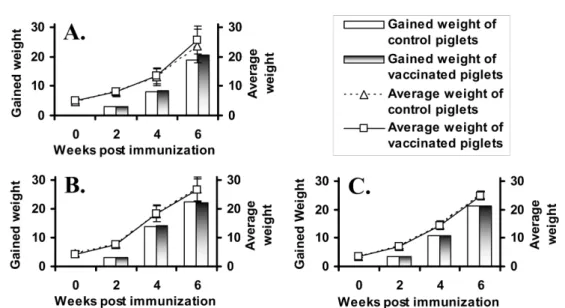
Body weight and clinical signs
The body weight was measured in the piglets of both vaccinated and non-vaccinated groups for 6 weeks after the vaccination. Clinical signs were observed in the experimental animals, piglets, sows and gilts in every week for 6 weeks after the vaccination. Clinical signs were determined by the presence or absence of survival, decrease in movement and feed conversion and typical symptoms of AR such as nasal discharge, eye discharge, pyrexia, anorexia, atrophy, sneeze, pink eye, hypokinesia and dyspnea, erythema, rhinorrhagia, necrosis and redness of injected site, etc.
Turbinate score
At slaughtering of the experimental pigs, heads of the pigs were collected and all snouts were sectioned transversely at the first premolar tooth region. Turbinate lesions of the rostral face were subjectively scored through gross examination by a group of collaborative investigators. Each snout was visually scored from the method of previous report [5]. Left and right turbinate atrophy and deviation of the nasal septum were each graded separately on a scale of 0 (normal) to 3 (complete atrophy). Normal turbinate was received a grade of 0.
Slight, but obvious, atrophy was graded as 1, moderate
atrophy of not less than half the turbinates, especially
the dorsal and ventral scrolls was graded as 2, and
severe atrophy of the dorsal and ventral scrolls was graded as 3. No, slight, moderate and severe deviation of the septum were graded from 0 to 3, respectively.
The three scores from each snout were then added together and divided by 3 to determine final visual scores for each pig, ranging from 0 to 3. Group means were calculated.
Statistical analysis
Statistical analysis was performed using the computer- based MS-Excel 2004 program. All results were expressed as the mean
±SD. Statistical differences between the groups were analyzed with two-tailed, non-paired Student’s t test assuming unequal variance.
Differences were considered significant if probability values of p < 0.05 were obtained.
Results
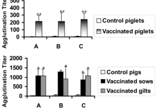
Anti- B. bronchiseptica antibody titer
Anti- B. bronchiseptica antibody titer was detected by MAT using B. bronchiseptica antigens. The titers against B. bronchiseptica in serum of the vaccinated piglets significantly increased from 4 weeks post vaccination. The titers of all farms at 6 weeks after the vaccination were significantly higher than those of the control piglets ( p < 0.01) (Fig. 1). High antibody titers against B. bronchiseptica in the sows and gilts were detected in 2 weeks after the vaccination and maintained more than six weeks even though the antibody titers in each farm were not same. The vaccinated sows and gilts in all farms showed significantly higher agglutination titers than those of the non-vaccinated pigs at 6 weeks post immunization ( p < 0.05) (Fig. 1).
Anti- P. multocida type A antibody titer
Anti- P. multocida type A antibody titer was detected by MAT using P. multocida type A antigens. The antibody titers against P. multocida type A of the vaccinated piglets in the farms A and B were significantly higher than those of the control piglets after the vaccination ( p < 0.05) (Fig. 2). Anti- P.
multocida type A antibody titers in the sows and gilts showed similar pattern to those of B. bronchiseptica in most farms. In serum samples of the vaccinated sows and gilts at 6 weeks post vaccination, P. multocida type A specific titers were significantly higher than those of the control groups ( p < 0.05) (Fig. 2).
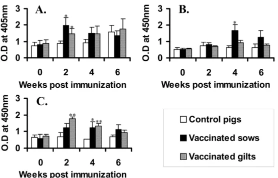
Anti- P. multocida type D antibody titer
The antibody titer against P. multocida type D was determined by ELISA. Most of the vaccinated piglets showed increase of the antibody titer with time after the vaccination. However, there were no significant differences between the vaccinated and control piglets in all farms (data not shown). In the results of pregnant pigs, the most of the vaccinated sows and gilts showed significantly higher antibody titers against P. multocida type D than those of the control pigs even though the patterns of increase were different (Fig. 3). In case of the farm A, the control pigs showed increase of Fig. 2. Anti- P. multocida type A antibody titers in serum of the experimental pigs at 6 week post immunization by MAT. A, B and C indicate the antibody titers in farms A, B and C, respectively. Significant difference between control and vaccinated pigs was expressed as
*p < 0.05.
Fig. 1. Anti- B. bronchiseptica antibody titers in serum of
the experimental pigs at 6 week post immunization by
MAT. A, B and C indicate the antibody titers in farms A,
B and C, respectively. Significant differences were
expressed as
**p < 0.01 and
*p < 0.05.
antibody response against P. multocida type D after 4 week. The increase seems to be due to natural infection of the bacterin since there was no vaccination history.
PMT- specific neutralizing antibody titer The PMT- specific neutralizing antibody titer was detected as its inhibitory ability to the cytopathic effects induced by PMT in Vero cells. At 6 weeks after the vaccination, PMT- specific neutralizing antibody titer (
≥1 : 64) was significantly higher in the vaccinated piglets of the farm A than that of the non- vaccinated piglets ( p < 0.05) (Fig. 4). However, the vaccinated piglets in other farms showed moderate increase of neutralizing antibody titer (
≥1 : 32) against PMT even though there was not significance. The vaccinated sows in the farm A could generate the highest PMT-specific neutralizing antibody titers after 6 weeks (1 : 128). The titers were significantly higher than those of the control pigs ( p < 0.05) (Fig. 4). The vaccinated sows in the farm B and C showed high levels of PMT- specific neutralizing antibody titer (
≥1 : 64) even though the value was lower than that of the vaccinated sows in the farm A. In the farm C, the neutralizing antibody titers in both of the vaccinated sows and gilts were significantly higher than those of
the non-vaccinated pregnant pigs ( p < 0.05) (Fig. 4).
Body weight and clinical signs
The significant increase in body weight of the vaccinated piglets was not observed in all farms even though there was increase in one farm (Fig. 5). Also, Fig. 3. Anti- P. multocida type D antibody titers by ELISA. A, B and C indicate the antibody titers in the farms a, b and c, respectively. Significant differences between the control and vaccinated pigs were expressed as
**p < 0.01 and
*
p < 0.05.
Fig. 4. P. multocida toxin-specific serum neutralizing
antibody titers at 6 week post immunization. The SN-titer
was expressed as the end-point dilution of serum that could
inhibit the cytotoxicity of four fold MTD of authentic PMT
on Vero cells. A, B and C indicate the neutralizing antibody
titer in farms A, B and C, respectively. Significant
difference between the control and vaccinated pigs was
expressed as
*p < 0.05.
it was very difficult to distinguish the difference in the degree of clinical signs regardless of the vaccination.
Turbinate scores
In the scoring of gross pathological lesions in nasal turbinate produced low turbinate atrophy scores ranging from 0 to 1 in the vaccinated pigs. The average score of turbinate atrophy in the vaccinated group was 0.67
±0.34. In contrast, the non-vaccinated pigs showed mild to severe turbinate atrophy with average scores of 1.78
±