울릉도 민속식물 추출물의 항염증 효과
김현준, 이동준, 구자정*, 최 경1, 박광우, 강신호2, 문 철3, 이평재2
국립수목원 산림자원보존과, 1국립수목원 연구기획팀, 2세명대학교 자연약재과학과, 3세명대학교 임상병리학과
Anti-inflammatory Effect of Extracts from Folk Plants in Ulleung Island
Hyun-Jun Kim, Dong-Jun Lee, Ja-Jung Ku*, Kyung Choi1, Kwang-Woo Park, Shin-Ho Kang2, Cheol Moon3 and Pyeong-Jae Lee2
Division of Plant Conservation, Korea National Arboretum of the Korea Forest Service, Pocheon 487-821, Korea
1Division of Research Planning and Coordination, Korea National Arboretum of the Korea Forest Service, Pocheon 487-821, Korea
2Department of Natural Medicine Resources, Semyung University, Jecheon 390-711, Korea
3Department of Clinical Laboratory Science, Semyung University, Jecheon 390-711, Korea
Abstract - We investigated the effect of extracts(50 µg/mL) from 31 folk plants in Ulleung Island on nitric oxide(NO) production in lipopolysaccharide (LPS)-stimulated RAW264.7 cells. Actinidia arguta(leaf, twig), Hovenia dulcis(leaf), Camellia japonica(leaf, twig), Sambucus sieboldiana var. pendula(leaf, twig) and Fallopia sachalinensis(root) showed significant NO inhibition activity(42%~88% NO inhibition rate). Of them, we selected 3 samples(A. arguta, H. dulcis and C. japonica) showing more than 50% NO inhibition activity and little effect on cell viability. We found that the extracts suppressed the NO and prostaglandin E2(PGE2) produced by LPS-activated RAW264.7 cells in dose-dependent manners.
Key words -Actinidia arguta, Hovenia dulcis, Camellia japonica, Nitric oxide, Prostaglandin E2
*
교신저자(E-mail) :[email protected]서 언
최근 세계 각국에서는 자국의 자생식물에 대한 전통지식 을 기반으로 활용하는 민속식물에 관한 관심이 고조되고 있다. 특히 2010년 일본 나고야에서 개최된 생물다양성협 약에서 유전자원과 전통지식의 접근 및 이익분배에 관한 나고야의정서가 채택되는 등, 전통지식을 공유하고, 나아 가 민속식물의 유용성 탐색을 통한 소재식물의 발굴 및 개 발이 절실히 필요한 실정이다. 이에 관한 국내의 연구는 경 기도 내륙 남부, 경상북도 내륙 및 울릉도, 경상남도 내륙, 강원도 남부, 강원도 북부 등 한반도 전역에서 민속식물 이 용 정보를 수집 및 발굴하였다(Choi et al., 2010; Chung et al., 2010; Chung et al., 2011; Im et al., 2011;
Jeong et al., 2012; Kim et al., 2012). 따라서 이를 활용 한 건강기능 제품 및 의약품분야에 폭넓게 활용 될 수 있는
기능성 소재를 탐색하는 것이 필요한 실정이다. 기능성 분 야 연구는 매우 다양화되고 있는데, 그 중 염증으로 인한 질병을 예방 또는 치료하는 데 목적을 둔 항염증 소재 연구 도 한 부분을 차지하고 있다.
염증 반응은 외상이나 세균의 침입으로 인해 손상된 조 직에 대한 생체 조직의 국소적 방어반응이다. 염증 반응이 일어나면 여러 가지 염증매개인자들이 만들어지는 데 이로 인하여 통증, 발열, 홍반, 부종 및 기능 상실 등의 증상이 나타난다(Sarkar and Fisher, 2006; Fierro and Serhan, 2001; Park et al., 2007). 대식세포는 숙주의 항상성 유 지와 같이 여러 종류의 숙주 반응에 관여하는 것으로 알려 져 있으며, nitric oxide(NO), prostaglandin E2(PGE2), 염증성 cytokine 등을 분비한다(Higuchi et al., 1990).
이 중 NO는 NO synthase(NOS)에 의해 L-arginine으로 부터 생성되며 농도에 따라 세포 기능유지에 중요한 작용 을 하기도 하고 세포독성을 일으키기도 한다(Moncada et al., 1991; Nathan and Xie, 1994). NOS는 endothelial
Original Research Article
NOS(eNOS), neuronal NOS(nNOS), inducible NOS (iNOS)가 있는데 이중 eNOS, nNOS는 칼슘에 농도 의존 적이고 자극에 대한 반응이 아닌 구성성분으로 일시적이며 소량 발현된다. 반면 iNOS는 칼슘농도에 상관없이 염증자 극에 의해 지속적인 다량 생성이 유도됨으로써 염증반응에 기여한다(Lowensterin and Snyder, 1992). iNOS는 일반 적으로 대식세포에서 염증성 cytokine 자극에 의해 유도되 며, 특히 Lipopolysaccharide(LPS) 또는 세균의 lipopep- tide를 처리했을 때 급격히 생성이 증가하는 것으로 알려져 있다(Sunyer et al., 1997). Prostaglandin은 arachidonic acid로부터 cyclooxygenase(COX)의 작용에 의해 합성되 어 염증반응과 면역반응을 촉진한다(Kim et al., 2004).
COX는 isoform인 COX-1과 COX-2가 있는데 COX-1은 정상상태에서 발현하여 위장관 보호, 신장 기능조절과 같 은 항상성 유지에 관여하며, COX-2는 염증이나 oxidative stress, 기타 면역 반응시 mitogen이나 cytokine에 의하 여 세포내 발현이 증가한다(Kwon, 2001). COX 활성에 의 한 주요 산물은 PGE2로, 염증질환, 자기면역질환, 종양성 질환의 병리에서 중요한 역할을 한다(Chou et al., 2003). 염 증반응에서 발현이 증가되는 대표적인 pro-inflammatory cytokine은 tumor necrosis factor(TNF)-α, interleukin (IL)-1β, IL-6 등이 있으며, 대식세포 등은 이들을 분비 하여 다양한 염증 반응을 매개하며 NO와 PGE2생성을 유 도한다(McDaniel et al., 1996).
따라서 본 연구에서는 Choi et al.(2010)에 의해 보고된 울릉도 민속식물을 대상으로 기능성 소재를 발굴하고 이를 활용하기 위해 울릉도 민속식물 추출물로 LPS에 의해 활 성화된 RAW 264.7 대식세포에서 항염증 활성을 검색하 고, 우수한 그룹을 선별하여 NO 및 PGE2 생성량에 미치는 영향을 조사하였다.
재료 및 방법
실험재료
본 연구는 울릉도 민속식물로 조사 된 109분류군 중 31 분류군을 선정하여 진행하였다(Table 1). 선정된 분류군의 생체시료 채집은 2011년 4월부터 11월까지 총 8차례에 걸 쳐 울릉군 서면 태하리 서달령, 태하령, 향목령, 북면 나 리, 천부리 섬목, 울릉읍 도동리 등에서 수행하였으며, 증 거표본은 국립수목원 표본관(KH)에 보관하였다. 식물체는
부위별로 세척 및 절단과정을 거친 뒤 음건하여 48점의 건 조시료를 확보하였다. 건조된 시료는 분쇄한 뒤 실험에 사 용하였다.
추출
건조된 식물체를 분쇄하여, 분쇄시료 100 g에 80%
MeOH 500 mL을 첨가하여 24시간 상온침지추출하고, 추 출물을 ADVANTEC paper(No. 6, ADVANTEC Co., Ltd., Tokyo, Japan)로 여과하였다. 위 과정을 3회 반복 실시하였다. 여과된 추출물은 감압농축기에서 농축한 후 동결 건조하여 시료로 사용하였다.
세포배양 및 시료처리
실험에 사용한 마우스 대식세포주 RAW264.7 세포는 Korean Cell Line Bank(KCLB)로부터 분양받아 사용하였 으며, 세포배양을 위해 10% fetal bovine serum(FBS, Lonza), 100 U/mL penicillin(Lonza), 100 mg/mL strep- tomycin(Lonza)을 함유한 dulbecco’s Modified Eagle’s Medium(DMEM, Lonza, Walkersville, MD, USA) 배지 를 사용하여 5% CO2가 공급되는 incubator에서 37℃로 배양하였다. 시료는 20 mg/mL(50% DMSO) 농도로 stock solution을 준비한 후 희석하여 사용하였다. DMSO의 최 종농도는 0.125%를 넘지 않았으며 이 농도에서 본 연구의 세포실험에 영향을 주지 않았다.
Cell viability
세포 생존은 3-(4,5-dimethylthiazol-2-ly)-2,5- diphenyl tetrazolium bromide(MTT, SIGMA, St, Louis, MO, USA)를 이용하여 측정하였다. RAW264.7 대식세포 를 3.0×10⁴ cells/well의 농도로 96 well plate에 분주하 여 24시간 배양한 후 다양한 농도의 시료를 처리하여 24시 간 배양하였다. 배양 후 MTT(5 mg/mL) 시약을 각 well에 20 µL 첨가하여 4시간 동안 37℃ incubator에서 반응시 킨 후 MTT 시약이 함유된 배지를 제거하였다. 각 well에 100 µL DMSO로 결정을 녹인 다음 540 nm에서 흡광도를 측정하였다. Cell viability는 대조군에 대한 퍼센트로 표 시하였다.
Nitric oxide 측정
RAW264.7 대식세포를 3.0×10⁴ cells/well로 96 well
Table 1. List of 31 taxa in folk plants of Ulleung Island (Choi et al., 2010)
Scientific name Korean name Local name and Usefulness Acer pictum subsp. mono (Maxim.) Ohashi 고로쇠나무 고로쇠, 고래솔나무 (Ed, Me) Actinidia arguta (Siebold & Zucc.) Planch. ex Miq. 다래 참다래 (Ed)
Anthriscus sylvestris (L.) Hoffm. 전호 전어 (Ed, Me)
Aralia cordata var. continentalis (Kitag.) Y.C.Chu 독활 땅두릅 (Ed, Me)
Aralia elata (Miq.) Seem. 두릅나무 누릅나무 (Ed, Me)
Aster glehni F.Schmidt 섬쑥부쟁이 부지깽이 (Ed)
Athyrium acutipinnulum Kodama ex Nakai 섬고사리 고비, 괴비, 깨비, 참고비 (Ed)
Camellia japonica L. 동백나무 동백깜부기 (Ed, Me, Ot)
Cirsium nipponicum (Maxim.) Makino 물엉겅퀴 엉거꾸, 엉겅쿠, 엉겅꾸, 엉겅퀴,
엉겅꾸뿌리, 울릉도엉겅퀴 (Ed, Me)
Dystaenia takesimana (Nakai) Kitag. 섬바디 돼지풀 (Ed, Me, Ot)
Fallopia sachalinensis (F.Schmidt) Ronse Decr. 왕호장근 유아때, 요왓대 (Ed, Me)
Ficus carica L. 무화과나무 무화 (Ed)
Hovenia dulcis Thunb. 헛개나무 헐깨, 호릿개, 호리깨나무 (Ed, Me)
Juniperus chinensis L. 향나무 향나무 (Ed, Me, Fu)
Kalopanax septemlobus (Thunb.) Koidz. 음나무 응게나무, 엉게나무 (Ed, Me, Ot)
Leonurus japonicus Houtt. 익모초 육모초 (Me)
Ligustrum obtusifolium Siebold & Zucc. 쥐똥나무 깨동나무 (Ed, Me)
Lilium lancifolium Thunb. 참나리 개나리 (Ed)
Lonicera insularis Nakai 섬괴불나무 딱총나무 (Ot)
Maianthemum dilatatum (Wood) A.Nelson & J.F.Macbr. 큰두루미꽃 닭똥집풀 (Ot)
Phytolacca insularis Nakai 섬자리공 장목, 장목뿌리 (Me)
Portulaca oleracea L. 쇠비름 쇠비듬 (Me)
Prunus armeniaca var. ansu Maxim. 살구나무 살구나무 (Ed)
Prunus mume Siebold & Zucc. 매실나무 매실 (Ed, Me)
Prunus takesimensis Nakai 섬벚나무 벚나무 (Ed)
Rhododendron brachycarpum D.Don ex G.Don 만병초 만뱅초 (Ed, Me)
Rhus javanica L. 붉나무 뿔나무 (Me)
Sambucus sieboldiana var. pendula (Nakai) T.B.Lee 말오줌나무 말오줌때, 마즈름때, 마지름때, 말오줌나무 (Ed, Me, Ot)
Sorbus commixta Hedlund 마가목 마구마, 마구막나무, 울릉도마가목
(Ed, Me)
Vitis flexuosa Thunb. 새머루 새머루 (Ed)
Zanthoxylum piperitum (L.) DC. 초피나무 재피나무 (Ed, Me)
Usefulness: Ed: Edible, Me: Medicinal, Fu: Fuel, Ot: Others
plate에 분주하여 24시간 배양한 후에 시료와 LPS(1 µg/mL)를 처리하여 24시간 배양하였다. 배양액 50 µL에 동일한 양의 Griess 시약(1% sulfanilamide / 0.1% naph-
thylethylene diamine dihydrochloride / 2% phosphoric acid, SIGMA)을 혼합하여 15분간 반응시킨 후 540 nm에 서 흡광도를 측정하였다. Sodium nitrate로 표준검량곡선
을 작성하여 NO2-농도를 계산하였다.
Prostaglandin E2 측정
RAW264.7 세포를 1.5×10⁵ cells/well로 24 well plate 에 분주하여 24시간 배양한 후에 시료와 LPS(1 µg/mL)를 처리하여 24시간 배양하였다. 배양액을 걷어 13,000 rpm 에서 5분간 원심 분리하여 상층액을 모아 PGE2 측정에 사 용하였다. PGE2 량은 ELISA kit(R&D Systems, Minnea- polis, MN, USA)를 제조사에서 제시한 방법으로 실험하 였다.
통계
모든 실험은 3회 반복 측정하였고, 그 결과는 평균 (mean) ± 표준편차(standard deviation: SD)로 나타냈 으며 유의성(P<0.05, 0.01)검증은 Student’s t-test 분석 법을 이용하여 결정하였다.
결과 및 고찰
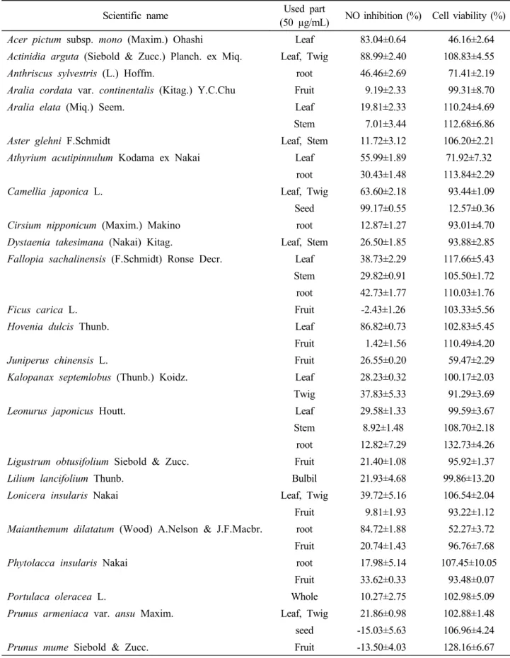
울릉도 민속식물 31분류군 49점에 대한 항염증 검색은 추출물의 최종농도를 50 µg/mL로 하여 LPS로 유도된 RAW 264.7 대식세포에서의 NO 생성량과 세포생존율을 측정하였다. 그 결과 다래(Actinidia arguta) 잎·가지 88.99±2.40%, 헛개나무(Hovenia dulcis) 잎 86.82±
0.73%, 동백나무(Camellia japonica) 잎·가지 63.60±
2.18%, 말오줌나무(Sambucus sieboldiana var. pendula) 잎·가지 46.75±3.74%, 왕호장근(Fallopia sachalinensis) 뿌리 42.73±1.77%, 순으로 NO 생성을 억제하고 세포생존 율은 93%~110%를 나타냈다. 이는 NO 생성 억제가 단순 한 세포의 사멸에 의한 효과가 아니라 추출물의 고유한 효 과라는 점을 의미한다. 동백나무 종자 99.17±0.55%, 큰두 루미꽃(Maianthemum dilatatum) 뿌리 84.72 ±1.88%, 고로쇠나무(Acer pictum subsp. mono) 잎 83.04±0.64%
로 NO 생성을 억제하는 것으로 나타났으나 세포생존율이 각각 12.57±0.36%, 52.27±3.72%, 46.16±2.64%로 나타 나 NO 생성억제는 세포독성의 영향으로 판단된다(Table 2).
1차 검색결과, 추출물 최종농도 50 µg/mL에서 50% 이 상의 NO 생성 억제효과를 보이고 세포 생존에 영향이 적은 다래 잎·가지, 헛개나무 잎, 동백나무 잎·가지 추출물을 대 상으로 LPS로 유도된 RAW 264.7 대식세포에 다양한 농
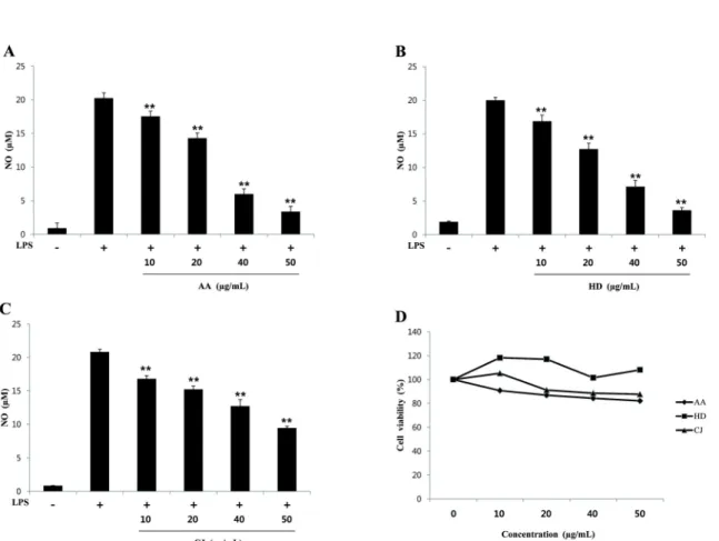
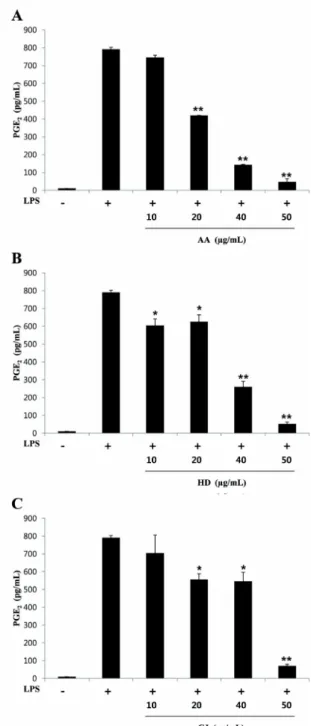
도 (10, 20, 40, 50 µg/mL)로 처리하여 NO 생성량과 세 포생존율, PGE2 생성량을 측정하였다. NO 생성은 농도별 (10, 20, 40, 50 µg/mL)로 각각 다래 잎·가지는 17.53, 14.28, 5.98, 3.38 µM(IC50=29.21 µg/mL), 헛개나무 잎 은 16.85, 12.70, 7.15, 3.63 µM(IC50=27.34 µg/mL), 동 백나무 잎·가지는 16.81, 15.23, 12.70, 9.43 µM (IC50=45.68 µg/mL)로 나타나 농도 의존적으로 유의하게 억제하였다(Fig. 1A~C). 세포생존율은 모든 농도에서 80%이상의 생존율을 보여 RAW264.7 대식세포에 독성을 나타내지 않는 것을 확인하였다(Fig. 1D). PGE2 생성량 측 정결과, LPS 처리군의 PGE2의 양은 790.74 pg/mL이었 고, 20, 40, 50 µg/mL 농도에서 다래 잎·가지는 420.15, 142.36, 46.83 pg/mL(IC50=21.06 µg/mL)로 PGE2 생성 량을 농도 의존적으로 유의성 있게 억제하였다. 헛개나무 잎은 10, 20, 40, 50 µg/mL 농도에서 604.27, 626.06, 259.74, 52.16 pg/mL (IC50=33.47 µg/mL)의 PGE2 생성 량을, 동백나무 잎·가지는 20, 40, 50 µg/mL 농도에서 555.91, 546.31, 70.53 pg/mL(IC50=43.90 µg/mL)의 PGE2 생성량을 보이며 유의적으로 억제효과를 나타냈다 (Fig. 2). 이는 추출물의 처리로 LPS에 의해 진행되는 염 증과정에서 중요한 매개인자인 NO의 형성과 PGE2의 일련 의 감소가 일어나므로 전체적인 염증의 진행이 억제되었다 고 판단된다.
본 실험을 통해 항염 활성이 높게 나타난 민속식물에 대 한 지금까지 보고에 의하면, 다래는 수피에 함유된 catechin(IC50=26.61 µg/mL), (-)-epicatechin(IC50=25.30 µg/mL), quercetin(IC50=30.46 µg/mL), quercetin-3- O-β-D-glucopyranoside(IC50=28.03 µg/mL), quercetin -3-O-galactopyranoside(IC50=27.24 µg/mL)화합물이 NO 생성억제 활성을 보여 본 연구결과와 유사한 억제효과 를 보였다(Lim et al., 2005). 헛개나무 잎에는 지방산계 열인 linoleic acid, plamitric acid와 glutamic acid, leucine, arginine 등의 아미노산류 함량이 열매보다 높은 것으로 보고되었으며, 잎과 줄기의 열수 추출물에서는 vanillic acid, ferulic acid 등이 보고되어 있다. 생리활성 실험으로는 간 독성에 대한 보호효과, 항돌연변이원성 및 암세포 성장억제효과, 항산화 및 항미생물 활성 등이 보고 되었다(Jeong and Shim, 1999; Cho et al., 2000; Park and Chang, 2007; Park et al., 2009; Ahn et al., 2010). 헛개나무 잎의 NO 및 PGE2 생성 억제 효능은 보고
Table 2. Effect of methanol extracts on cell viability and NO production in LPS-stimulated RAW 264.7 cells
Scientific name Used part
(50 µg/mL) NO inhibition (%) Cell viability (%) Acer pictum subsp. mono (Maxim.) Ohashi Leaf 83.04±0.64 46.16±2.64 Actinidia arguta (Siebold & Zucc.) Planch. ex Miq. Leaf, Twig 88.99±2.40 108.83±4.55
Anthriscus sylvestris (L.) Hoffm. root 46.46±2.69 71.41±2.19
Aralia cordata var. continentalis (Kitag.) Y.C.Chu Fruit 9.19±2.33 99.31±8.70
Aralia elata (Miq.) Seem. Leaf 19.81±2.33 110.24±4.69
Stem 7.01±3.44 112.68±6.86
Aster glehni F.Schmidt Leaf, Stem 11.72±3.12 106.20±2.21
Athyrium acutipinnulum Kodama ex Nakai Leaf 55.99±1.89 71.92±7.32
root 30.43±1.48 113.84±2.29
Camellia japonica L. Leaf, Twig 63.60±2.18 93.44±1.09
Seed 99.17±0.55 12.57±0.36
Cirsium nipponicum (Maxim.) Makino root 12.87±1.27 93.01±4.70
Dystaenia takesimana (Nakai) Kitag. Leaf, Stem 26.50±1.85 93.88±2.85 Fallopia sachalinensis (F.Schmidt) Ronse Decr. Leaf 38.73±2.29 117.66±5.43 Stem 29.82±0.91 105.50±1.72 root 42.73±1.77 110.03±1.76
Ficus carica L. Fruit -2.43±1.26 103.33±5.56
Hovenia dulcis Thunb. Leaf 86.82±0.73 102.83±5.45
Fruit 1.42±1.56 110.49±4.20
Juniperus chinensis L. Fruit 26.55±0.20 59.47±2.29
Kalopanax septemlobus (Thunb.) Koidz. Leaf 28.23±0.32 100.17±2.03
Twig 37.83±5.33 91.29±3.69
Leonurus japonicus Houtt. Leaf 29.58±1.33 99.59±3.67
Stem 8.92±1.48 108.70±2.18 root 12.82±7.29 132.73±4.26 Ligustrum obtusifolium Siebold & Zucc. Fruit 21.40±1.08 95.92±1.37
Lilium lancifolium Thunb. Bulbil 21.93±4.68 99.86±13.20
Lonicera insularis Nakai Leaf, Twig 39.72±5.16 106.54±2.04
Fruit 9.81±1.93 93.22±1.12 Maianthemum dilatatum (Wood) A.Nelson & J.F.Macbr. root 84.72±1.88 52.27±3.72 Fruit 20.74±1.43 96.76±7.68
Phytolacca insularis Nakai root 17.98±5.14 107.45±10.05
Fruit 33.62±0.33 93.48±0.07
Portulaca oleracea L. Whole 10.27±2.75 102.98±5.09
Prunus armeniaca var. ansu Maxim. Leaf, Twig 21.86±0.98 102.88±1.48 seed -15.03±5.63 106.96±4.24
Prunus mume Siebold & Zucc. Fruit -13.50±4.03 128.16±6.67
Table 2. Continued
Scientific name Used part
(50 µg/mL) NO inhibition (%) Cell viability (%)
Prunus takesimensis Nakai Leaf, Twig 19.87±2.77 105.48±0.62
Fruit 16.97±1.33 97.27±4.42 Rhododendron brachycarpum D.Don ex G.Don Leaf 33.32±0.34 94.45±9.57
Rhus javanica L. Twig 36.77±1.72 94.61±1.14
Sambucus sieboldiana var. pendula (Nakai) T.B.Lee Leaf, Twig 46.75±3.74 96.08±4.13 Fruit -20.44±2.60 114.27±3.88
Sorbus commixta Hedlund Leaf 21.34±2.72 91.41±1.99
Twig 37.13±3.34 96.36±5.18 Fruit 36.47±4.13 91.07±6.55
Vitis flexuosa Thunb. Leaf, Twig 4.80±3.76 128.09±2.04
Zanthoxylum piperitum (L.) DC. Leaf, Twig 18.07±5.53 125.76±3.81
Fig. 1. Effect of methanol extracts from AA, HD and CJ on LPS-stimulated NO production in RAW 264.7 cells and cell viability. RAW 264.7 cells(3.0×10⁴ cells/well) were treated with different concentrations of AA, HD, and CJ extract in the presence of LPS(1 µg/mL) for 24h. NO in the medium was determined by Griess assay(A~C). Cell viability was determined by MTT assay(D). Three independent experiments were performed, and the data are presented as mean±SD *P <0.05, **P <0.01, compared with cells treated with LPS alone. AA: leaf, twig of Actinidia arguta, HD:
leaf of Hovenia dulcis, CJ: leaf, twig of Camellia japonica.
Fig. 2. Effect of methanol extracts from AA, HD, and CJ on LPS-stimulated PGE2 production in RAW 264.7 cells.
RAW 264.7 cells(1.5×10⁵ cells/well) were treated with different concentrations of AA, HD and CJ extract in the presence of LPS(1 µg/mL) for 24h. PGE2 in medium was determined by ELISA. Three independent experiments were performed, and the data are presented as mean±SD
*P <0.05, **P <0.01, compared with cells treated with LPS alone. AA: leaf, twig of Actinidia arguta, HD: leaf of Hovenia dulcis, CJ: leaf, twig of Camellia japonica.
된 바 없으며, 수피 추출물 80 µg/mL 농도에서 NO 생성 을 10% 이하로 억제하여 잎 추출물 효과가 월등히 좋은 것 으로 확인되었다(Ryu et al., 2003). 이는 식물부위에 따 른 함유 성분이 다르게 나타나는 결과라 생각된다. 동백나 무 잎은 tannin계열인 camelliatannins D, F, G, H와 camellianoside, okicamelliaside 등의 성분이 알려졌고, 항산화, 항미생물, 항암, 항균활성 등에 효과가 있는 것으 로 보고되었다(Han et al., 1994; Hatnno et al., 1995;
Onodera et al., 2006, 2010; Han, 2005; Lee et al., 2005; Kim et al., 2010). 동백나무 수피의 경우 triterpene saponin계열인 camellioside A, B, E, F, G, H 화합물이 분리·동정되었으며, 이들 화합물이 RAW264.7 대식세포에서 NO 생성(IC50: 4.96~18.25 µM)을 억제하였 다는 보고가 있다(Thao et al., 2010). 또한 동백나무 종자 추출물이 RAW264.7 대식세포에서 NF-kB와 AP-1의 활 성을 억제하여 NO, PGE2, TNF-α의 생성을 억제하는 것 으로 나타났다(Kim et al., 2012). 본 연구 결과 동백나무 잎·가지 추출물도 NO 및 PGE2 생성량을 유의적으로 억제 하여 항염 효과가 있는 것으로 판단된다. 따라서 이에 대한 성분 및 작용기전에 대한 추후 연구가 필요하다고 사료된 다.
이상의 결과를 통해 울릉도 민속식물로부터 안전하고 효 능이 우수한 항염증소재 후보를 발굴하였다. 따라서 향후 이들 소재에 대한 항염 활성 기반의 유효성분이 규명되어 져야 할 것이며, 유효성분의 기전연구를 통해 동맥경화, 고 혈압, 암, 당뇨, 관절염 등과 같은 만성 염증성 질환의 치 료와 예방에 효과적으로 적용할 수 있을 것으로 기대된다.
적 요
울릉도 민속식물 31분류군 49점 추출물을 대상으로 최 종농도 50 µg/mL로 LPS에 의해 유도된 RAW 264.7 대식 세포의 NO 생성량을 조사하였다. 그 결과, 다래(Actinidia arguta) 잎·가지, 헛개나무(Hovenia dulcis) 잎, 동백나무 (Camellia japonica) 잎·가지, 말오줌나무(Sambucus sieboldiana var. pendula) 잎·가지, 왕호장근(Fallopia sachalinensis) 뿌리 순으로 항염 활성이 우수하게 나타났 다. 이 중에서 다래 잎·가지, 헛개나무 잎, 동백나무 잎·가 지 추출물을 LPS로 유도된 Raw 264.7 대식세포에서 최종 농도가 10, 20, 40, 50 µg/mL로 처리하여 NO 생성량과
세포생존율, PGE2 생성량을 측정하였다. 그 결과 다래 잎·
가지(IC50=29.21 µg/mL), 헛개나무 잎(IC50=27.34 µg/mL), 동백나무 잎·가지(IC50=45.68 µg/mL) 모두 NO 생성을 농도 의존적으로 유의성 있게 억제하였다. 또한 PGE2 측정결과 다래 잎·가지(IC50=21.06 µg/mL), 헛개나 무 잎(IC50=33.47 µg/mL), 동백나무 잎·가지(IC50=43.90 µg/mL)에서도 유의성 있는 억제효과를 보였다. 본 연구 결과 다래 잎·가지, 헛개나무 잎, 동백나무 잎·가지의 추출 물은 염증 유발의 중요 인자인 NO 및 PGE2 생성을 저해하 여 우수한 항염증 효과를 보였다. 이들 소재에 대한 염증 억제 유효성분 규명 및 그 작용기전을 추가적으로 연구함 으로써 만성 염증질환의 예방과 치료에 효과적으로 사용할 수 있을 것으로 기대된다.
인용문헌
Ahn, B.S., J.W. Kim, H.T. Kim, S.D. Lee and K.W. Lee. 2010.
Antioxidant effects of Hovenia dulcis in the streptozotocin- induced diabetic rats. J. Vet. Clin. 27:336-373 (in Korean).
Cho, J.Y., J.H. Moon and K.H. Park. 2000. Isolation and identification of 3-methoxy-4-hydroxybenzoic acid and 3- methoxy-4-hydroxycinnamic acid from hot water extracts of Hovenia dulcis Thunb, and confirmation of their antioxidative and antimicrobial activity. Korean J. Food Sci. Technol.
32:1403-1408 (in Korean).
Choi, K., J.J Ku, K.W. Park, H.R. Jeong, H.J. Kim and P.S.
Shin. 2010. Folk Plants in Korean Peninsula Ⅵ.: Gyeongnam
& Gyeongbuk Provinces. Pocheon, Korea National Arbortum, Korea. pp. 1-424 (in Korean).
Chou, T., F. Earl and E. Shen. 2003. Chitosan inhibits PGE2
formation and COX-2 induction in lipopolysaccharide-treated RAW 264.7 macrophages. Biochem. Biophys. Res. Com.
308:403-7.
Chung, G.Y., M.S. Park, B.M. Nam, K.N. Hong, J. Jang and C.H. Lee. 2010. The regional folk plants in inland of Gyeongsangbuk-do (Ⅰ). Korean J. Plant Res. 23:465-479 (in Korean).
Chung, G.Y., M.S. Park, B.M. Nam, D.H. Jeong, C.H. Lee, Y.H. Shin, H.J. Kim and S.H. Kang. 2011. The folk plants in southern region of Gangwon-do. Korean J. Plant Res.
24:379-394 (in Korean).
Fierro, I.M. and C.N. Serhan. 2001. Mechanisms in anti- inflammation and resolution: the role of lipoxins and
aspirintriggered lipoxins. Braz. J. Med. Biol. Res. 34:555- 566.
Han, L., T. Hatano, T. Yoshida and T. Okuda. 1994. Tannins of theaceous plants V. camelliatannins F, G and H, three new tannins from Camellia japonica L. Chem. Pharm. Bull.
42:1399-1409.
Han, Y.S. 2005. Antimicrobial effects of Camellia japonica L.
leaves extract on food-orne phathogenic microorganisms.
Korean J. Food Sci. Technol. 37:113-121 (in Korean).
Hatano, T., L. Han, S. Taniguchi, T. Okuda, Y. Kiso, T. Tanake and T. Yoshida. 1995. Camelliatannin D, a new inhibitor of bone resorption, from Camellia japonica. Chem. Pharm.
Bull. 43:2033-2035.
Higuchi, M., N. Higashi, H. Taki and T. Osawa. 1990. Cytolytic mechanism of activated macrophages. Tumor necrosis factor and L-arginine-dependent mechanism act as synergistically as the major cytolytic mechanism of activated macrophages.
J. Immunol. 144:1425-1431.
Im, H.T., H.H. Hong, H.D. Son, M.S. Park, B.M Nam, B.K.
Kwon, C.H Lee and G.Y. Chung. 2011. The usage of regional folk plants in Gyeongsangnam-do. Korean J. Plant Res. 24:419-429 (in Korean).
Jeong, C.H and K.H. Shim. 1999. Chemical components in leaf fruit stalk of Hovenia dulcis Thunb. Korean J. Postharvest Sci. Technol. 6:469-471 (in Korean).
Jeong, H.R., K. Choi and G.W. Park. 2012. The regional folk plants in southern inland area of Gyeonggi-do. Korean J.
Plant Res. 25:523-542 (in Korean).
Kim, J.H., C.H. Jeong and K.H. Shim. 2010. Antioxidative and anticancer activities of various solvent fractions from the leaf of Camellia japonica L. Korean J. Food Preserv.
17:267-274 (in Korean).
Kim, J.Y., K.S. Jung and H.G. Jeong. 2004. Suppressive effects of the kahweol and catestol on cycloocygenase-2 expression in macrophages. FEBS Letters 569:321-326.
Kim, K.A., J.S. Han, K.S. Cheon, J.H. Jang, G.H. Ok and K.O.
Yoo. 2012. Folk plants in the inland of northern area in Gangwon-do. Korean J. Plant Res. 25:48-62 (in Korean).
Kim, S.B., E.S Jung, S.W. Shin, M.H. Kim, Y.S. Kim, J.S. Lee and D.H. Park. 2012. Anti-inflammatory activity of Camellia japonica oil. BMB reports. 45:177-182.
Kwon S.K. 2001. SAR of COX-2 inhibitors. Biomolecules &
Therapeutics 9:69-78
Lee, S.Y., E.J. Hwang, G.H. Kim, Y.B. Choi, C.Y. Lim and S.M. Kim. 2005. Antifungal and antioxidant activities of
extracts from leaves and flowers of Camellia japonica L.
Korean J. Med. Crop Sci. 13:93-100 (in Korean).
Lim, H.W., J.G. Shim, H.K. Choi and M.W. Lee. 2005.
Phenolic compounds from barks of Actinidia arguta Planchon growing in Korea and its anti-oxidative and nitric oxide production inhibitory activities. Kor. J. Pharmacogn.
36:245-251 (in Korean).
Lowenstein, C.J. and S.H. Snyder. 1992. Nitric oxide, a novel biologic messenger. Cell 70:705-707.
McDaniel, M.L., G. Kwon, J.R. Hill, C.A. Marshall and J.A.
Corbett. 1996. Cytokines and nitric oxide in islet inflammation and diabetes. Proc. Soc. Exp. Biol. Med. 211:24-32.
Moncada, S., R.M. Palmer and E.A. Higgs. 1991. Nitric oxide:
physiology, pathophysiology, and pharmacology. Pharmacol.
Rev. 43:109-142.
Nathan, C. and Q.W. Xie. 1994. Nitric oxide synthases: roles, tolls and controls. Cell 78:915-918.
Onodera, K., K. Hanashiro and T. Yasumoto. 2006. Came- llianoside, a novel antioxidant glycoside from the leaves of Camellia japonica. Biosci. Biotechnol. Biochem. 70:1995- 1998.
Onodera, K., K. Tsuha, M. Yasumoto-Hirose, K. Tsuha, K.
Hanashiro, H. Naoki and T. Yasumoto. 2010. Okicame- lliaside, an extraordinarily potent anti-degranulation glucoside isolated from the leaves of Camellia japonica. Biosci.
Biotechnol. Biochem. 74:2532-2534.
Park, J.S., E.J. Lee, J.C. Lee, W.K. Kim and H.S. Kim. 2007.
Anti-inflammatory effects of short chain fatty acids in
IFN-gamma-stimulated RAW 264.7 murine macrophage cells: involvement of NF-KappaB and ERK signaling pathways. Int. Immunopharmacol. 7:70-77.
Park, S.H. and E.Y. Chang. 2007. Antimutagenic and cytotoxic effects of Hovenica dulcis Thumb leaves extracts. J. Korean Soc. Food Sci. Nutr. 36:1371-1376 (in Korean).
Park, S.H., E.Y. Chang, J.S. Chang and K.Y. Yoon. 2009.
Protective effect of Hovenica dulcis Thumb leaves extract on hepatic injury induced by benzo(α)pyrene in mice. J.
Korean Soc. Food Sci. Nutr. 38:509-573 (in Korean).
Ryu, J.H., H. Ahn, J.Y. Kim and Y.K. Kim. 2003. Inhibitory activity of plant extracts on nitric oxide synthesis in LPS-activated macrophages. Phytother. Res. 17:485-489.
Sarkar, D and P.B. Fisher. 2006. Molecular mechanisms of aging-associated inflammation. Cancer Lett. 236:13-23 Sunyer, T., L. Rothe, X. Jiang, F. Anderson, P. Osdoby and P.
Collin-Osdoby. 1997. Ca2+ or phorbol ester but not inflammatory stimuli elevate inducible nitric oxide synthase messenger ribonucleic acid and nitric oxide (NO) release in avian osteoclasts: autocrine NO mediates Ca2+-inhibited bone resorption. Endocrinology 138(5):2148-2162.
Thao, N.T.P., Hung, T.M., Cuong, T.D., Kim, J.C., Kim, E.H., Jin, S.E., Na, M., Lee, Y.M., Choi, J.S and B.S. Min. 2010.
28-Nor-oleanane-type triterpene saponins from Camellia japonica and their inhibitory activity on LPS-induced NO production in macrophage RAW264.7 cells. Bioorg. Med.
Chem. Lett. 20:7435-7439.
(Received 12 November 2012 ; Revised 18 February 2013 ; Accepted 25 February 2013)