• 검색 결과가 없습니다.

The activation of PPAR-α and Wnt/β-catenin by Paeonia lactiflora root supercritical carbon dioxide extract

N/A
N/A
Protected

Academic year: 2021

Share "The activation of PPAR-α and Wnt/β-catenin by Paeonia lactiflora root supercritical carbon dioxide extract"

Copied!
7
0
0

로드 중.... (전체 텍스트 보기)

전체 글

(1)

ISSN 2288-1069 (Online)

http://dx.doi.org/10.12925/jkocs.2019.36.4.1136

The activation of PPAR-α and Wnt/β-catenin by Paeonia lactiflora root supercritical carbon dioxide extract

Bora Kim

Division of Biomedicinal Chemistry and Cosmetics, Mokwon University, Daejeon, 35349, Republic of Korea

(Received November 26, 2019; Revised December 13, 2019; Accepted December 17, 2019)

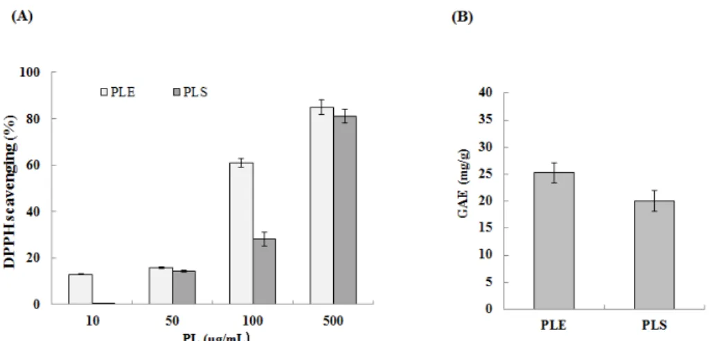
Abstract : The root of Paeonia lactiflora has been used in Chinese medicine. We conducted to check the comparative qualities of ethanol solvent extraction (PLE) and supercritical carbon dioxide extraction (PLS) of P. lactiflora root. PLE had higher antioxidant and polyphenol contents than PLS. But, PLS were significantly increased peroxisome proliferator-activated receptor (PPAR)-α. In addition, PLS inhibited the adipocyte differentiation of 3T3-L1 cells. When treated with the extract at a concentration of 100 μg/mL, the Wnt/β-catenin pathway reporter luciferase activity of HEK 293-TOP cells increased approximately by 3-folds compared to that of the untreated control group. These results indicate that P. lactiflora supercritical carbon dioxide extract may serve as a cosmeceutical for improving skin barrier function and the treatment of obesity.

Keywords : Paeonia lactiflora, Supercritical carbon dioxide extraction, Peroxisome proliferators activated receptors, Wnt/β-catenin, Adipocyte differentiation

1. Introduction

Medicinal plants are used in traditional medicine and synthesise hundreds of biological compounds for functions including defence against insects, fungi, diseases, and herbivorous mammals. Despite profound therapeutic benefits of many biological compounds, most medicinal plants have been studied mostly for the development of drugs and there are only a few reports on cosmeceutical agents.

Additionally, more effective and less toxic medicinal compounds are still required.

Ethanol, water and supercritical carbon dioxide



Corresponding author

(E-mail: [email protected])

are generally considered as safe solvents.

Especially, supercritical carbon dioxide (CO

2

) extraction method is an eco-friendly and has been successfully used to isolate the essential oil from natural products[1].

PPAR-α promotes the synthesis of

cholesterol and ceramides in keratinocytes

during epidermal differentiation[2]. In terms of

keratinocyte differentiation and epidermal

permeability barrier, PPAR-α agonists have

been known extensively, and it has been

studied that treatment with PPAR ligands

promotes differentiation of the murine

epidermis[3]. Therefore, a search for new

agonists is necessity to determine whether

activators of PPAR-α can alter the rate of

keratinocyte differentiation. Wnt/β-catenin

(2)

signaling pathway plays a role in adipose cell communication and the inhibition of adipogenesis[4]. The understanding of molecular and cellular mechanisms regulating adipogenesis is essential for designing rational therapies for treatment of obesity[5].

Paeonia lactiflora is a species of herbaceous perennial flowering plant in the family Paeoniaceae, native to central and eastern Asia.

Also, the root of its plant has been used in Chinese medicine[6]. Several studies have reported that the root of P. lactiflora shows positive biological effects, such as neuroprotective activity[7], anti-inflammation effects[6], and skin-depigmentation[8].

However, the effects of its root extract as a skin therapeutic agent for skin barrier function and as a nutraceutical product for obesity treatment have not been studied. In this study, we examined PPAR-α activity and adipogenesis activity with P. lactiflora root extract.

2. Experimental

2.1. Sample preparation

The P. lactiflora root was purchased from the Jeonnam herbal farming cooperative (Hwasun-gun, Jeollanam-do, Korea). For supercritical co-solvent modified carbon dioxide extraction (PLS), the system and required components were acquired at 250 bar 50℃ 150 min at a flow rate of ethyl alcohol 3 mL/min for 90min from Nano bio research center (Janseong-gun, Jeollanam-do, Korea).

The extraction was performed by a method previously described[9]. In case of PLE, P.

lactiflora root was soaked in 99.9% ethyl alcohol for 7 days and then was dried by speed vacuum at 50℃.

2.2. Cell culture and materials

3T3-L1 preadipocytes, which were obtained from Yonsei University, were cultured by a method previously described[10]. Human

normal keratinocyte and HEK 293-TOP cells were purchased from American Type Culture Collection (ATCC, USA) and cultured by a method previously described[11].

2.3. Cell viability

Cell viability was measured using the 3-(4,5-dimethylthiazol-2-yl)-2,5-diphenyltetra zolium bromide (MTT) assay. Human normal keratinocytes were treated with different concentrations of P. lactiflora root (PL) for 48 h at 37°C, followed by the addition of 50 μ L of 2 mg/mL MTT (Sigma-Aldrich, St.

Louis, MO, USA) solution to respective wells, and then incubation for 3 h at 37°C. The procedure was conducted by the method previously described[12].

2.4. DNA constructs, transient transfection, and PPRE transactivation assay

PPAR-α transcription activity was performed using the PPAR response element (PPRE) transactivation method[13] with slight modifications. PPAR-α transcription activity was performed using the PPRE transactivation method (Kim et al ., 2006) with slight modifications. A commercial PPAR-α expression vector was purchased from Promega (Mannheim, Germany) and transformed into Escherichia coli competent cells. PPAR-α DNA constructs were extracted using a DNA preparation kit (Qiagen, Hilden, Germany).

The reporter construct (SA Bioscience, Hilden, Germany) and PPAR-α expression vector were co-transfected into CV-1 cells by using Lipofectamine (Invitrogen, Carlsbad, CA, USA).

Transactivation assays were performed using the Dual-Luciferase Reporter Assay System (Promega, Mannheim, Germany), and normalized luciferase activity was determined.

2.5. Luciferase assay

HEK 293-TOP cells (3 x 10

4

) were seeded

into 96-well plates and incubated in a

medium with 10% FBS for one day. The

procedure was carried out as previously

(3)

described[11]. HEK 293-TOP cells (3 x 10

4

) were seeded into 96-well plates and incubated in medium with 10% FBS for one day. Total cell lysates were extracted with 25 μl 1x reporter lysis buffer (Promega, Madison, WI) per each well and luciferase activities were measured by adding 25 μl luciferin (USB, Cleveland, OH) per each well using a Microplate Luminometer (BMG Labtech, Offenburg, Germany).

2.6. Adipocyte differentiation and Oil Red O staining

The 3T3-L1 preadipocytes were cultured in Dulbecco's modified Eagle's medium (DMEM) with high glucose, 110 mg/L pyruvate supplemented with heat inactivated 10% (v/v) calf serum (Gibco, CA, USA), 100 μg/mL penicillin, 100 μg/mL streptomycin in a CO

2

incubator at 37 °C. To induce adipocyte differentiation, 3T3-L1 cells were cultured with DMEM plus 10% (v/v) heat inactivated fetal bovine serum (FBS) (Gibco) containing 520 μM isobutylmethylxanthine, 1 μM dexamethasone and 167 nM insulin. After 2 days, the media was then changed, and 167 m M of insulin. On day 4, medium was replaced with DMEM containing 10% FBS, and changed with fresh identical medium every 2 days. To measure the anti-differentiation effects of each drug, 3T3-L1 preadipocytes were induced to differentiate in the presence of different concentrations of LiCl, plant extracts.

Adipocyte cell layers were washed with PBS, fixed with 4% paraformaldehyde in PBS, stained with the Oil Red O dye solution for 1 hour, and then washed with distilled water.

Cells were checked by bright-field optical microscope (Nikon TE-200U, Tokyo, Japan).[10]

2.7. Antioxidant assay

The 2,2-diphenyl-1-picrylhydrazyl (DPPH) assay was performed to determine the antioxidant capacity of LC. The SC

50

(concentration required to obtain a 50%

antioxidant effect), a commonly used parameter to indicate the antioxidant capacity, was also measured[14].

2.8. Total polyphenol content assay Total phenolic content and other oxidation substrates were examined using the Folin Ciocalteau (FC) reagent. We used a previously described method[15]. The FC assay relies on the transfer of electrons in alkaline medium from phenolic compounds to phosphomolybdic/

phosphotungstic acid complexes, which are determined at 765 nm. Antioxidant reducing capacity is expressed as gallic acid equivalents (GAE).

2.9. Statistical analysis

All data are presented as the mean ± standard error of the mean (S.E.M). Statistical analyses were conducted with GraphPad Prism 5.0 (GraphPad Software, Inc. San Diego, CA, USA). Comparisons between multiple groups were performed using the one-way analysis of variance with Bonferroni post-hoc test.

P-value of less than 0.05 was considered significant.

3. Results and Discussion

3.1. Effect of PL on cell viability

We examined the effect of PL on cell viability. After 24 h treatment of Human normal keratinocytes with PL (10, 50, and 100 µg/mL), cell viability was assessed by the MTT assay. Cell viability was not affected by 100 µg/mL of PLE (Figure 1). In case of PLS, there was no cell toxicity up to 10 µg/mL.

3.2. Antioxidant effect and total polyphenol content of PL

The radical scavenging activities of PL increased in a concentration-dependent manner. The radical scavenging activity SC

50

of PLE was 141.7 µg/ml and that of PLS was

265.2 µg/ml, therefore PLE has better

(4)

Fig. 2. (A) Antioxidant effect on PL. PL indicate P. lactiflora root extract. (B) Total polyphenol content of PLE and PLS GAE indicate gallic acid equivalent in mg/g. Values are presented as means ± standard error of the mean (SEM).

antioxidant activity. The polyphenol content of PLE was 25.27 GAE mg/g and that of PLS was 20.02 GAE mg/g. So, This seems to be due to the higher polyphenol content of PLE (Fig. 2A, B).

Fig. 1. Effect of PL on cell viability. Ethanol solvent extraction and supercritical carbon dioxide extraction from P.

lactiflora root indicate PLE and PLS respectively. No treatment was used as a negative control. Values are presented as means ± standard error of the mean (SEM).*p < 0.05, **p <

0.01 compared to the untreated control group.

3.3. Transactivation of PPRE

As shown in Fig. 3, the concentration of PL largely influenced PPAR-α ligand binding activity in the treated group, with 50 µg/mL of PLS showing an increased binding compared to that in the untreated control group. In addition, the PLE-treated groups showed low levels of transactivation activity compared to the WY14643-treated groups.

These results show that PLS has the potential as a novel PPAR-a agonist. Therefore, PLS may serve as potential compounds for improving skin barrier function.

3.4. Effect of PLS on the adipocyte differentiation and Wnt/β-catenin activity

The reporter activity was increased approximately by 3-folds with the treatment of PLS at concentrations of 100 µg/mL, compared to that of the non-treated control group (Fig. 4). Additionally, PLS inhibited adipocyte differentiation of 3T3-L1 cells in a concentration dependent manner (Fig. 5).

These results indicate that PLS induces

differentiation of 3T3-L1 cells, showing the

role of the Wnt/β-catenin activator in the

adipocyte differentiation. Wnt/β-catenin

(5)

signaling has been known to modulate additional developmental processes of adipocytes and to play a role in the suppression of differentiation of pre-adipocyte cells of 3T3-L1 cells[16]. At present, there are some reports about the interaction between PPAR-α and Wnt/β-catenin signaling.

PPAR-α activation, with its agonist clofibrate, inhibits Wnt/β-catenin signaling in renal fibrosis and pancreatic cancer cells[17]. Also, Wnt/β-catenin activation promotes PPAR-α expression in Alzheimer disease[18]. Totally, our study indicate that PLS promotes PPAR- α and inhibits adipocyte differentiation in relation to the Wnt/β-catenin signaling pathway. Here, we report that PLS can be used in the development of cosmeceutical products for skin barrier dysfunction such as atopic dermatitis (AD) and obesity. Therefore, natural substances derived from plants influencing adipogenesis are receiving attention for the treatment of obesity.

Fig. 3. Transactivation of peroxisome proliferator-ctivated receptor (PPAR)- esponsive element (PPRE) by different concentrations of PLE and PLS.

HEK293 cells were transfected with PPRE luciferase construct. WY14643 was used as a positive control and no treatment was used as a negative control. Values are presented as means

± standard error of the mean (SEM).

*p < 0.01 compared to the untreated control group.

Fig. 4. Effect of PLS on Luciferase reporter activity. HEK293 cells containing pTOPFlash reporter gene in its chromosome were cultured and treated with different LC at 1, 10 or 100 μ g/mL concentration, and cellular extract was prepared after 24 h as described in the Materials and methods. PLS indicate supercritical carbon dioxide extraction from P.

lactiflora root. *p < 0.05, **p < 0.01 compared to the untreated control group.

4. Conclusion

This study shows that supercritical carbon dioxide extraction (PLS) of Paeonia lactiflora root can increase peroxisome proliferator- activated receptor (PPAR)-α and inhibite the adipocyte differentiation of 3T3-L1 cells.

When treated with the extract at a

concentration of 100 μg/mL, the Wnt/β

-catenin pathway reporter luciferase activity of

HEK 293-TOP cells was increased

approximately by 3-folds compared to that of

the untreated control group. This study

suggests that PLS have potential as

cosmeceutical agents for improving skin barrier

functions for atopic dermatitis and the

treatment of obesity.

(6)

Fig. 5. Effect of PLS on the adipocyte differentiation of 3T3-L1 preadipocyte cells.

3T3-L1 cells were induced to differentiation for 7 days with or without 20 mM LiCl. Intracellular lipids of 3T2-L1 cells were visualized by Oil Red O staining as described in the Materials and methods. Original magnification: x400. PLS indicate supercritical carbon dioxide extraction from P. lactiflora root.

Acknowledgements

The author is grateful to Enprani Co. Ltd and Translational Research Center for Protein Function Control in Yonsei University for their support of this project and technical assistance.

References

1. K. Tyskiewicz, M. Konkol, E. Roj,

“Supercritical Carbon Dioxide (scCO

2

) Extraction of Phenolic Compounds from Lavender (Lavandula angustifolia) Flowers:

A Box-Behnken Experimental

Optimization”, Molecules , Vol. 23, No.10 10.3390/molecules23102625 (2018).

2. M. Rivier, I. Safonova, P. Lebrun, C. E.

Griffiths, G. Ailhaud, S. Michel,

“Differential expression of peroxisome proliferator-activated receptor subtypes during the differentiation of human keratinocytes”, Journal of Investigative Dermatology , Vol. 111, No.6 pp.

1116-1121 (1998).

3. L. G. Kömuves, K. Hanley, A. M.

Lefebvre, M. Q. Man, D. C. Ng, D. D.

Bikle, M. L. Williams, P. M. Elias, J.

Auwerx, K. R. Feingold, “Stimulation of PPAR-a promotes epidermal keratinocyte differentiation in vivo”, Journal of Investigative Dermatology , Vol. 115, No.3 pp. 353-360 (2000).

4. J. Liu, S. R. Farmer, “Regulating the Balance between Peroxisome Proliferator- activated Receptor γ and β-Catenin Signaling during Adipogenesis”, Journal of Biological Chemistry , Vol. 279, No.43 pp.

45020-45027 (2004).

5. G. Fruhbeck, J. Gomez-Ambrosi, F. J.

Muruzabal, M. A. Burrell, “The adipocyte:

a model for integration of endocrine and metabolic signaling in energy metabolism regulation”, American Journal of Physiology-Endocrinology and Metabolism , Vol. 280, No.6 pp. 827-847 (2001).

6. D. He, S. Dai, “Anti-inflammatory and

(7)

immunomodulatory effects of Paeonia lactiflora Pall., a Traditional chinese herbal medicine”, Frontier in Phamacology , Vol.

2, No.10 pp. 1-5 (2011).

7. S. H. Kim, M. K. Lee, K. Y. Lee, S. H.

Sung, J. Kim, Y. C. Kim, “Chemical constituents isolated from Paeonia lactiflora roots and their neuroprotective activity against oxidative stress in vitro”’ Journal of Enzyme Inhibition and Medicinal Chemistry , Vol. 24, No.5 pp. 1138-1140 (2009).

8. J. Qiu, M. Chen, J. Liu, X. Huang, J.

Chen, L. Zhou, J. Ma, P. Sextius, A. M.

Pena, Z. Cai, S. Jeulin, “The skin-depigmenting potential of Paeonia lactiflora root extract and paeoniflorin: in vitro evaluation using reconstructed pigmented human epidermis”, International Journal of Cosmetic Science , Vol. 38, No.5 pp 444-451 (2016).

9. B. Kim, S. M. Lee, T. Hwang, H. Kim,

“Anti-oxidative and skin barrier effects of natural plants with a supercritical extract”, Korean Journal of Food Preservation , Vol.

20, No.5 pp. 597-601 (2013).

10. S. Lee, B. Kim, M. Oh, J. Yoon, H. Kim, K. Lee, J. Lee, K. Choi, “ Persicaria hydropiper (L.) spach and its flavonoid components, isoquercitrin and isorhamnetin, activate the Wnt/-βcatenin pathway and inhibit adipocyte differentiation of 3T3-L1 cells”, Phytotherapy Research, Vol. 25, No.11 pp.

1629-1635 (2011).

11. B. Kim, Y. Choi, H. Kim, “ Eruca sativa and its flavonoid components, quercetin and isorhamnetin, improve skin barrier function by activation of peroxisome proliferator-activated receptor (PPAR)-α and suppression of inflammatory cytokines”, Phytotherapy Research, Vol.

28, No.9 pp. 1359-1366 (2014).

12. B. Kim, H. Kim, “Novel peptide inhibits inflammation by suppressing of protease activated receptor-2”, European Journal of

Pharmacology , Vol. 832, No.1 pp. 25-32 (2018).

13. S. H. Kim, G. W. Nam, H. K. Lee, S. J.

Moon, I. S. Chang, “The effects of Musk T on peroxisome proliferator-activated receptor [PPAR]-a activation, epidermal skin homeostasis and dermal hyaluronic acid synthesis”, Archives of Dermatological Research , Vol. 298, No.6 pp. 273-282 (2006).

14. K. V. Sharma, R. Sisodia, “Evaluation of the free radical scavenging activity and radioprotective efficacy of Grewia asiatica fruit”, Journal of Radiological Protection , Vol. 29, No.3 pp. 429-443 (2009).

15. S. Dall’Acqua, R. Cervellati, M. C. Loi, G. Innocenti, “Evaluation of in vitro antioxidant properties of some traditional Sardinian medicinal plants: Investigation of the high antioxidant capacity of Rubus ulmifolius ”, Food Chemistry , Vol. 106, No.2 pp. 745-749 (2008).

16. J. Y. Ahn, H. J. Lee, S. N. Kim, T. Y.

Ha, “Curcumin‐inducedsuppression of adipogenic differentiation is accompanied by activation of Wnt/β‐catenin signaling”, American Journal of Physiology Cell Physiolology, Vol. 298, No.6 pp. 1510–

1516 (2010).

17. J. Xu, W. Zhu, J. Song, Y. Jiao, J. Luo, C. Yu, J. Zhou, J. Wu, M. Chen, W.

Ding, “Activation of PPARa by clofibrate sensitizes pancreatic cancer cells to radiation through the Wnt/b-catenin pathway”, Oncogene , Vol. 37, No.7 pp.

953-962 (2018).

18. A. Vallée, Y. Lecarpentier, “Alzheimer

Disease: Crosstalk between the Canonical

Wnt/Beta-Catenin Pathway and PPARs

Alpha and Gamma”, Frontiers in

neuroscience , Vol. 10, No.1 pp. 1-12

(2016).

수치

Fig.  1.  Effect  of  PL  on  cell  viability.  Ethanol  solvent  extraction  and  supercritical  carbon  dioxide  extraction  from  P
Fig. 3. Transactivation  of  peroxisome  proliferator-ctivated  receptor  (PPAR)-  esponsive  element  (PPRE)  by  different  concentrations  of  PLE  and  PLS
Fig.  5.  Effect  of  PLS  on  the  adipocyte  differentiation  of  3T3-L1  preadipocyte  cells

참조

관련 문서

When fiber posts were cemented into the root canal using total-etch adhesives, the bond strength and adaptation between post and root canal dentin were affected

In this study, it can be interpreted that the combined increase of the trust and the alternative attraction makes the intention of conversion higher rather

The limit is therefore the carbon char as a carrier utilizing only 27% of available hydrogen but sequestering 91kg of carbon dioxide (as measured experimentally) per million

Null hypotheses of this study was there is no difference in root canal length determination between conventional and heat-treated Ni-Ti files by 2

This study shows that the root cause of suicide is based on social and legal exclusion and proposes that suicide prevention measures should be aimed at social cohesion

In this study, it is shows that the stream of CO 2 is more effective in the reduction of unreacted TEGDMA and the increase of surface microhardness than that of N 2

- the right side : to move deoxygenated blood that is loaded with carbon dioxide from the body to the lungs - the left side : to receive oxygenated blood that has.. had most

The aim of this study was to evaluate the sealing ability in Type II root canal obturation by comparing the presence of voids between single cone technique using Endoseal MTA