750
F OREST S OCIETY
Development of Zygotic Embryos and Seedlings is Affected by Radiation Spectral Compositions from Light Emitting Diode (LED)
System in Chestnut ( Castanea crenata S. et Z.)
So-Young Park
1* and Man-Jo Kim
21
Forest biotechnology Division, Korea Forest Research Institute, Suwon 441-847, Republic of Korea
2
Special purposed tree Division, Korea Forest Research Institute, Suwon 441-847, Republic of Korea
Abstract : Among the environmental conditions employed in micropropagation, light quality plays an important role in growth, specially morphogenesis and photosynthesis. The effect of radiation quality (350- 740 nm) on the development and growth of zygotic embryos and in vitro plantlets of open-pollinated chestnut ( Castanea crenata S. et Z.) were studied. Two types of explants were exposed for 4 weeks to cool white (W, as control), monochromatic red (R, peak emission 650 nm), monochromatic blue (B, peak emission 440 nm), red+blue (R+B, 1:1), or red+far-red (R+Fr, 1:1, far-red peak emission 720 nm) radiation from a light-emitting-diode (LED) system. While the zygotic embryos showed positive photoblastic behavior, their germination was inhibited by blue radiation. Hypocotyl elongation and root development were promoted by red radiation. The emergence of primary leaf and its expansion were faster under blue than under red radiation. In the plantlets, red and red+far-red radiation significantly increased the formation and growth of the root, whereas blue light reduced rooting. Therefore, radiation quality appears to influence some steps in the development of zygotic embryos and plantlets in the chestnut.
Key words : chestnut, light quality, LED, embryo, growth
Introduction
Light is an essential and primary energy source for plants (Stuefer and Huber, 1998). Light quality, mediated by phytochrome, affects the growth and development of a wide range of plant species in vitro. Fluorescent lamps are generally used as a conventional light source for growing plantlets in vitro; however, the light from these lamps contain unwanted wavelengths that are inadequate in promoting growth. A light-emitting diode (LED) sys- tem has been considered as a better alternative with improved features such as a smaller mass and volume, a longer life, and an effective single wavelength for mor- phogenesis and photosynthesis. In recent years, the use of LEDs as a radiation source for plants has attracted considerable interest because of its vast potential for developmental and photomorphogenetic studies as well as for its commercial applications.
Plant development is strongly influenced by light, which is mediated through photoreceptors known as phytochromes (which detect red and far-red light), cryp- tochromes (blue and UV-B), and phototropins (blue and
UV-A). All these photoreceptors are involved in one or more processes of photomorphogenesis (Ascencio-Cabral et
al ., 2008). The growth and development of herbal plants are influenced by the wavelength of incident light (Nhut
et al. , 2003; Heo et al. , 2006; Moon et al. , 2006), but the effect of light wavelength on tree growth and develop- ment remains unclear. Moreover, the effect of light quality on the development of embryos and plantlets in hardwood species, especially chestnut, is not as well known as those in herbs (Aphalo and Lehto, 1997; 2001).
Since the 1980’s, European and American chestnut have been studied for their disease resistance by employ- ing in vitro culture techniques. However, studies on Korean chestnuts are rare because of the recalcitrant behavior of these species in vitro.
The aim of the present study was to investigate the devel- opmental and growth responses of Korean chestnut embryos and plantlets grown in vitro under 5 different types of mono- chromic or mixed radiations emitted through an LED system.
Materials and Methods 1. Plant material
For the experiment, seeds of chestnut which were har- vested from Hwaseung, October 2005, and kept in 4
oC
*Corresponding author
E-mail: [email protected]
for 4-month were used for this experiment. The embryos were dissected from the surface sterilized fruit. The embryos around 3-5 mm in size (before cotyledons were developed) were cultured onto Woody Plant medium (WPM; Lloyd and McCown, 1981) containing 3% (w/v) sucrose and 2.5 g/L gelrite. To investigate the embryo development under different light quality, embryos cul- tured onto medium were placed under various light irra- diation treatments.
To investigate the effect of light quality on plantlet growth, in vitro plantlets (two internodes with 1~2 leaves) were used. Five plantlets were cultured in a magenta culture box containing 60 mL WPM, and the culture vessels were placed under the different light treatments.
In all experiment, the pH of the medium was adjusted to 5.5 and all media were autoclaved for 20 min at 115°C (1.37×10
5Pa).
2. Culture conditions
Cultures were incubated in a tissue culture room main- tained at 25±1°C under a 16-h photoperiod with various light irradiance at 40 mmol m
−2s
−1photosynthetic photon flux (PPF). After 2 months of culture, several growth fac- tors, e.g., number of leaves, internodal length and num- ber of nodes were investigated.
3. LED system
The LED system (GF-320s; Good Feeling, Korea) used in this study comprised of LED sticks, a panel, and a main controller for light intensity. In the experiment, cultures were placed either under various 1:1 combinations of radiation (RB, red+blue; RFr, red+far-red) from the LEDs, red (R), blue (B), or cool white fluorescent lamps (Kumho FL40D, Korea) (F) as the control. The energy ratio (%) in spectral distributions of LED used in the experiment
was 1:1 or 1:2 in mixed radiations. As spectral energy sources, the emission of blue LED was 440 nm, red was at 650 nm, and far-red was at 730 nm (Figure 1).
4. Histological observation
Stem collected from cultures were fixed in a fixative solution (2.5% glutaraldehyde and 1.6% paraformalde- hyde buffered with 0.05M phosphate buffer) for 24-h at room temperature. Samples were dehydrated in an alco- hol series and then embedded in Technovit 7100 (Kulzer, Germany) according to Yeung (1999). Serial 3 µ m sec- tions were cut with disposable tungsten knives on an Autocut rotary microtome (RM 2165, Leica, Wetzlar GmbH, Gemany). The sections were stained with toluidine blue O for five minutes .The sections were examined using a Leica DMR light microscope and recorded with digital camera (Leica DC 300F) and IM 50 software.
Results and Discussion
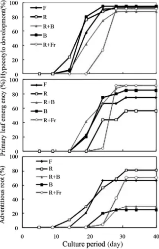
In our study, we investigated the effects of light qual- ity, i.e., F, R, B, RFr, and RB, on both zygotic embryo development (experiment 1) and plantlet growth (exper- iment 2) using an LED system. After 4 weeks of embryo culture, the survival rate, adventitious root and primary leaf emergence, and hypocotyl development were assessed.
In experiment 1, the rate of survival and adventitious root emergence were significantly greater in embryos grown under R irradiation, but the primary leaf emer- gence was delayed (Figure 2). Compared to the other growth factors, hypocotyl development was not signifi- cantly affected by light quality (Figure 3). Morphologi- cal characteristics of seedlings originating from R+B irradiation were intermediate to those of seedlings from R and B monochrome irradiation (Figure 4). The mor- phology and developmental characteristics of embryos
Figure 1. Spectral distributions in relative energy of the LEDs and fluorescent lamps. (R: Red, B: Blue, R+B:
Red+Blue, F: Fluorescent lamps)
Figure 2. Survival rate of zygotic embryos under different spectral radiations during entire culture period in chestnut.
(F: Fluorescent lamps, R: Red, R+B: Red1+Blue1, B: Blue,
R+Fr: Red1+Far-red1)
grown under R radiation indicated that, R irradiation increases the survival rate of embryos and stimulates root development, but prohibits leaf development. Monochrome B irradiation, in contrast, was found to decrease the sur-
vival rate (about 48%) and strongly prohibit adventitious root formation (about 25%) (Figure 2-4).
In contrast to our results that suggested that the devel- opment of zygotic embryos was positively or negatively affected by certain different wavelengths of light, Park et
al. (2010) reported that all light wavelengths positively stimulated the formation of somatic embryos from the explants in Doritaenopsis . The following may be the 3 reasons for this difference in the results. (1) Light at a specific wavelength plays a different role in each devel- opmental stage, i.e., embryo formation and development.
(2) The influence of light may vary from species to spe- cies. (3) The origin of zygotic embryos differs from that of somatic embryos, even though both seem to share their physiological traits.
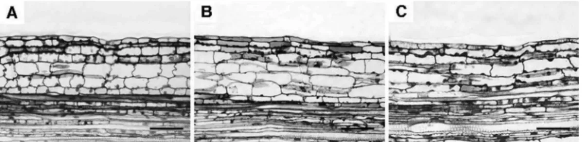
In experiment 2, seedlings with 1-2 leaves were cultured, similar to that in zygotic embryo culture, under 5 dif- ferent light irradiations. The stem was significantly elon- gated by R irradiation, and this tendency was accelerated by the R and Fr combination (R+Fr) (Table 1). The number of nodes was increased in plantlets grown under R+Fr irradiation, but the others did not significantly affect in the node number of plants. It was observed that only internodes were elongated under the R and R+Fr treatment (Table 1). To determine the reason why the internodes were elongated, stems from plants grown under each treatment were collected and observed under the microscope. Epidermis and cortex cells from the stem grown under the R irradiation were around 2 layers thicker than those of plant stems grown under a fluo- rescent lamp (Figure 4A and B). Interestingly, B irradi- ation also influenced stem elongation and resulted in an increased internodal length (Table 1), which was con- firmed by microscopic observation (Figure 4).
This finding is in contrast with the observations in experiment 1 in which B irradiation strongly prohibited stem elongation in embryo culture. Seedling growth, i.e., height, nodes and leaf number, and leaf area were mark- Figure 3. Developmental characteristics of zygotic embryos
under different spectral radiations during entire culture period in chestnut. (F: Fluorescent lamps, R: Red, R+B:
Red1+Blue1, B: Blue, R+Fr: Red1+Far-red1)
Figure 4. Developmental characteristics of zygotic embryos under different spectral radiations during entire culture period in chestnut. (A: Fluorescent lamps, B: Blue, C:
Red1+Blue1, D: Red, E: Red1+Far-red1)
Table 1. Effects of radiation quality on growth of in vitro plantlets in chestnut.
Plant height (cm)
Internodal length
z(mm)
No of nodes (per plant)
No of leaves
(per plant) Leaf area (cm
2) F 2.4 c
y5.9 bc 3.9 b 2.6 b 0.9 c
B 2.5 c 4.2 c 4.0 b 2.8 b 1.0 c
R+B 2.3 c 2.8 d 3.9 b 2.9 b 1.6 b
R 3.9 b 6.7 b 4.1 b 2.9 b 1.5 b
R+Fr 6.6 a 13.3 a 4.9 a 3.7 a 2.1 a
y
Mean values followed by same letters are not significantly dif- ferent according to Duncan’s multiple range test at p ≤ 0.05.
z