Veterinary Science
Surgical treatment for different forms of hernias in sheep and goats
Fahd A. Al-Sobayil, Ahmed F. Ahmed*
Department of Veterinary Medicine, College of Agriculture and Veterinary Medicine, Qassim University, Qassim, Saudi Arabia
Sheep and goats are frequently presented with different forms of hernias to veterinary clinics. The aim of this study is to investigate the outcome of the surgical treatment of abdominal, umbilical, inguinal and scrotal hernias in sheep and goats. Fifty-eight clinical cases (sheep = 44, goat = 14) were presented to the Veterinary Teaching Hospital, College of Agriculture and Veterinary Medicine, Qassim University, Saudi Arabia from September, 2003 to September, 2006. These animals had abdominal (sheep = 30, goat = 10), umbilical (sheep = 6, goat = 4), inguinal (sheep = 7) and scrotal (sheep = 1) hernias. All the cases of hernias in sheep and goats were subjected to full study including the history of the case, classification of hernias, the size of the hernial ring, surgical repair of the hernias, adhesions between the hernial sacs in each case, the postoperative care and follow up of the cases. The results revealed that gender had an effect on the incidence of hernia. The incidence of abdominal hernias was higher in females and the incidence of inguinal hernia was higher in males. There was a positive correlation between the history of hernia and the degree of adhesion. For the sheep, 26 out of 30 cases of abdominal hernia had good outcomes and the healing was excellent. There were postoperative complications in 4 ewes. For the goats, there were slight swellings at the site of operation in 2 out of 10 cases of abdominal hernia, while the remaining 8 cases had good outcomes. There was one case of umbilical hernia with an umbilical abscess that had broken down with sepsis formation at the surgical site. In conclusion, the success rates of surgical treatment for all types of hernias were very high and there were no significant differences in the success rates among the different types of hernias in both sheep and goats. The types of suture materials and the types of hernias had no significant effect on the outcome of the surgical treatment.
Key words: goats, hernia, sheep, surgical treatment
Introduction
Sheep and goats are frequently presented with different forms of hernias to veterinary clinics. Abdominal hernias may occur when the abdominal wall is severely traumatized and these hernias may be high or low in the flank, along the costal arch or between the last few ribs [12]. They are usually caused by violent force, such as from the impact of blunt objects, but they may result from overstretching of the abdominal muscles [27]. Various corrective procedures have been described elsewhere [2,5,8,9,11,13,14,22,24,26].
Umbilical hernias may be congenital or acquired, and they are seen in foals, calves, pups and pigs [7,26]. Many small umbilical hernias may appear to resolve spontaneously, but large or strangulated umbilical hernias will require surgical correction. Inguinal hernia is relatively common in bulls, rams and boars. Scrotal hernia is merely an extension of an inguinal hernia. Congenital inguinal hernia is rare in bulls, but it may result in evisceration at castration. Acquired inguinal hernias occur in mature bulls and rams [25,26].
The aim of the present study is to investigate the outcome of surgical treatment for abdominal, umbilical, inguinal and scrotal hernias in sheep and goats.
Materials and Methods
Animals
The present study was carried out on 58 clinical cases (44 sheep and 14 goats) that were presented to the Veterinary Teaching Hospital, College of Agriculture and Veterinary Medicine, Qassim University, Saudi Arabia from September, 2003 to September, 2006. These animals had abdominal (sheep = 30, goat = 10), umbilical (sheep = 6, goat = 4), inguinal (sheep = 7) and scrotal (sheep = 1) hernias.
The sheep were 18 males and 26 females, and they were classified as 3 local breeds (Nagdi = 34, Naimi = 9 and Sakni = 1). The ages of the sheep ranged from 1 month to 6 years. The histories of the cases indicated that the hernias were noticed at 10 days to up to 1 year before presentation to the hospital and the majority occurred 3 to 6 months before presentation.
Goats were 3 males and 11 females and they were
*Corresponding author
Tel: +966 3801480; Fax: +966 3801360
E-mail: [email protected], [email protected]
classified as 2 breeds (Syrian = 9, Baladi = 5). The goats’
ages ranged from 3 months to 6 years. Hernias were noticed in the goats at 1 month up to 8 months before admission to the clinic.
All the cases of hernias in the sheep and goats were subjected to full study, including the history of the case, classification of the hernias, the size of the hernial ring, surgical repair of the hernias, adhesions between the hernial sacs in each case (adhesions were graded from 1 to 4 with 1 as slight adhesions and 4 as severe adhesions), the postoperative care and the follow up of the cases, which was done by direct contact with or via phone calls to the owners.
The data of the cases is summarized in Tables 1, 2 and 3.
Surgical treatment
Food was withheld for 24 h prior to surgery in each case.
Surgical repair was conducted by aseptically preparing the site of operation after intramuscularly tranquilizing the fractious animals with 2% xylazine hydrochloride (Rompun 2%; Bayer, Turkey) at a dose rate of 0.05 mg/kg. The animal was restrained in the dorsal or lateral recumbent position, according to the type and position of the hernia.
In cases of abdominal and umbilical hernias, circular infiltration anesthesia was done using 2% lidocaine (Norbrook Laboratories, UK) at a dose rate of 10 mg/kg. An elliptical skin incision was performed and the adhesions between the parietal peritoneum and skin were freed with using both blunt and sharp dissection. The hernial ring was exposed and freshened before its suturing by simple interrupted or interrupted horizontal mattress sutures with using No. 2 chromic catgut (Ethicon, UK), polydioxanone (PDS; Ethicon, UK) or silk (Lukens Medical, USA) sutures.
The subcutaneous tissue was then sutured by catgut or PDS, and the excessive skin was removed before its suture with using polypropylene (Ethicon, UK) or silk suture.
In cases of inguinal and scrotal hernias, linear infiltration anesthesia was applied at the site of the operation, which was lateral to the scrotum or the udder. A linear skin incision was made followed by sharp and blunt dissection to expose the hernial contents. The contents were reduced into the abdominal cavity through the inguinal canal and the external inguinal ring was narrowed by application of interrupted chromic catgut stitches (in 1 ram to keep the testis according to the owner’s request). The testicles were removed whenever they appeared atrophied, and this was followed by complete closure of the external inguinal ring using catgut, PDS or silk sutures (in 7 cases).
Each animal was given postoperative therapy with penicillin- streptomycin at a dose rate of 30,000 IU/kg for the penicillin and 10 mg/kg streptomycin for 5 days (Norbrook Laboratories, UK) and a prophylactic dose of anti-tetanus serum 1,500 IU subcutaneously.
Statistical analysis
The data was analyzed by a computer program and utilizing the SAS technique. Analysis of variance was used as the statistical method to evaluate the effects of breed, age, gender, the history of hernia, the size of the hernial ring, the type of the hernia, the degree of adhesions, the type of suture material and the outcome on the other variables. Multiple comparisons of means were determined using Tukey's method. Pearson correlation coefficient analysis was used to study the relationship between the different variables. The significant level was set at
p< 0.05.
Results
Sheep
The sheep in the present study had 4 types of hernias;
abdominal = 30, umbilical = 6, inguinal = 7 and scrotal = 1 (Fig. 1 & 2). The size of the hernial ring ranged from a finger breadth up to more than 2-hands breadth. All the cases were reducible hernias except for 1 ram that had a nonreducible abdominal hernia (Tables 1 & 2).
Fig. 1.
A huge abdominal hernia in a 2-year-old Nagdi ewe (A) and an abdominal hernia just cranial to the udder in a 5-year-old female Baladi goat (B).
Fig. 2.
An umbilical hernia in a 5-month-old Nagdi ram (A) and
a scrotal hernia in a 3-year-old Naimi ram (B).
All the cases of hernias in the sheep were treated surgically (Fig. 3). Cesarean section was performed for 2 ewes before herniorrhaphy. The gravid uterus was the hernial content (hysterocele). Rumenotomy was done in 1 case to remove ruminal foreign bodies. In those 2 instances, open reduction was performed for which the parietal peritoneum was opened.
Goats
The hernias in the goats were 10 abdominal and 4 umbilical hernias (Fig. 1). The size of the hernial ring ranged from 2-fingers breadth up to 2-hands breadth. All cases were reducible hernias. Two kids had umbilical abscesses along with their umbilical hernias (Table 3).
Outcomes
For the sheep, 26 out of 30 cases of abdominal hernia had good outcomes and their healing was excellent. There were postoperative complications in 4 ewes; abdominal hernia reocurred 1 month later, slight swelling
in situ, muscular weakness at the site of operation and an abomasal fistula at the site of operation (Table 1).
The cases of umbilical hernias in sheep were surgically corrected without postoperative complications. However, a case of inguinal hernia, for which castration was not carried out upon the owner’s request, had postoperative complications in the form of inflammation and swelling of the testis and scrotum of the affected side. This case necessitated castration at 2 weeks after the first operation. The other cases of
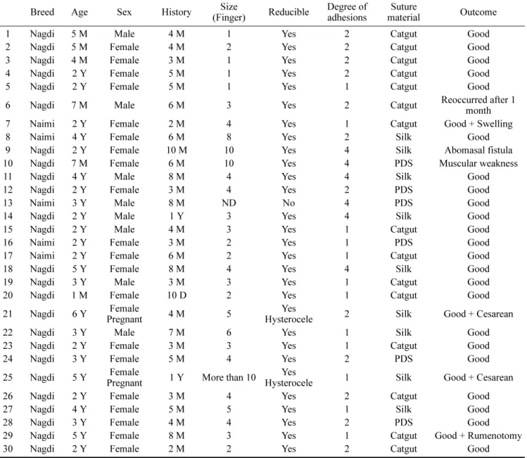
Table 1.
Cases of abdominal hernias in sheep
Breed Age Sex History Size
(Finger) Reducible Degree of
adhesions Suture
material Outcome
1 Nagdi 5 M Male 4 M 1 Yes 2 Catgut Good
2 Nagdi 5 M Female 4 M 2 Yes 2 Catgut Good
3 Nagdi 4 M Female 3 M 1 Yes 2 Catgut Good
4 Nagdi 2 Y Female 5 M 1 Yes 2 Catgut Good
5 Nagdi 2 Y Female 5 M 1 Yes 1 Catgut Good
6 Nagdi 7 M Male 6 M 3 Yes 2 Catgut Reoccurred after 1
month
7 Naimi 2 Y Female 2 M 4 Yes 1 Catgut Good + Swelling
8 Naimi 4 Y Female 6 M 8 Yes 2 Silk Good
9 Nagdi 2 Y Female 10 M 10 Yes 4 Silk Abomasal fistula
10 Nagdi 7 M Female 6 M 10 Yes 4 PDS Muscular weakness
11 Nagdi 4 Y Male 8 M 4 Yes 4 Silk Good
12 Nagdi 2 Y Female 3 M 4 Yes 2 PDS Good
13 Naimi 3 Y Male 8 M ND No 4 PDS Good
14 Nagdi 2 Y Male 1 Y 3 Yes 4 Silk Good
15 Nagdi 2 Y Male 4 M 3 Yes 1 Catgut Good
16 Naimi 2 Y Female 3 M 2 Yes 1 PDS Good
17 Naimi 2 Y Female 6 M 2 Yes 1 Catgut Good
18 Nagdi 5 Y Female 8 M 4 Yes 4 Silk Good
19 Nagdi 3 Y Male 3 M 3 Yes 1 Catgut Good
20 Nagdi 1 M Female 10 D 2 Yes 1 Catgut Good
21 Nagdi 6 Y Female
Pregnant 4 M 5 Yes
Hysterocele 2 Silk Good + Cesarean
22 Nagdi 3 Y Male 7 M 6 Yes 1 Silk Good
23 Nagdi 2 Y Female 3 M 3 Yes 1 Catgut Good
24 Nagdi 3 Y Female 5 M 4 Yes 2 PDS Good
25 Nagdi 5 Y Female
Pregnant 1 Y More than 10 Yes
Hysterocele 1 Silk Good + Cesarean
26 Nagdi 2 Y Female 3 M 4 Yes 2 Catgut Good
27 Nagdi 4 Y Female 5 M 5 Yes 1 Silk Good
28 Nagdi 3 Y Female 4 M 4 Yes 2 PDS Good
29 Nagdi 5 Y Female 8 M 3 Yes 1 Catgut Good + Rumenotomy
30 Nagdi 2 Y Female 2 M 2 Yes 2 Catgut Good
M = month (s), Y = year (s), ND = not determined externally, PDS = polydioxanone.
inguinal and scrotal hernias had good healing without complications (Table 2).
For the goats, there were slight swellings at the site of operation in 2 out of 10 cases of abdominal hernia, while the remaining 8 cases had good outcomes. A case of umbilical hernia with an umbilical abscess had broken down with sepsis formation at the surgical site. The umbilical hernia reoccurred in this case (Table 3).
For the sheep, their age had no significant effect on the other parameters except its effect on the type of suture materials used (
p= 0.006). Their gender only had an effect on the incidence of hernia. The incidence of abdominal hernias was higher in the females and the incidence of
inguinal hernia was higher in the males. The history of hernia had no significant effect on the other variables except for its effect on the type of suture materials (
p= 0.003). The size of hernia showed a significant effect only on the type of suture materials used (
p< 0.001) and the outcome of surgery (
p= 0.04). The other variables showed no significant effect on each other.
For the goats, there was no significant effect among the different variables except the effect of gender on the incidence of hernia. Both the abdominal and the umbilical hernias occurred more often in the females than in the males.
In this study, it was found that there was a positive correlation between the history of hernia and the degree of
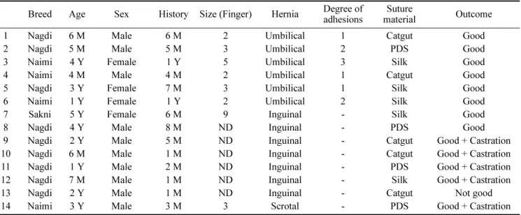
Table 2.
Cases of umbilical, inguinal and scrotal hernias in sheep
Breed Age Sex History Size (Finger) Hernia Degree of
adhesions Suture
material Outcome
1 Nagdi 6 M Male 6 M 2 Umbilical 1 Catgut Good
2 Nagdi 5 M Male 5 M 3 Umbilical 2 PDS Good
3 Naimi 4 Y Female 1 Y 5 Umbilical 3 Silk Good
4 Naimi 4 M Male 4 M 2 Umbilical 1 Catgut Good
5 Nagdi 3 Y Female 7 M 3 Umbilical 1 Silk Good
6 Naimi 1 Y Female 1 Y 2 Umbilical 2 Silk Good
7 Sakni 5 Y Female 6 M 9 Inguinal - Silk Good
8 Nagdi 4 Y Male 8 M ND Inguinal - PDS Good
9 Nagdi 2 Y Male 5 M ND Inguinal - Catgut Good + Castration
10 Nagdi 6 M Male 1 M ND Inguinal - Catgut Good + Castration
11 Nagdi 1 Y Male 2 M ND Inguinal - PDS Good + Castration
12 Nagdi 7 M Male 1 M ND Inguinal - Silk Good + Castration
13 Nagdi 2 Y Male 1 M ND Inguinal - Catgut Not good
14 Naimi 3 Y Male 3 M 3 Scrotal - PDS Good + Castration
M = month (s), Y = year (s), ND = not determined externally, PDS = polydioxanone.
Table 3.
Cases of abdominal and umbilical hernias in goat
Breed Age Sex History Size (Finger) Hernia Degree of
adhesions Suture
material Outcome
1 Syrian 3 M Male 1 M 3 Abdominal 4 Catgut Good
2 Baladi 5 Y Female 8 M 4 Abdominal 3 PDS Good + Slight swelling
3 Syrian 3 Y Female 2 M 2 Abdominal 1 Silk Good
4 Baladi 2 Y Female 1 M 10 Abdominal 4 Silk Good
5 Baladi 5 Y Female 5 M 4 Abdominal 3 Silk Good
6 Baladi 6 Y Female 7 M 5 Abdominal 3 Silk Good
7 Syrian 3 M Male 1 M 4 Abdominal 4 PDS Good + Slight swelling
8 Syrian 3 Y Male 7 M 3 Abdominal 3 Silk Good
9 Syrian 4 Y Female 3 M 3 Abdominal 2 Catgut Good
10 Baladi 1 Y Female 2 M 3 Abdominal 2 PDS Good
11 Syrian 1.5 Y Female 3 M 2 Umbilical 1 Catgut Good
12 Syrian 3 M Female 3 M 3 Umbilical 3 + abscess Silk Good
13 Syrian 4 M Female 4 M 7 Umbilical 3 + abscess Silk Recurred + Sepsis
14 Syrian 3 M Female 3 M 4 Umbilical 1 Silk Good
M = month (s), Y = year (s), PDS = polydioxanone.