Detection of Coronary Restenosis by Serial Doppler Echocardiographic Assessment of Coronary Flow Velocity Reserve after Percutaneous Intervention
10
0
0
전체 글
(2) Jae-Hyeong Park, et al. Methods. due to coronary artery disease,7) the necessity of functional tests for assessing the blood flow impairment has been suggested.. The study population. In the mid 1990s, the direct intravascular measurement of the coronary flow velocity during a coronary angio-. The study population was comprised of 36 patients who successfully underwent coronary angioplasty in the. gram by Doppler guide wire became available,8) and measurement of the coronary flow velocity reserve (CFVR). left anterior descending coronary artery between March and August 2002. Patients without sinus rhythm, those. was calculated by the ratio of the coronary flow velocity under hyperemia and under baseline conditions. As the. with a previous history of acute myocardial infarct or bronchospasm, over 30% residual stenosis after coronary. coronary flow velocity reserve is a crucial diagnostic indication of the overall coronary flow, including coro-. angioplasty or who refused to undergo the follow-up coronary angiogram were excluded from the study.. nary restenosis and microvascular impairment, its possible application to the diagnosis of coronary stenosis and restenosis after coronary angioplasty has been reported.9)10). The measurement of coronary flow reserve by transthoracic Doppler echocardiography Echocardiography was performed by applying a high. Recently, the technical improvement in trans thoracic Doppler echocardiography allows the visualization of co-. frequency transducer (5 MHz) using Acuson Sequoia 512 digital ultrasound system (Acuson, California). In. ronary flow in the distal left anterior descending coronary artery and the non-invasive assessment of the coronary. color Doppler, the velocity range was adjusted to between 12-24 cm/second. With correct filtering, the artifact of. flow velocity reserve,11-14) which is highly correlated to the coronary flow velocity reserve measured by invasive. low frequency wall movement was minimized. Echocardiographic images from the 4th or 5th intercostal space. means.16)18)19) This permits the measurement of the coronary flow velocity reserve, which has previously been. in midclavicular line of the left decubitus were obtained. After obtaining the longitudinal cross-section of the lo-. invasively measured by applying the Doppler guide wire in the catheterization laboratory, in the echocardiography. wer interventricular sulcus, the transducer was turned to the left, and the distal left anterior descending coronary. laboratory simply without additional costs and thus its clinical application has become more convenient.15)16) Ho-. artery visualized under the guide of color flow mapping. After focusing the sample volume on the colored region. wever, as the coronary flow velocity reserve varies according to coronary stenosis and microvascular impairment,. of the distal left anterior descending coronary artery, the coronary blood flow velocity was measured by a 5 MHz. a single measurement may cause problems for the diagnosis of stenosis.. pulsed Doppler wave. Efforts have been made to align the direction of the beam of ultrasound and the color. Here, assuming that the decrease in the coronary flow velocity reserve, within the relatively short 6-month. signal of the distal left anterior artery as parallel as possible. After recording the baseline spectral Doppler sig-. follow-up period, is due to coronary restenosis rather than the exacerbation of microvascular impairment, ef-. nals, while infusing adenosine for over 2-3 minutes (140 μg/kg/min), the spectral Doppler signals during the hype-. forts were made to improve the accuracy of the diagnosis of restenosis by repeatedly measuring the coronary flow. remia were recorded. The Doppler signal of the baseline and the hyperemia period in the left anterior descending. velocity reserve in patients having undergone coronary angioplasty and thus minimize the effect of microvascular. coronary artery were recorded continuously for three beats. Attention was paid not to dislodge the transducer. impairment.. during the adenosine infusion. The maximum and average diastolic coronary flow velocities during the baseline and. 661.
(3) Echo Diagnosis of Coronary Restenosis by CFVR maximum hyperemia periods were measured. Coronary. the analysis of variable values of the continuous variables. flow velocity reserve was defined as the value of the average diastolic coronary flow velocity during the ma-. was performed by paired t-tests. To assess the standard error among the investigators, two specialists analyzed. ximum hyperemia period divided by the baseline period. Here, as the value may become smaller if the baseline. the baseline coronary flow velocity reserve value. The correlation of the stenosis of the internal diameter on the. coronary flow velocity reserve was measured after coronary angioplasty, due to the increase of the baseline coro-. angiogram to the follow-up coronary flow velocity reserve and the variable value of coronary flow velocity. nary flow volume, to minimize such an effect, the baseline coronary flow velocity reserve was measured 24 hours. reserve was analyzed by Pearson’s correlation. The statistical analyses were performed using the software Windows®. after coronary angioplasty. The follow-up coronary flow reserve was measured one day prior to the coronary an-. SPSS® 11.0. A p-value less than 0.05 were considered statistically significant in all statistical analyses.. giogram on the in-patients admitted for coronary angiograms. All patients received drugs for angina pectoris. Results. during a transthoracic Doppler echocardiogram. After signing a consent form, the coronary flow velocity reserve. In all 36 patients, the coronary flow velocity reserve. was measured by thoracic Doppler echocardiogram.. was measured 1 day and 6 months after the coronary angioplasty. The coronary flow velocity reserve, the. Quantitative coronary angiogram. change in coronary flow velocity and coronary angiography were compared.. In all patients, the coronary angiogram was performed by conventional methods. The quantitative analysis of the coronary angiography was performed blindly by expe-. The clinical characteristic of patients. rienced investigators. The calibration was performed by applying the diameter of the guide catheter or the diagno-. The mean age of the study population was 57±11 years (ranged from 30 to 75 years), with 23 males (64%).. stic catheter. After administration of nitroglycerin, the coronary angiogram was performed.. In regard to the risk factors of coronary diseases, 12 (33%), 20 (56%), 13 (36%) and 12 cases (33%) had. The coronary angiographic pictures were analyzed by a quantitative analyzer (Ancor 2.0, Siemens, Erlangen,. diabetes, high blood pressure, smoking and high serum cholesterol, respectively. On electrocardiogram, left ven-. Germany). The internal diameter of the reference vessel, the internal diameter of lesions, and the diameter of. tricular hypertrophy was detected in 7 cases (21%). During coronary angioplasty, coronary intervention was. stenosis were measured. The length of a lesion was determined on a coronary angiographic picture that had. limited to the left anterior descending coronary artery in 30 patients, and on multiple vessels in 6 patients. At the. the least foreshortening by measuring the distance from the proximal shoulder to the distal shoulder. Over 50%. follow-up coronary angiogram, significant restenosis, over 50% of the internal diameter of the left anterior de-. restenosis of the internal diameter was defined as significant restenosis.. scending coronary artery, was detected in 9 patients (25%). Comparing the value of blood pressure, the serum chole-. Statistical analysis. sterol level or the blood sugar level, measured at the baseline and 6 months later, no statistically significant. Continuous variables were presented as the mean± standard deviation. In the comparison of groups, the. differences were detected in these two groups (Table 1).. continuous variables were analyzed using the Student ttest and nominal variables by the χ2 test In each group,. Coronary flow velocity reserve. 662. In a comparison of patients with and without restenosis,. Korean Circulation J 2004;34(7):660-669.
(4) Jae-Hyeong Park, et al the baseline value of the maximum diastolic velocity in. nary angioplasty, the coronary flow velocity under the. patients without restenosis (n=27), as assessed by thoracic Doppler echocardiogram on the next day after coro-. maximum hyperemia and the value of coronary flow velocity reserve were 49.0±44.6 cm/sec, 101.8±56.0 cm/. Table 1. Patient baseline characteristics. sec and 2.9±1.1, respectively. These values were not significantly different from patients with restenosis, where. Characteristics Age. these values were 60.7±20.0 cm/sec, 116.5±48.0 cm/ sec and 3.3±0.7, respectively.. 57±11 (30-75). Male sex (%). 23 (64%). However, in the follow-up transthoracic Doppler echocardiogram, the diastolic coronary flow velocity values. Risk factors for coronary artery disease Diabetes. 12 (33%). Hypertension. 20 (56%). Smoking. at the baseline in patients without and with restenosis were 32.9±13.2 cm/sec and 47.4±18.8 cm/sec, respec-. 13 (36%). Hypercholesterolemia. 12 (33%). LVH by ECG criteria. 07 (27%). tively, and the difference was statistically significant (p< 0.05). The follow-up coronary flow velocity reserves in. Site of PCI LAD/multivessel. patients without and with restenosis were 3.3±0.9 and 2.0±0.7, respectively, and the difference was significant. 20/6 Baseline. Follow up. Systolic blood pressure (mmHg) Diastolic blood pressure (mmHg). 125.6±11.5 127.0±15.3. Heart rate (/min)*. 072.4±17.6 063.2±11.9. (p<0.001, Table 2). To obtain the standard error among the investigators, the baseline coronary flow velocity reserve was obtained by two specialists. The standard error of investigators was estimated to be 3.2%.. 078.4±11.9 078.0±10.8. Appling a follow-up coronary flow velocity reserve of less than 2.0 as the criteria, the sensitivity and specificity. Serum glucose level (mg/dL) 134.9±45.2 127.0±40.5 Serum total cholesterol level 167.0±38.9 167.9±33.8 (mg/dL) Values are mean±SD. *: p<0.05 vs. baseline and followup values. LVH: left ventricular hypertrophy, PCI: percutaneous coronary intervention, LAD: left anterior descending coronary artery, ECG: electrocardiography. of the diagnosis of restenosis in the left anterior descending coronary artery lesion were 56 (5/9) and 100% (27/27), respectively. The positive and negative predictive values were 100 (5/5) and 68% (27/31), respectively.. Table 2. Transthoracic Doppler parameters and coronary flow velocity reserve in the groups with and without restenosis Restenosis (-) (n=27). Restenosis (+) (n=9). Baseline. Hyperemia. Baseline. Hyperemia. Heart rate (bpm). 73.4±16.8*. 081.2±15.9. 69.1±21.0*. 084.5±23.6. Vmaxsystole (cm/s). 21.0±13.8*. 052.4±33.5. 15.7±06.4*. 064.5±20.4. Vmaxdiastole (cm/s). 49.0±44.6*. 101.8±56.0. 60.7±20.0*. Baseline. CFVR0. 002.9±01.1. 116.5±48.0 03.3±00.7. Follow-up Heart rate (bpm). 63.4±11.4*. 076.2±13.3. 62.2±12.8*. Vmaxsystole (cm/s). 19.1±08.8*. 061.5±25.9. 29.7±11.3*. 60.0±14.0. Vmaxdiastole (cm/s). 32.9±13.2*. 105.9±38.6. 47.4±18.8*. 84.2±28.0. CFVR1†. -03.3±00.9. 74.2±18.7. 02.0±00.7. Change of CFVR† -0.30±0.46 0.42±0.23 Values are mean±SD. TVI: time velocity integral, CFVR: coronary flow velocity integral, Vmax: maximal velocity. *: p<0.05, †: p< 0.001 vs. group with restenosis and without restenosis. 663.
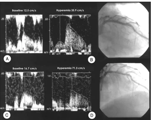
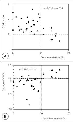
(5) Echo Diagnosis of Coronary Restenosis by CFVR. Baseline 12.5 cm/s. Hyperemia 35.9 cm/s. A. B Baseline 16.7 cm/s. Hyperemia 71.3 cm/s. C. D. Figure 1. Baseline transthoracic Doppler (A) and coronary angiogram (B) results after the procedure, and follow up Doppler (C) and angiographic (D) results six months after the procedure, in a patient who had undergone stenting of the left anterior descending coronary artery. Baseline coronary flow velocity reserve and follow up results were 2.88 (35.9/12.5) and 4.27 (71.3/16.7), respectively. The follow up coronary angiography revealed no restenosis (diameter of stenosis was 6.1% by quantitative coronary angiographic analysis).. In such an assessment, the shortcoming was low sensitivity. Appling a follow-up coronary flow velocity reserve. significant reduction of coronary flow velocity reserve, the sensitivity and specificity of significant decrease of. of less than 2.5 as the criteria, the sensitivity and specificity were 89 (8/9) and 81% (22/27), respectively. The. CFVR were 100 (9/9) and 93% (25/27), respectively. Additionally, the follow-up coronary flow velocity re-. positive and negative predictive values were 62 (8/13) and 96% (22/23), respectively. Although the sensitivity. serve was significantly and inversely correlated to the degree of restenosis of the internal diameter shown on. was increased, the shortcoming was the decreased specificity.. the coronary angiogram. The decrease in the CFVR was significantly correlated to the stenosis of the internal dia-. Figures 1 and 2 show the transthoracic Doppler and coronary angiogram results of a patient without and with. meter shown on the coronary angiogram (Figure 4). In 15 patients with diabetes or left ventricular hypertro-. restenosis, respectively. Regarding the significant difference in the CFVR ana-. phy, which are known to induce microvascular impairment, in the diagnosis of restenosis, the assessment of. lyzed by the ROC curve, the best sensitivity and specificity were obtained when 10% was applied as the criterion. the significant reduction of coronary flow velocity reserve was more accurate than when a follow-up coronary flow. of a significant decrease in the coronary flow velocity reserve (Figure 3).. velocity reserve of less than 2.5 was applied as the standard criteria (Table 4).. When measured repeatedly, for the assessment of the. 664. Korean Circulation J 2004;34(7):660-669.
(6) Jae-Hyeong Park, et al. Baseline 20.0 cm/s. Hyperemia 77.5 cm/s. A. B Baseline 67.1 cm/s. Hyperemia 87.6 cm/s. C. D. Figure 2. Transthoracic Doppler (A) and coronary angiogram (B) results after the procedure in another patient who had undergone stenting of the left anterior descending coronary artery. The baseline coronary flow velocity reserve was 3.87 (77.5/20.0). Follow up transthoracic Doppler (C) and coronary angiogram (D) results six months after the procedure. The follow up coronary flow velocity reserve was 1.31 (67.1/87.6) and the coronary angiogram showed significant restenosis (diameter of stenosis was 77.5% by quantitative coronary angiographic analysis). Table 3. Diagnostic accuracy for detection of restenosis Criteria. Sensitivity. Specificity. Accuracy. CFVR1 <2.0. 056% (5/9) 100% (27/27) 89% (32/36). CFVR1 <2.5. 089% (8/9) 081% (22/27) 83% (30/36). Decrease of CFVR 100% (9/9) 093% (25/27) 94% (34/36) CFVR: coronary flow velocity reserve. Discussion. guide wire or non-invasive methods, such as echocardiogram, magnetic resonance imaging and positron emission tomogram, etc.8)20)21) Recently, by applying a high frequency transducer over 5 MHz in the assessment of coronary flow, the direct measurement of the coronary flow velocity has become possible. Especially, for the left anterior descending coronary artery, which runs through the cardiac interventri-. Here, after left anterior descending coronary artery. cular sulcus, and its distal part that is separated from the thorax by 1-4 cm, a transducer with the frequency range. angioplasty, it was confirmed that by reducing the effect of microvascular impairment with repeated measurements. 5-12 MHz placed between the 4th and 5th intercostal space in midclavicular line of the left decubitus, reveals. of the coronary flow reserve on applying transthoracic Doppler echocardiogram, the accuracy of the diagnosis. the coronary blood flow moving forward in over 95% cases on reacting to the low speed range color Doppler. of restenosis can be improved. The coronary flow velocity reserve is known to repre-. program installed in the machine. The angle of the Doppler sample volume to the blood flow direction was ad-. sent the overall coronary flow velocity condition, including epicordial coronary stenosis and microvascular impair-. justed as parallel as possible so that the coronary flow velocity, which is primarily diastolic by on pulsed Dopp-. ment. It has been measured by invasive coronary Doppler. ler, can be measured. However, in the right coronary. 665.
(7) Echo Diagnosis of Coronary Restenosis by CFVR. 6. 1.00. r=-0.390, p=0.028 CFVR1 value. Change of CFVR >0.1. Sensitivity. 0.75. 4. 0.50 2. 0.25 0 0. A. 1.00 0. 0.25. 0.50. 0.75. 50. 100. Deameter stenosis (%). 1.00. 1-specificity. artery and the left circumflex artery, measurement of blood flow velocity is difficult in many cases because the distal part is separated from the thorax and its running. r=0.410, p=0.02 Change of CFVR. Figure 3. ROC curve for obtaining the optimal cut off value for the change of CFVR. ROC: receiver operating curve, CFVR: coronary flow velocity reserve.. 1.0. 0.1 0.0. 1.0. direction does not concur to the ultrasound beam in many cases. In comparison of the assessment of coronary flow velocity reserve in the distal left anterior descending artery by an intracoronary Doppler guide wire and transthoracic Doppler echocardiogram, the highly correlated relationship of these two tests has been confirmed in. -2.0 0. B. 50. 100 Deameter stenosis (%). numerous studies, which has facilitated its clinical application.15)16)18) Such a test method is anticipated to be. Figure 4. Relation between diameter of stenosis (%) and follow up coronary flow velocity value (CFVR1) (A) and change of CFVR value (B). CFVR: coronary flow velocity reserve.. applicable for the assessment of angina pectoris patients showing normal coronary angiogram, in the evaluation. coronary flow velocity in response to the oxygen require-. following percutaneous coronary angioplasty, functional assessment prior to interventional procedures for angina. ment of the myocardium. When the hyperemia of coronary flow velocity is induced by exercise, a vasodilator or. pectoris that is difficult to accurately evaluate by coronary angiogram and the early diagnosis of the accelerated. other stresses, small vessels are dilated. If small vessels were already somewhat dilated, due to existing coronary. atherosclerosis after a heart transplant. In addition, the advantage of this procedure is its lower cost than assess-. arteriosclerosis, the increase in the coronary flow velocity reserve, which is the velocity value, in response to stress. ment by magnetic resonance imaging or positron emission tomography.. is limited. Hence, the flow velocity reserve is decreased in cases with over 50% stenosis. By applying this, steno-. Under normal physiological condition, 95% of coronary resistance is determined by the intra-myocardial resistant vessel; particularly, the 50-500 μm intra-myocardial. sis over 50% can be diagnosed. Recently, such a procedure has been reported as non-invasive and accurate in. resistant vessel plays an important role of controlling. 666. predicting restenosis after coronary angioplasty. Ruscazio et al. reported that applying a coronary flow velocity. Korean Circulation J 2004;34(7):660-669.
(8) Jae-Hyeong Park, et al Table 4. Diagnostic accuracy in patients with diabetes or left ventricular hypertrophy Criteria CFVR1 <2.0. Sensitivity. Specificity. Accuracy. 050% (2/4) 100% (11/11) 087% (13/15). CFVR1 <2.5 100% (4/4) 063% (07/11) 073% (11/15) Decrease of 100% (4/4) 100% (11/11) 100% (15/15) CFVR CFVR: coronary flow velocity reserve. velocity reserve value that is significantly lower than the baseline value may be helpful in the diagnosis of restenosis. Our results have demonstrated that when the decrease in the coronary flow velocity reserve was significant, the sensitivity and specificity were 100 and 93%, respectively. Applying a follow-up coronary flow velocity reserve of. reserve, as measured by transthoracic Doppler echocar-. less than 2.0 as the criteria, the sensitivity and specificity of the diagnosis of restenosis in the left anterior descen-. diogram, of less than 2.0 as the standard, the sensitivity and specificity were 78 and 93%, respectively, in the. ding coronary artery lesion were 56 (5/9) and 100% (27/27), respectively. The positive and negative predictive. diagnosis of significant restenosis.10) Our data are in agreement with this report. Our results showed that. values were 100 (5/5) and 68% (27/31), respectively. Using the standard 2.5, the diagnosis of restenosis could. applying a coronary flow velocity reserve value of 2.0 as the standard, the sensitivity and specificity were 56 and. be more accurately made. In such an assessment, the sensitivity and specificity were 89 and 81%, respectively.. 100%, respectively. Applying 2.5 as the standard, the sensitivity and specificity were 89 and 81%, respectively.. Furthermore, in the diagnosis of restenosis in patients with left ventricular hypertrophy or diabetes that could. In addition, Nagel et al.22) reported that in the diagnosis of restenosis, by measuring the coronary flow velocity. cause microvascular impairment, the prediction was more accurate when a decrease in the coronary flow velocity. reserve using magnetic resonance imaging (MRI), applying 1.2 as the standard, the sensitivity and specificity. reserve was applied as the standard. In this study, 10% was considered as a significant. were 83 and 94%, respectively. These studies predicted coronary restenosis by a single. decrease in the follow-up coronary flow velocity reserve value, as it must be larger than the 3.2% standard error. measurement during a coronary angiogram. The factors influencing the coronary flow velocity are the heart rate,. of the investigators. In addition, when 10% was applied as the standard to the ROC curve analysis of the change. the change in arterial pressure, left ventricular hypertrophy and microvascular angina.24-30) It has been reported. in the coronary flow velocity reserve value, the sensitivity and the specificity were improved.. that in patients with an increased heart rate or preload, myocardiac hypertrophy or microvascular angina, even. With regard to the heart rate and arterial pressure that may affect the coronary flow velocity reserve, their in-. without coronary stenosis, the coronary flow velocity reserve can be decreased. Thus, the prediction of the. fluence was ruled out, as a significant difference in patients with and without restenosis was not detected.. degree of coronary stenosis by a single measurement may cause problems.. Similarly, in comparison of the serum cholesterol or serum sugar levels of the baseline and follow-up periods,. As the measurement of coronary flow velocity reserve by transthoracic Doppler echocardiogram has the advan-. their differences were not statistically significant. It is anticipated that the most suitable clinical indications. tages of lower cost and noninvasiveness, it can readily be repeatedly measured. The change in the coronary flow. of the coronary flow velocity reserve value obtained by transthoracic Doppler echocardiogram are; 1) the diagno-. velocity reserve, when measured repeatedly within a short period, may be due to coronary restenosis rather than a. sis of coronary restenosis, where the conventional noninvasive procedure has limitations. 2) the cases requiring Doppler guide wire, which are invasive and expensive;. change in the left ventricular hypertrophy or exacerbation of microvascular impairment. Thus, the coronary flow. in other words, in cases requiring the evaluation of the. 667.
(9) Echo Diagnosis of Coronary Restenosis by CFVR hemodynamic significance of the lesions as the degree of coronary stenosis and clinical conditions, disagree. The limitation of our research is that, firstly, the measurement of coronary flow velocity reserve, by transthoracic Doppler echogram, is only anatomically suitable to the left anterior descending coronary artery. Its application to other coronary arteries is not suitable. However, the left anterior descending coronary artery is the vessel where most clinically important percutaneous coronary angioplasty is performed. Thus, the diagnosis of restenosis in the left anterior descending coronary artery may be clinically important. Secondly, it is desirable to analyze the significance by performing trials on a large number of patients, rather than the limited small number of patients in this study. This research was supported by grant from clinical perfomance. Improvement project in Asan medical center, south korea. REFERENCES 1) Macaya C, Serruys PW, Ruygrok P, Suryapranata H, Mast G,. Klugmann S, et al. Continued benefit of coronary stenting versus balloon angioplasty: one-year clinical follow-up of Benestent trial. J Am Coll Cardiol 1996;27:255-61. 2) Meier B, King SB 3rd, Gruentzig AR, Douglas JS, Hollman J, Ischinger T, et al. Repeat coronary angioplasty. J Am Coll Cardiol 1984;4:463-6. 3) Popma JJ, Califf RM, Topol EJ. Clinical trials of restenosis after coronary angioplasty. Circulation 1991;84:1426-36. 4) Serruys PW, Luijten HE, Beatt KJ, Geuskens R, de Feyter PJ, van den Brand M, et al. Incidence of restenosis after successful coronary angioplasty: a time-related phenomenon: a quantitative angiographic study in 342 consecutive patients at 1, 2, 3, and 4 months. Circulation 1988;77:361-71. 5) Kent KM, Ewels CJ, Krucoff MW. Noninvasive assessment of patients following coronary artery angioplasty. Cardiovasc Clin 1988;19:199-208. 6) Rosanio S, Tocchi M, Stouffer GA. Use of stress testing to evaluate patients with recurrent chest pain after percutaneous coronary revascularization. Am J Med Sci 1998;316:46-52. 7) Topol EJ, Nissen SE. Our preoccupation with coronary luminology: the dissociation between clinical and angiographic findings in ischemic heart disease. Circulation 1995;92: 2333-42. 8) Doucette JW, Corl PD, Payne HM, Flynn AE, Goto M, Nassi M, et al. Validation of a Doppler guide wire for intravascular measurement of coronary artery flow velocity. Circulation 1992;85:1899-911. 9) Gould KL, Lipscomb K. Effects of coronary stenoses on coronary flow reserve and resistance. Am J Cardiol 1974;. 668. 34:48-55. 10) Ruscazio M, Montisci R, Colonna P, Caiati C, Chen L, Lai. G, et al. Detection of coronary restenosis after coronary angioplasty by contrast-enhanced transthoracic echocardiographic Doppler assessment of coronary flow velocity reserve. J Am Coll Cardiol 2002;40:896-903. 11) Fusejima K. Noninvasive measurement of coronary artery blood flow using combined two-dimensional and Doppler echocardiography. J Am Coll Cardiol 1987;10:1024-31. 12) Ross JJ Jr, Mintz GS, Chandrasekaran K. Transthoracic twodimensional high frequency (7.5 MHz) ultrasonic visualization of the distal left anterior descending coronary artery. J Am Coll Cardiol 1990;15:373-7. 13) Kenny A, Shapiro LM. Transthoracic high-frequency twodimensional echocardiography, Doppler and color flow mapping to determine anatomy and blood flow patterns in the distal left anterior descending coronary artery. Am J Cardiol 1992;69:1265-8. 14) Kenny A, Wisbey CR, Shapiro LM. Measurement of left anterior descending coronary artery flow velocities by transthoracic Doppler ultrasound. Am J Cardiol 1994;73:1021-2. 15) Caiati C, Zedda N, Montaldo C, Montisci R, Iliceto S. Contrast-enhanced transthoracic second harmonic echo Doppler with adenosine: a noninvasive, rapid and effective method for coronary flow reserve assessment. J Am Coll Cardiol 1999;34:122-30. 16) Hozumi T, Yoshida K, Akasaka T, Asami Y, Ogata Y, Takagi T, et al. Noninvasive assessment of coronary flow velocity and coronary flow velocity reserve in the left anterior descending coronary artery by Doppler echocardiography: comparison with invasive technique. J Am Coll Cardiol 1998;32: 1251-9. 17) Lambertz H, Tries HP, Stein T, Lethen H. Noninvasive assessment of coronary flow reserve with transthoracic signalenhanced Doppler echocardiography. J Am Soc Echocardiogr 1999;12:186-95. 18) Caiati C, Montaldo C, Zedda N, Montisci R, Ruscazio M, Lai G, et al. Validation of a new noninvasive method (contrastenhanced transthoracic second harmonic echo Doppler) for the evaluation of coronary flow reserve: comparison with intracoronary Doppler flow wire. J Am Coll Cardiol 1999;34: 1193-200. 19) Caiati C, Montaldo C, Zedda N, Bina A, Iliceto S. New noninvasive method for coronary flow reserve assessment: contrast-enhanced transthoracic second harmonic echo Doppler. Circulation 1999;99:771-8. 20) Iliceto S, Marangelli V, Memmola C, Rizzon P. Transesophageal Doppler echocardiography evaluation of coronary blood flow velocity in baseline conditions and during dipyridamoleinduced coronary vasodilation. Circulation 1991;83:61-9. 21) Redberg RF, Sobol Y, Chou TM, Malloy M, Kumar S, Botvinick E, et al. Adenosine-induced coronary vasodilation during transesophageal Doppler echocardiography: rapid and safe measurement of coronary flow reserve ratio can predict significant left anterior descending coronary stenosis. Circulation 1995;92:190-6. 22) Nagel E, Thouet T, Klein C, Schalla S, Bornstedt A, Schnackenburg B, et al. Noninvasive determination of coronary. Korean Circulation J 2004;34(7):660-669.
(10) Jae-Hyeong Park, et al blood flow velocity with cardiovascular magnetic resonance in patients after stent deployment. Circulation 2003;107: 1738-43. 23) Demer LL, Gould KL, Goldstein RA, Kirkeeide RL, Mullani NA, Smalling RW, et al. Assessment of coronary artery disease severity by positron emission tomography: comparison with quantitative arteriography in 193 patients. Circulation 1989;79:825-35. 24) Domenech RJ, Goich J. Effect of heart rate on regional coronary blood flow. Cardiovasc Res 1976;10:224-31. 25) McGinn AL, White CW, Wilson RF. Interstudy variability of coronary flow reserve: influence of heart rate, arterial pressure, and ventricular preload. Circulation 1990;81:1319-30. 26) Mancini GB, McGillem MJ, De Boe SF, Gallagher KP. The diastolic hyperemic flow versus pressure relation: a new index of coronary stenosis severity and flow reserve. Circula-. tion 1989;80:941-50. 27) Rossen JD, Winniford MD. Effect of increases in heart rate. and arterial pressure on coronary flow reserve in humans. J Am Coll Cardiol 1993;21:343-8. 28) Wangler RD, Peters KG, Marcus ML, Tomanek RJ. Effects of duration and severity of arterial hypertension and cardiac hypertrophy on coronary vasodilator reserve. Circ Res 1982; 51:10-8. 29) Pichard AD, Gorlin R, Smith H, Ambrose J, Meller J. Coronary flow studies in patients with left ventricular hypertrophy of the hypertensive type: evidence for an impaired coronary vascular reserve. Am J Cardiol 1981;47:547-54. 30) Cannon RO 3rd, Schenke WH, Quyyumi A, Bonow RO, Epstein SE. Comparison of exercise testing with studies of coronary flow reserve in patients with microvascular angina. Circulation 1991;83(5 Suppl):III77-81.. 669.
(11)
수치


관련 문서
