• 검색 결과가 없습니다.

Correlation between skeletal and dental changes after mandibular setback surgery-first orthodontic treatment: Cone-beam computed tomography-generated half-cephalograms

N/A
N/A
Protected

Academic year: 2022

Share "Correlation between skeletal and dental changes after mandibular setback surgery-first orthodontic treatment: Cone-beam computed tomography-generated half-cephalograms"

Copied!
7
0
0

로드 중.... (전체 텍스트 보기)

전체 글

(1)

Correlation between skeletal and dental changes after mandibular setback surgery-first orthodontic treatment: Cone-beam computed tomography- generated half-cephalograms

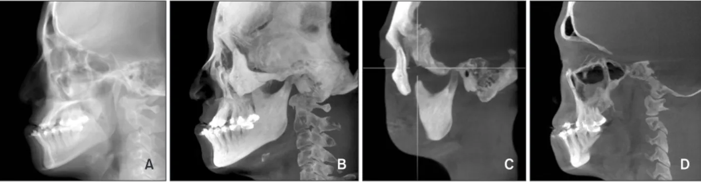
Objective: To investigate skeletal and dental changes after application of a mandibular setback surgery-first orthodontic treatment approach in cases of skeletal Class III malocclusion. Methods: A retrospective study of 34 patients (23 men, 11 women; mean age, 26.2 ± 6.6 years) with skeletal Class III deformities, who underwent surgery-first orthodontic treatment, was conducted. Skeletal landmarks in the maxilla and mandible at three time points, pre-treatment (T0), immediate-postoperative (T1), and post-treatment (T2), were analyzed using cone-beam computed tomography (CBCT)-generated half-cephalograms.

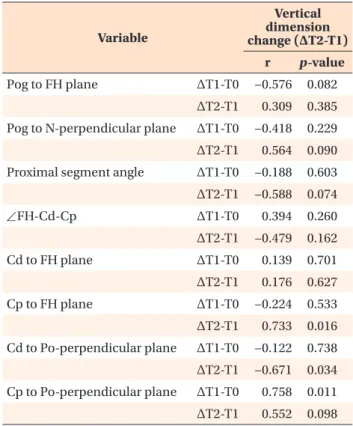
Results: The significant T0 to T1 mandibular changes occurred −9.24 ± 3.97 mm horizontally. From T1 to T2, the mandible tended to move forward 1.22

± 2.02 mm, while the condylar position (Cd to Po-perpendicular plane) shifted backward, and the coronoid process (Cp to FH plane) moved vertically. Between T1 and T2, the vertical dimension changed significantly (p < 0.05). Changes in the vertical dimension were significantly correlated to T1 to T2 changes in the Cd to Po-perpendicular plane (r = −0.671, p = 0.034), and in the Cp to FH plane (r = 0.733, p = 0.016), as well as to T0 to T1 changes in the Cp to Po- per pendicular plane ( r = 0.758, p = 0.011). Conclusions: Greater alterations in the vertical dimension caused larger post-treatment (T2) stage skeletal changes.

Stu dying the mandibular position in relation to the post-surgical vertical dimension emphasized the integral importance of vertical dimension control and proximal segment management to the success of surgery-first orthodontic treatment.

[Korean J Orthod 2015;45(2):59-65]

Key words: Three-dimensional cephalometrics, Computed tomography, Class III orthognathic surgery

Chang-Hoon Rhee

a

Youn-Kyung Choi

b

Yong-Il Kim

b,c

Seong-Sik Kim

c

Soo-Byung Park

c

Woo-Sung Son

c

a

Private Practice, Busan, Korea

b

Department of Orthodontics, Biomedical Research Institute, Pusan National University Hospital, Busan, Korea

c

Department of Orthodontics, Dental Research Institute, Pusan National University Dental Hospital, Yangsan, Korea

Received April 24, 2014; Revised September 18, 2014; Accepted September 24, 2014.

Corresponding author: Yong-Il Kim.

Assistant Professor, Department of Orthodontics, Dental Research Institute, Pusan National University Dental Hospital, Geumoro20, Mulgeum-eup, Yangsan 626-787, Korea.

Tel +82-55-360-5153 e-mail [email protected]

*The Research Fund Program of Research Institute for Basic Sciences, Pusan National University, Korea, 2014, Project No. RIBS-PNU-2014, and the Busan Gyeongnam Ulsan branch of the Korean Association of Orthodontists supported this study.

© 2015 The Korean Association of Orthodontists.

The authors report no commercial, proprietary, or financial interest in the products or companies described in this article.

This is an Open Access article distributed under the terms of the Creative Commons Attribution Non-Commercial License (http://creativecommons.org/licenses/by-nc/3.0) which permits unrestricted non-commercial use, distribution, and reproduction in any medium, provided the original work is properly cited.

pISSN 2234-7518 • eISSN 2005-372X

http://dx.doi.org/10.4041/kjod.2015.45.2.59

(2)

INTRODUCTION

Recently, several reports on orthodontic surgery with- out pre-operative treatment have been published.

1−5

This mandibular setback surgery-first approach offers advantages over conventional orthodontic-surgical treatments, including enhanced patient cooperation, efficient and effective decompensation, and reduced total treatment time.

3

Liou et al.

4

maintained that the phenomenon of accelerated postoperative orthodontic tooth movement achieved by the surgery-first approach shortened the treatment period, and partially resolved the issue of dental decompensation, which minimized the number of postoperative orthodontic treatments.

However, disadvantages of the surgery-first approach have been reported. Baek et al.

5

noted that considerable clinical experience is required for accurate assessment of skeletal discrepancies and the prediction of overall postoperative orthodontic treatment results. Further, postoperative occlusal instability can cause skeletal in- stability and associated postoperative orthodontic treat- ment difficulties.

According to previous surgery-first studies, surgery-first orthodontic treatment postoperative occlusal instability results primarily from premature contact of the extruded upper second molar. Premature cusp-to-cusp premolar contact is often associated with postoperative surgical occlusion. Additionally, premature contact induces post operative occlusal instability, increased vertical di- mension, and postoperative forward mandibular move- ment.

5,6

Hwang et al.

7

suggested that in addition to occlusal factors, proximal segments can affect post- operative skeletal stability. Therefore, a practical planning prerequisite for surgery-first orthodontic treatment is the analysis of both skeletal (including proximal segment) and dental (including vertical dimension) changes. For accurate analysis, the cone-beam computed tomography (CBCT) superimposition method and CBCT-generated

cephalograms with ray-cast and maximum intensity projection (MIP) images could be used for simultaneous acquisition of skeletal and dental information.

8,9

The aim of the present study was to investigate post- surgery-first-treatment skeletal and dental changes in skeletal Class III malocclusion, by superimposition of CBCT volumes representing the pre-treatment (T0), immediate-postoperative (T1), and post-treatment (T2) stages. Likewise, an additive correlation analysis of skeletal and dental changes was performed. The operative hypothesis was the existence of a correlation between vertical dimension changes and skeletal changes after surgery-first orthodontic treatment.

MATERIALS AND METHODS

Subjects

To test the proposed hypothesis, a retrospective study was conducted using data from 34 patients (23 men, 11 women; mean age: 26.2 ± 6.6 years) that presented to the Department of Orthodontics, Pusan National University Dental Hospital (Yangsan, Korea)between January 2010 and July 2012. The exclusion criteria were extraction history, severe facial asymmetry, cleft lip and/

or palate, and temporomandibular joint disorder. The inclusion criteria for mandibular setback surgery-first orthodontic treatment were skeletal Class III malocclu- sion with no need for extractions. Accordingly, the patients selected suffered from skeletal Class III defor- mities that were corrected using a surgery-first without orthodontic treatment approach. The surgery-first orthodontic treat ment protocol was as follows: First, a surgical occlusal setup was performed, taking into consideration the final position of the upper and lower anterior teeth. Second, a single surgeon performed a mandibular setback sa gittal split ramus osteotomy (SSRO) with rigid internal fixation achieved by miniplates on all of the patients. Next, inter-maxillary fixation was

A B C D

Figure 1. Generation of cone-beam computed tomography generated half-cephalograms. Raycast images. A, Maximum

intensity projection (MIP) image. B, Coronid process and condyle MIP image. C, Maxillary and mandibular molar MIP

image. D, Maxillary and mandibular molar MIP image.

(3)

applied for one week, followed by physiotherapy invol- ving muscle and mouth-opening exercises using vertical elastics. One month after surgery, active orthodontic treatment was resumed. Last, the surgical stent was adjusted during the postoperative orthodontic treatment, and temporary anchorage de vices were used to improve premature occlusion. The Ins titutional Review Board of Pusan National University Hos pital reviewed and approved this study (E-2011069).

Data acquisition

To evaluate statistically significant differences, all of the measurements were analyzed at three time points on CBCT-generated Half-Cephalograms (DCT pro;

Vatech Co., Hwaseong, Korea). The Half-Cephalograms were obtained by reformatting the CBCT raw data to three-dimensional (3D) images using 3D imaging software (OnDemand 3D; Cybermed Co., Seoul, Korea).

After superimposing the CBCT data on the anterior cranial base according to the maximization of mutual information theory, ray-cast images and MIP images were acquired from right side of the CBCT data (Figure 1).

10

The measurement points and reference planes are defined in Tables 1 and 2. To eva luate the correlation between vertical dimension changes and skeletal changes, vertical dimension was defined as the distance from the furcation of the upper first molar to that of lower first molar. All of the CBCT-generated Half-Cephalograms were digitized using cephalometric analysis software (VCeph version 6.0; Osstem Implant Co., Seoul, Korea) at three time points including pre- treatment (T0), immediate-postoperative (T1),and post- treatment (T2).

Statistical analysis

All statistical analyses were performed using a statis- tical software package program (SPSS ver. 12.0; SPSS, Chicago, IL, USA). The same investigator repeated all of the measurements two weeks after initial data co- llection, and the mean of the two values was used in the statistical analysis. The systematic intra-examiner error between the two measurements was determined by a paired t-test, and the magnitude of the corresponding error was assessed using the intraclass correlation coefficient (ICC). Repeated-measures analysis of vari- ance was used to determine the skeletal and dental changes at T0, T1, and T2, and multiple comparisons were performed with the Bonferroni’s test. Spearman corre lation coefficients were calculated to assess the relationship between the vertical dimension and skeletal changes after the surgery-first orthodontic treatment.

RESULTS

The means and standard deviations of the CBCT-ge- nerated Half-Cephalometric measurements taken at T0, T1, and T2 for all of the subjects are summarized in Table 3. Analysis of the systematic intra-examiner error revealed no statistically significant differences, and the ICC indicated robust reproducibility (ICC range:

0.945−0.989).

Changes from the pre-treatment to postoperative stage (T0 to T1)

The postoperative measurement changes from T0 to T1 are listed in Tables 3 and 4. The skeletal change at the Pog point manifested as −9.24 ± 3.97 mm ho- rizontal and −0.33 ± 3.00 mm vertical mandibular mo- vement. Further, the horizontal skeletal changes at the

Table 1. Reference points and planes

Landmark Definition

Point

Condylion (Cd) Superior point of mandibular condyle head

Coronoid process (Cp) Tip of coronoid process Upper 1st incisor tip (U1tip) Upper 1st incisor crown tip Lower 1st incisor tip (L1tip) Lower 1st incisor crown tip

Upper 1st molar point (U6contact) Mesial contact point of upper 1st molar Lower 1st molar point (L6contact) Mesial contact point of lower 1st molar Plane

Frankfurt-Horizontal (FH) plane Plane formed by right Po and both sides of Or

Midsagittal Reference (MSR) plane Perpendicular to FH plane and passing through Na and Ba

N-perpendicular plane Perpendicular to FH and MSR planes and passing through Na

Po-perpendicular plane Perpendicular to FH and MSR planes and passing through Po

(4)

Table 2. Skeletal and dental change measurement variables

Variable Definition

Skeletal measurement

A point to FH plane (mm) Distance from A point to FH plane

A point to N-perpendicular plane (mm) Distance from A point to N-perpendicular plane B point to FH plane (mm) Distance from B point to FH plane

B point to N-perpendicular plane (mm) Distance from B point to N-perpendicular plane Proximal segment measurement

Proximal segment angle (

o

) Angle formed by FH plane and posterior border of ramus ∠Cd-Cp-FH plane (

o

) Angle formed by FH plane and line of Cd and Cp Cd to FH plane (mm) Distance from Cd to FH plane

Cp to FH plane (mm) Distance from Cp to FH plane

Cd to Po-perpendicular plane (mm) Distance from Cd to Po-perpendicular plane Cp to Po-perpendicular plane (mm) Distance from Cp to Po-perpendicular plane Dental change

Vertical dimension (inter-furcation distance) (mm)

Distance from furcation of upper 1st molar to that of lower 1st molar

U6

contact

to FH plane (mm) Distance from upper 1st molar mesial contact point to FH plane

U6

contact

to N-perpendicular plane (mm) Distance from upper 1st molar mesial contact point to N-perpendicular plane L6

contact

to FH plane (mm) Distance from lower 1st molar mesial contact point to FH plane

L6

contact

to N-perpendicular plane (mm) Distance from lower 1st molar mesial contact point to N-perpendicular plane Refer to Table 1 for the definition of points and planes.

Table 3. Pre-treatment (T0), immediate-postoperative (T1), and post-treatment (T2) CBCT-generated Half-Cephalometric measurements for all subjects*

Variable 1-Jaw

p-value Multiple comparison, groups

T0 T1 T2

Skeletal change

B point to FH plane 78.09 (7.08) 77.69 (5.63) 76.96 (5.32) 0.489 Pog to FH plane 91.00 (7.65) 90.67 (6.37) 89.01 (5.62) 0.097

Pog to N perpendicular plane 9.50 (6.04) 0.27 (7.68) 1.48 (7.51) 0.000 T0 > T1, T2 Proximal segment angles 84.56 (6.61) 82.48 (5.92) 82.58 (6.83) 0.068

∠FH-Cd-Cp 15.72 (7.07) 14.35 (6.75) 12.40 (7.05) 0.000 T0, T1 > T1, T2

Cd to FH plane 2.15 (1.46) 3.18 (1.13) 2.37 (1.01) 0.126

Cp to FH plane 9.59 (4.97) 10.25 (4.86) 8.30 (5.11) 0.003 T1, T0 > T2, T0 Cd to Po-perpendicular plane 11.96 (1.64) 12.85 (1.67) 11.81 (1.52) 0.002 T0, T1 > T0, T2 Cp to Po-perpendicular plane 48.08 (3.01) 48.31 (3.31) 46.80 (6.37) 0.407

Dental changes

U6

contact

to FH plane 47.60 (1.10) 47.54 (1.14) 46.99 (0.99) 0.059

L6

contact

to FH plane 56.05 (1.47) 55.57 (1.34) 53.95 (1.29) 0.005 T0, T1 > T2 Vertical dimension of inter-furcation 18.39 (2.02) 18.87 (1.89) 17.29 (1.76) 0.029 T0, T1 > T0, T2 Values are presented as mean (standard deviation).

*Repeated-measures analysis of variance test and Bonferroni’s multiple correction comparison test were conducted.

T0, Preoperative; T1, immediate-postoperative; T2, seven months after surgery.

A positive value indicates forward and downward movement in linear measurements, and an increase in angular

measurements.

(5)

Pog point were statistically significant (p < 0.05). No statistically significant differences were detected in the proximal segment changes (Cd and Cp to FH plane/Po- perpendicular plane; p > 0.05). Similarly, no significant postoperative dental changes were detected in the ver- tical dimension (p > 0.05).

Changes from the postoperative to post-treatment stage (T1 to T2)

No statistically significant differences were detected in the skeletal changes from T1 to T2 (p > 0.05). However, the mandible tended to move forward 1.22 ± 2.02 mm.

Regarding the angular change of the proximal segment, the condylar position (Cd to Po-perpendicular plane) moved backward, and the coronoid process (Cp to FH plane) moved vertically. The dental changes in the vertical dimension were statistically significant (p < 0.05), and included the upward movement of the lower 1st molar, and a 1.62 ± 1.35 mm reduction from L6 to FH (Table 3 and 4).

Spearman correlation analysis (factors influencing vertical dimension changes during the postoperative orthodontic treatment stage)

T1 to T2 vertical dimension changes (ΔT2-T1) were significantly correlated with T1 to T2 Cd to Po-perpen- dicular plane changes (r = −0.671, p = 0.034), T1 to T2 Cp to FH plane changes (r = 0.733, p = 0.016), and T0 to T1 Cp to Po-perpendicular plane changes (r = 0.758, p = 0.011; Tables 3 and 4).

DISCUSSION

Our aim in the present study was to search for corre- lations between dental changes (vertical dimension) and skeletal changes (proximal segment) after the application of a surgery-first orthodontic approach to treat skeletal Class III malocclusion. We initially hypothesized that a correlation existed between vertical dimension and skeletal changes. Consequently, the study results revealed statistically significant correlations between T1 to T2 vertical dimension changes and T1 to T2 Cd to Po-perpendicular plane changes, T1 to T2 Cp to FH plane changes, and T0 to T1 Cp to Po-perpendicular plane changes.

There have been several reports of the surgery-first approach in orthognathic surgery cases,

1−6,11

but the pro per setup for the surgical model remains unclear.

Liou et al.

6

suggested that the maxilla and mandible be arranged in a proper molar relationship and with a positive overbite, which has been applied to non-ex- traction cases of Class I malocclusion.

11

Baek et al.

5

insisted that in order to obtain a normal post-surgical overbite and overjet, it was better to increase the extent

of mandibular setback than to perfectly incline the lower incisor. For accurate prediction and simulation, the establishment of the appropriate setup and surgical mo dels is critical.

8,12

Nonetheless, in their surgical model setup, orthodontists empirically increase the vertical di- mension and the extent of mandibular setback move- ment due to the occlusal interference.

Unfortunately, there are no specific guidelines for any case. Likewise, it is unclear how to resolve the increased vertical dimension resulting from premature contact in premolar or molar areas during the post-treatment stage. In the present study, the vertical dimension tended to increase at the immediate-postoperative (T1) stage, and decrease at the post-treatment (T2) stage.

At T2, the mandible tended to move forward. Similarly, Baek et al.

5

reported significant backward mandibular repositioning due to surgery, but significant forward relapse during postoperative orthodontic treatment.

Additionally, Hwang et al.

13

stressed that the increased extent of the vertical dimension changes affected T1 to T2 Pog point movement. For effective treatment planning, the extent of the increased vertical dimension and mandibular setback movement should be considered in surgical treatment objective.

Table 4. Correlation coefficient analysis between selected variables and vertical dimension changes

Variable

Vertical dimension change (ΔT2-T1)

r p-value

Pog to FH plane ΔT1-T0 −0.576 0.082

ΔT2-T1 0.309 0.385 Pog to N-perpendicular plane ΔT1-T0 −0.418 0.229 ΔT2-T1 0.564 0.090 Proximal segment angle ΔT1-T0 −0.188 0.603 ΔT2-T1 −0.588 0.074

∠FH-Cd-Cp ΔT1-T0 0.394 0.260

ΔT2-T1 −0.479 0.162

Cd to FH plane ΔT1-T0 0.139 0.701

ΔT2-T1 0.176 0.627

Cp to FH plane ΔT1-T0 −0.224 0.533

ΔT2-T1 0.733 0.016

Cd to Po-perpendicular plane ΔT1-T0 −0.122 0.738

ΔT2-T1 −0.671 0.034

Cp to Po-perpendicular plane ΔT1-T0 0.758 0.011

ΔT2-T1 0.552 0.098

Refer to Tables 1 and 2 for the measurement points and

reference planes.

(6)

However, consideration should also be given to other factors that influence increased vertical dimension change that emerged in the present Spearman corre- lation analysis, including T1 to T2 Cd to Po-per- pendicular plane changes, T1 to T2 Cp to FH plane changes, and T0 to T1 changes in Cp to Po-perpen- dicular plane. During the period from the T1 to T2 stage, the condylion (Cd) and Cp points were signi- ficantly altered, even though the Pog point was not.

Specifically, the Cd and Cp points, but not the Pog point, were significantly correlated to vertical dimension changes. Hence, the results indicated that the position of the proximal segment should be controlled.

Prior reports have emphasized that surgery-first postoperative instability stems from occlusal instability of the premature occlusal contacts and from proximal segment changes. However, a surgical stent can be used to control occlusal instability after surgery-first orthodontic treatment. In fact, several authors have recommended this strategy as a means of preventing occlusal interference, and have asserted that skeletal relapse caused by proximal and distal segment positional changes can be overcome by rigid fixation.

1−6,8,11

However, Kim et al.

14

reported proximal segment dis- placement during the healing-period, despite the application of rigid fixation systems. Therefore, even when rigid fixation is applied to mandibular setback SSRO, the proximal segment should be securely mana- ged for skeletal stability, as well as in surgery-first or- thodontic treatment cases. After careful management of skeletal stability and proximal segment position in the T1 stage, a surgical stent could be utilized as an effective tool for resolving premature occlusal contacts in the premolar or molar region, and for maintaining the T2 stage position of the Pog point. To this end, Hwang et al.

7

maintained that the intended manual condylar position could improve skeletal stability in the short term.

However, the study had several design limitations.

First, there was no comparison to a control group. Se- cond, most of the cases involving an average of 2 mm crowding that were not extraction cases were in- vestigated. Consequently, additional multi-centered studies that include extraction cases and control groups is recommended. Likewise, the changed vertical di- mension or “transitional” occlusion was established postoperatively in the surgery-first approach, and orthodontics were an adjunctive postoperative treatment used to transfigure transitional occlusion to solid final occlusion.

6

However, in surgery-first orthodontic treat- ment, in order to improve postoperative instability and achieve skeletal stability, not only surgical occlusion but also proximal segment management should be con- sidered.

CONCLUSION

A correlation existed between vertical dimension changes and skeletal changes after mandibular setback surgery-first orthodontic treatment. Larger post-surgical vertical dimension changes were related to a greater number of skeletal changes during the post-treatment stage. Examining the mandibular position in relation to the post-surgical vertical dimension served to emphasize the integral importance of vertical dimension control and proximal segment management to the success of surgery-first orthodontic treatment.

REFERENCES

1. Park KR, Kim SY, Park HS, Jung YS. Surgery-first approach on patients with temporomandibular joint disease by intraoral vertical ramus osteotomy.

Oral Surg Oral Med Oral Pathol Oral Radiol 2013;

116:e429-36.

2. Hernández-Alfaro F, Guijarro-Martínez R, Molina- Coral A, Badía-Escriche C. “Surgery first" in bimaxi- llary orthognathic surgery. J Oral Maxillofac Surg 2011;69:e201-7.

3. Nagasaka H, Sugawara J, Kawamura H, Nanda R.

“Surgery first" skeletal Class III correction using the Skeletal Anchorage System. J Clin Orthod 2009;

43:97-105.

4. Liou EJ, Chen PH, Wang YC, Yu CC, Huang CS, Chen YR. Surgery-first accelerated orthognathic surgery:

postoperative rapid orthodontic tooth movement. J Oral Maxillofac Surg 2011;69:781-5.

5. Baek SH, Ahn HW, Kwon YH, Choi JY. Surgery-first approach in skeletal class III malocclusion treated with 2-jaw surgery: evaluation of surgical move- ment and postoperative orthodontic treatment. J Craniofac Surg 2010;21:332-8.

6. Liou EJ, Chen PH, Wang YC, Yu CC, Huang CS, Chen YR. Surgery-first accelerated orthognathic surgery:

orthodontic guidelines and setup for model surgery.

J Oral Maxillofac Surg 2011;69:771-80.

7. Hwang DS, Kim YI, Lee KM. Effect of intended ma- nual condylar positioning on skeletal and dental changes in Skeletal Class III deformities: CBCT- gene rated half-cephalograms. J Craniomaxillofac Surg 2014;42:7-12.

8. Kim SJ, Park SB, Kim YI, Cho BH, Hwang DS. The reliability of cone-beam computed tomography (CBCT) - generated frontal cephalograms. J Cranio- maxillofac Surg 2012;40:e331-6.

9. Hwang HS, Lee KM, Uhm GS, Cho JH, McNamara JA

Jr. Use of Reference Ear Plug to improve accuracy

of lateral cephalograms generated from cone-beam

computed tomography scans. Korean J Orthod

(7)

2013;43:54-61.

10. Lee JH, Kim MJ, Kim SM, Kwon OH, Kim YK. The 3D CT superimposition method using image fu- sion based on the maximum mutual information algorithm for the assessment of oral and maxillo- facial surgery treatment results. Oral Surg Oral Med Oral Pathol Oral Radiol 2012;114:167-74.

11. Kang SH, Kim MK, Park SY, Lee JY, Park W, Lee SH.

Early orthognathic surgery with three-dimensional image simulation during presurgical orthodontics in adults. J Craniofac Surg 2011;22:473-81.

12. Jeon J, Kim Y, Kim J, Kang H, Ji H, Son W. New bimaxillary orthognathic surgery planning and

model surgery based on the concept of six degrees of freedom. Korean J Orthod 2013;43:42-52.

13. Hwang DS, Kim YI, Lee JY, Lee ST, Kim TH, Lee JM, et al. Evaluation of skeletal stability following two- jaw surgery via surgery first orthodontic treatment in class iii malocclusion. J Korean Assoc Maxillofac Plast Reconstr Surg 2011;33:407-12.

14. Kim YI, Park SB, Jung YH, Hwang DS, Lee JY. Eva-

luation of intersegmental displacement according

to osteosynthesis method for mandibular setback

sagittal split ramus osteotomy using cone-beam

computed tomographic superimposition. J Oral

Maxillofac Surg 2012;70:2893-8.

수치

Figure 1. Generation of cone-beam computed tomography generated half-cephalograms. Raycast images
Table 1. Reference points and planes
Table 3. Pre-treatment (T0), immediate-postoperative (T1), and post-treatment (T2) CBCT-generated Half-Cephalometric  measurements for all subjects*
Table 4. Correlation coefficient analysis between selected  variables and vertical dimension changes

참조

관련 문서

The effects of recycling on the tensile bond strength of new and clinically used stainless steel orthodontic brackets: an in vitro study. Material testing of

First, the rhythmic gymnastics exercise program improves the skeletal muscle mass of adult women in office workers and reduces body fat percentage,

First, the general characteristics of the two groups were analyzed by frequency analysis and correlation analysis, which is technical statistics, and the

Major improvements on the contract are offered. First, a broader menu of gas supply services is called for including interruptible and priority services. In

Measurement of proximal contact tightness between the left first molar and second molar(dental implant) in the mandible... Clinical dental anatomy, histology,

A clinical and radiographic evaluation of fixed partial dentures (FPDs) prepared by dental school students: a retrospective study.. A retrospective analysis

For establishing appropriate treatment plans and prognosis, the estimated value of the correlation between the change in hard and soft tissues is required,

success rates of dental implants placed at the time of or after alveolar ridge augmentation with an autogenous mandibular bone graft and titanium mesh: a 3-to