• 검색 결과가 없습니다.

Iatrogenic Subclavian Artery Perforation Rescued by Operator-Modified Graft Stent

N/A
N/A
Protected

Academic year: 2022

Share "Iatrogenic Subclavian Artery Perforation Rescued by Operator-Modified Graft Stent"

Copied!
5
0
0

로드 중.... (전체 텍스트 보기)

전체 글

(1)

INTRODUCTION

Stenosis of the subclavian artery (SCA), although not com- mon, occurrs in a number of patients. Most etiologies are ath- erosclerotic, and patients may present with vertebrobasilar insufficiency (subclavian steal syndrome), angina due to re- versal of flow from an internal mammary arterial graft (coro- nary subclavian steal syndrome), and arm ischemia or weak- ness due to distal embolization or claudication. Treatment of these lesions has traditionally been through surgical bypass.

Because of the lower complication rate, high technical suc- cess rate and good long-term patency rate (>80% at 5 years), catheter-based treatment has become more and more popu- lar.

1

We present a case of patient with total situs inversus and

stenosis of the left SCA, who was treated with catheter-based revascularization with stent that was complicated with SCA perforation with hemothorax. Percutaneous stenting with a graft stent was used as a rescue, but this procedure was com- plicated with occlusion of the common carotid artery. The oc- clusion of the common carotid artery was finally re-opened by carotid stenting. We present a way to rescue perforation of large artery during percutaneous intervention.

CASE REPORT

This 73-year-old female, with a history of coronary artery dis- ease and situs inversus, had recently noticed left arm exercise weakness. The magnetic resonance angiogram and computer tomography of the upper limb vessels showed critical stenosis of the left-side SCA with calcification near the orifice (Fig. 1).

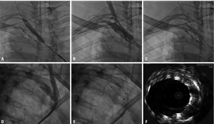
Percutaneous revascularization was done via right femoral artery approach, and severe left SCA stenosis was confirmed (Fig. 2A) (Supplementary Video 1, only online). Direct stenting in the left SCA was carried out with a balloon-expansible Ex- press LD 10×25 mm stent (Boston Scientific Corporation, Natick, MA, USA) (Fig. 2B) up to 8 atmospheres for 13 seconds.

However, severe shortness of breath and hypotension devel- oped after stenting. The blood pressure was down to 88/56, Chih-Hung Lai , Chung-Lin Tsai , Wei-Chun Chang , Chieh-Shou Su , and Wen-Lieng Lee

1

Divisions of Interventional Cardiology and Adult Cardiac Surgery, Cardiovascular Center, Taichung Veterans General Hospital, Taichung;

2

School of Medicine, National Yang-Ming University, Taipei, Taiwan, R.O.C.

Subclavian artery (SCA) perforation is a rare complication while performing SCA intervention. In our present report, a 73-year- old female, with stenosis of the left SCA and situs inversus, presented with exercise-induced left arm weakness. The SCA stenosis was treated with direct stenting with a balloon-expansible Express LD 10×25 mm stent. However, it caused iatrogenic SCA perfo- ration and hemothorax. The perforation was sealed by endovascular repair with operator-modified Endurant II graft stent, which complicated with occlusion of left common carotid artery. And, the carotid artery was rescued by another stent. The graft stent, which was originally designed for abdominal aortic aneurysm, can be modified to suitable length and take as a rescue stent of large vessel with iatrogenic perforation. Due to strong radial force of graft stent, preservation of large side branches should been watched out.

Key Words: Subclavian artery stenosis; subclavian artery perforation; graft stent; peripheral angioplasty, hemothorax

Received: July 30, 2015 Revised: December 8, 2015 Accepted: December 17, 2015

Corresponding author: Dr. Wen-Lieng Lee, Division of Interventional Cardiology, Cardiovascular Center, Taichung Veterans General Hospital, 1650 Taiwan Boule- vard Sect. 4, Taichung, Taiwan 40705, R.O.C.

Tel: 886-4-23592525, Fax: 886-4-23595046, E-mail: [email protected]

•The authors have no financial conflicts of interest.

© Copyright: Yonsei University College of Medicine 2017

This is an Open Access article distributed under the terms of the Creative Com- mons Attribution Non-Commercial License (http://creativecommons.org/licenses/

by-nc/3.0) which permits unrestricted non-commercial use, distribution, and repro-

duction in any medium, provided the original work is properly cited.

(2)

A B

Fig. 1. Magnetic resonance angiogram (MRA) and computer tomography (CT) of the upper limb vessels. (A) MRA shows severe stenosis in the proximal left subclavian artery (circle). (B) CT reveals calcification near the orifice of left subclavian artery.

Fig. 2. Angiography with anterior view of the left brachiocephaic trunk and left subclavian artery (SCA) showing proximal severe stenosis (these images are all mirrored image of this patient with situs inversus; that is, it look-like “right brachiocephaic trunk in images” actually are left ones). (A) Severe ste- nosis in the proximal SCA (arrow). (B) Direct stenting by an Express LD 10×25 mm stent. (C) SCA perforation with contrast extravasation (arrows). (D) Ret- rograde approach through the left brachial artery: covered by the graft stent (arrow). (E) Slow flow in the left common carotid artery (arrow) due to ostium occluded by the graft stent. (F) Intravascular ultrasound shows severe occlusion by the graft stent.

A

D

B

E

C

F

(3)

and the heart rate was up to 90. The oxygen saturation was down to 74%. Emergent intubation, fluid infusion, inotropic agent with norepinephrine were given. The immediate angi- ography showed vascular perforation in the stented segment of left SCA (Fig. 2C) (Supplementary Video 2, only online).

Emergency balloon inflation within the stent was done with an 8×40 mm balloon. The cardiovascular surgeon was also consulted. However, due to the high surgical risk of bypass, endovascular treatment was suggested. We decided to do a retrograde approach through the left brachial artery by surgi- cal cut-down. Since there was no suitable graft stent in our cath lab, we modified and cut the iliac extension of self- ex- panding Endurant II graft stent 10×82 mm (Medtronic, Inc., Minneapolis, MN, USA), which was originally designed to treat abdominal aortic aneurysms (AAA), to a suitable length (around 30 mm) (Fig. 3). After retrograde wiring, an operator-modified Endurant II graft stent was deployed in the stent segment (Fig.

2D) and the perforation was sealed successfully. After stenting of the graft stent, however, slow flow of left common carotid ar- tery (LCCA) was noted, and angiography confirmed the near total occlusion of the LCCA, occluded by the SCA graft stent (Fig. 2E) (Supplementary Video 3, only online). A firm wire (Con- quest pro, Asahi Inc, Seto, Japan) was successfully advanced outside the graft stent and into the LCCA. Despite sequent bal- loon dilations at the bifurcation site, the flow to the LCCA was still poor. Intravascular ultrasound also confirmed the severe compromise of the LCCA ostium which was caused by the graft stent (Fig. 2F). Therefore, we put a balloon-expansible carotid

stent, Express 7×37 mm (Boston Scientific Corporation), from the LCCA to the left brachiocephalic artery (Fig. 4A). TIMI III flow of the LCCA was restored after stenting (Fig. 4B-E) (Supple- mentary Video 4, only online). Intravascular ultrasound also confirmed proper expansion of the LCCA stent (Fig. 4F). The follow-up chest radiography revealed left-side hemothorax.

After thoracocentesis with drainage and proper care, the pa- tient was discharged. Till now, the patient has been free from symptoms for six months.

DISCUSSION

Percutaneous angioplasty has become an increasingly popu- lar therapy for treating symptomatic SCA stenosis. The techni- cal success rate of percutaneous therapy for SCA stenosis with stent is up to 91% to 100% and ranges from 83% to 94% in to- tally occluded SCA.

2

The complication rate has also been low.

No peri-procedural deaths have been reported. Stenting of SCA stenosis is also characterized by its proven efficacy, long- term patency, lack of a need for general anesthesia, and short- er healing hospital stay.

3

Therefore, when an intervention is indicated, a percutaneous approach is currently favored over surgical intervention.

There are several instructional points in our present case.

First, what kind of mechanism is involved in the SCA perfora- tion after stenting? Despite many advantages, there are still some possible procedural complications, including stroke, Fig. 3. The procedure of modification of graft stent. (A and B) After deployed out graft stent, the stent was cut between ring and ring and to a wanted length. (C) Using sutures to aid collapse of the ring of deployed stent. (D) With the compression of sutures, the stent could be resheathed into sheath. (E) Repeating the same way, the stent was resheathed ring by ring. (F) Resheathed modified stents ready to be deployed.

A

D

B

E

C

F

(4)

dissection, distal embolization, arterial thrombosis, and ac- cess site complication. SCA perforation caused by stenting has rarely been reported; however, as in our case, it may cause severe complications, such as hemothorax and hypotension.

Therefore, it is highly possible that perforation might be caused by an over-sized stent or/and too rapid inflation of the stent, especially in a calcified lesion. It would be better off if we had inflated the balloon slowly and correlated with the pa- tient’s discomfort. In addition to angiography, further image modality, such as intravascular ultrasound, could help choose more precise stent size.

Second, how to deal with an iatrogenic SCA perforation?

Due to massive blood loss at the time, immediate balloon in- flation was mandatory to seal the perforation. In our case, the cardiovascular surgeon suggested endovascular intervention, rather than surgical bypass, because of higher surgical risk and complication rate. Due to the lack of any usual cover stent with such big size, we had to modify the iliac extension of En- durant II stent graft, which was originally designed to treat AAA. Furthermore, because this graft stent was designed for AAA, the delivery system was too short to approach SCA le- sion by femoral approach. This made us to use the retrograde approach through the brachial artery by surgical cut-down.

However, it was not easy to place this modified stent in a pre- cise position, making the stent to accidentally occlude the LCCA. Because of the strong and firm force of the graft stent,

the balloon could not dilate the occlusion well, and we had to put a balloon-expanded stent in the LCCA to keep the ostium open. In our opinion, before stenting a graft stent, a protective wire should be put in every important branch that might be impinged by the stenting. In this way, locating the stent posi- tion would be more precise, and total occlusion of the side branches could be avoided.

We described herein our valuable experience in dealing with a rare complication of SCA perforation while stenting the SCA. Choosing an SCA stent of a suitable size or even an un- der-sized SCA stent should be considered, and gradual slow inflation of the stent may also help lessen the risk of perfora- tion. Except for open surgery, modified graft stent described in the present study is an alternative choice. While using this therapy, protective side branches wiring in addition to precise locating, should also be considered if the risk of jailing is high.

SUPPLEMENTARY DATA

Video 1. Anterior view of the left subclavian artery: proximal se- vere stenosis.

Video 2. Subclavian artery perforation after stenting.

Video 3. The graft stent occluded the ostium of the left com- mon carotid artery, resulting in a slow flow.

Video 4. Final angiography showed patency of kissing stents.

Fig. 4. Angiographic images during rewiring and stenting of the left common carotid artery (LCCA) (these images are all mirrored image of this patient with situs inversus; that is, the “right brachiocephaic trunk in images” actually are left ones). (A) Stenting from the LCCA to the left brachiocephalic artery.

(B and C) After stenting, frontal view of angiography after (B) and before (C) contrast injection. (D and E) After stenting, lateral view of angiography after (D) and before (E) contrast injection. (F) Intravascular ultrasound shows the ostium of the LCCA re-opened by the stent.

A

D

B

E

C

F

(5)

수치

Fig. 1. Magnetic resonance angiogram (MRA) and computer tomography (CT) of the upper limb vessels
Fig. 4. Angiographic images during rewiring and stenting of the left common carotid artery (LCCA) (these images are all mirrored image of this patient with  situs inversus; that is, the “right brachiocephaic trunk in images” actually are left ones)

참조

관련 문서