Received:December 22, 2020, Revised:April 30, 2021, Accepted:May 3, 2021 Corresponding to:Sang-Cheol Bae http://orcid.org/0000-0003-4658-1093
Department of Rheumatology, Hanyang University Hospital for Rheumatic Diseases, 222 Wangsimni-ro, Seongdong-gu, Seoul 04763, Korea. E-mail:[email protected]
Copyright ⓒ 2021 by The Korean College of Rheumatology. All rights reserved.
This is an Open Access article, which permits unrestricted non-commerical use, distribution, and reproduction in any medium, provided the original work is properly cited.
Clinical and Genetic Risk Factors Associated With the Presence of Lupus Nephritis
Jung-Min Shin, M.D.1, Dam Kim, M.D., Ph.D.2, Young-Chang Kwon, Ph.D.3, Ga-Young Ahn, M.D.4,
Jiyoung Lee, M.S.3, Youngho Park, Ph.D.5, Yeon-Kyung Lee, M.D.1, Tae-Han Lee, M.D.1, Dae Jin Park, M.D.1, Yeo-Jin Song, M.D.1, Eunji Ha, B.S.6,7, Kwangwoo Kim, Ph.D.6,7, So-Young Bang, M.D., Ph.D.1,3,
Chan-Bum Choi, M.D., Ph.D.1,3, Hye-Soon Lee, M.D., Ph.D.1,3, Sang-Cheol Bae, M.D., Ph.D., MPH1,3
1Department of Rheumatology, Hanyang University Hospital for Rheumatic Diseases, Seoul, 2Department of Rheumatology, Myongji Hospital, Hanyang University College of Medicine, Goyang, 3Hanyang University Institute for Rheumatology Research, Seoul, 4Division of Rheumatology, Department of Internal Medicine, Korea University Guro Hospital, Seoul, 5Department of Big Data Application, College of Social Economic &
Interdisciplinary Studies, Hannam University, Daejeon, Departments of 6Life and Nanopharmaceutical Sciences, 7Biology, Kyung Hee University, Seoul, Korea
Objective. To elucidate whether clinical features and the weighted genetic risk score (wGRS) were associated with the presence of lupus nephritis (LN). Methods. We retrospectively divided patients with systemic lupus erythematosus (SLE, n=1,078) into biopsy-proven LN (n=507) and non-LN groups (non-LN, n=571). Baseline clinical features, serologic markers, and the wGRS were collected. The wGRS was calculated from 112 non-human leukocyte antigen (non-HLA) loci and HLA-DRβ1 amino acid haplotypes for SLE. Associations among clinical features, wGRS, and the presence of LN were identified. Results. In the multi- variate analysis, patients with LN were younger at diagnosis (odds ratio [OR]=0.97, p<0.001), had more pleuritis (OR=2.44, p<0.001) and pericarditis (OR=1.62, p=0.029), had a higher detection rate of anti-double stranded deoxyribonucleic acid (anti-dsDNA antibodies, OR=2.22, p<0.001), anti-Smith antibodies (anti-Sm antibodies, OR=1.70, p=0.002), low level of complement (OR=1.37, p=0.043) and absence of antiphospholipid antibodies (aPL antibodies, OR=1.60, p=0.002), and had higher wGRS (OR=1.16, p=0.012). Mediation analysis suggested that anti-Sm antibodies and low complement could be medi- ators in the relationship between high wGRS and the presence of LN. Conclusion. Onset age, pleuritis, pericarditis, several sero- logic markers, and wGRS were associated with the presence of LN. Anti-Sm antibodies and low complement appeared to medi- ate the indirect relationship between wGRS and the presence of LN. (J Rheum Dis 2021;28:150-158)
Key Words. Systemic lupus erythematosus, Lupus nephritis, Genetic risk score, Associated factors
INTRODUCTION
Systemic lupus erythematosus (SLE) is a heterogeneous autoimmune disease well known for its multisystemic presentation, ranging from cutaneous manifestations to vital organ disorders [1]. Lupus nephritis (LN) is one fre- quent and severe organ manifestation of SLE [2] that af- fects 12%∼69% of patients with SLE [3]. Despite current advanced treatments, LN constitutes a major cause of re- nal failure and is associated with reduced long-term sur-
vival [3]. Early diagnosis and treatment with im- munosuppressive agents are important for improving outcomes associated with LN [4]. Thus, recognizing pa- tients at risk for LN is advantageous for early diagnosis and prompt intervention with immunosuppressive agents, thereby reducing the mortality risk.
Previous studies have suggested that several clinical and immunologic features, such as age, sex, hypertension, malar rash, anemia, anti-double stranded deoxyribonucleic acid antibodies (anti-dsDNA antibodies), anti-Smith an-
Figure 1. Flow diagram of the study design. SLE: systemic lu- pus erythematosus, wGRS: weight- ed genetic risk score, ACR:
American College of Rheum- atology, LN: lupus nephritis.
tibodies (anti-Sm antibodies), low complement, lupus anticoagulant [5-7], and genetic factors [8] are associated with LN. Familial and twins studies have elucidated the genetic associations of SLE [9], and large-scale meta- analyses of genome-wide association studies (GWASs) have determined a significant association between com- mon genetic variants and disease risk for SLE [10,11]. A genetic risk score (GRS) effectively calculates the esti- mated effect of genetic risk on disease susceptibility, ag- gregating the risk loci determined in GWASs into a single measurement [12]. The weighted genetic risk score (wGRS) reflects how frequently the risk alleles were found in patients’ genomes using an allelic odds ratio (OR) in a logarithmic scale [13]. A recent study demon- strated that the individual GRS for SLE can predict early disease onset and damage accrual including LN [8]. We investigated the ability of genetic risk to predict presence of LN using the wGRS calculated from the recently re- ported 112 non-human leukocyte antigen (non-HLA) loci and HLA-DRβ1 amino acid haplotypes [11].
Here, we investigated clinical and genetic features sig- nificantly associated with LN. Genetic risk was estimated using wGRS.
MATERIALS AND METHODS
Patients
All patients who fulfilled the American College of Rheumatology (ACR) classification criteria for SLE [14]
were enrolled. All subjects provided written informed consent for inclusion in the Hanyang BAE Lupus cohort (BAE Registry of Autoimmune Diseases for Epidemiology) [15], a Korean single-center prospective observational cohort for which clinical information was updated annu- ally from 1998∼2018. This study protocol was approved by the Institutional Ethics Review Board of Hanyang University Hospital (IRB no: HYUH2001-06-001).
All patients with SLE were retrospectively classified into two groups based on renal involvement. All patients with LN were confirmed by renal biopsy before enrollment or during the follow-up period. Those who never met the ACR renal disorder criteria [14,16] before enrollment and during the follow-up period were classified as pa- tients without LN (non-LN). Patients who met the ACR classification criteria for renal disorder but who lacked re- nal biopsy results were excluded from this study. A flow diagram of this study is summarized in Figure 1.
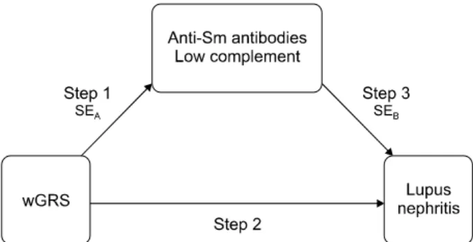
Figure 2. Conceptual model on three-step relationship be- tween the exposure, mediator, and outcome. Step 1: associa- tion between the wGRS and serologic markers calculated by logistic regression analysis, step 2: association between the wGRS and lupus nephritis (LN) calculated by logistic re- gression analysis, step 3: association between serologic mark- ers and LN calculated by logistic regression analysis, Sm:
Smith, SEA: associated standard error for step 1 calculated by Sobel’s test, SEB: associated SE for step 3 calculated by Sobel’s test, SEA for anti-Sm antibodies: 0.066, SEA for low comple- ment: 0.057, SEB for anti-Sm antibodies: 0.157, SEB for low complement: 0.141.
Clinical characteristics
Demographic and socioeconomic data were collected at enrollment. Clinical features such as the SLE Disease Activity Index-2000 [17], Systemic Lupus International Collaborating Clinics/ACR Damage Index (SDI) [18], complete blood count, chemistry, immunology, and uri- nalysis results were recorded and followed up at least annually.
Until 2004, histological classification of biopsy findings was based on the 1982 World Health Organization (WHO) classification for LN [19]; then, the International Society of Nephrology/Renal Pathology Society (ISN/RPS) classification was adopted [20]. Activity and chronicity indices [21,22] were also collected. If a patient with LN had undergone more than two renal biopsies, we selected the first renal biopsy result for analysis.
Calculation of the wGRS
The GRS was evaluated to elucidate its genetic effect on LN in patients with SLE. The GRS was weighted accord- ing to effect size based on the allelic odds ratio of each var- iant from previously reported SLE-risk loci [13], which constitute the 112 non-HLA loci of the most updated East Asian study [11] and the HLA-DRβ1 haplotypes in ami- no acid positions 11, 13, and 26 [23]. Finally, we used the wGRS of each patient to predict the presence of LN.
Statistical analyses
We used Student’s t-test for continuous variables and the chi-square test for categorical variables of demo- graphics, clinical characteristics, laboratory findings, and wGRS to compare the differences between the LN and non-LN groups. Logistic regression analysis was con- ducted to determine the associations among the demo- graphics, clinical features, wGRS, and the presence of LN.
All analyses were performed using the SAS 9.2 statistical software (SAS Institute, Cary, NC, USA). All tests were two-sided, and p-values<0.05 were considered statisti- cally significant.
Mediation analysis has been commonly used in sociol- ogy, epidemiology [24], and clinical fields to evaluate cor- onary artery disease and osteoarthritis [25,26]. This method has been adopted for its ability to identify the causal relationship between an exposure and an outcome through mediators. In this study, among all of the asso- ciated clinical features of LN determined in the logistic re- gression analysis, we focused on serologic markers such as anti-Sm antibodies and low complement, which are ob-
jective and less affected by inter- and intrarater reliability, and assumed that they were mediators. We defined a three-step relationship among the exposure, mediators, and outcome (Figure 2). Step one was to determine the association between the wGRS and serologic markers, step two was to determine the association between the wGRS and LN, and step three was to determine the asso- ciation between serologic markers and LN. If a variable was confirmed to be a mediator, all three steps should be statistically significant [27]. Furthermore, the sig- nificance of step three should be decreased with the addi- tion of a mediator [24]. We sequentially performed logis- tic regression analyses on all three steps and conducted Sobel’s test [27] to recertify the hypothesis. In Sobel’s test statistics, absolute values over ±1.96 were consid- ered statistically significant.
RESULTS
Differences in demographics and clinical manifestations
A total of 1,078 patients were enrolled in this study, and all patients were of a single ethnic origin. The study pop- ulation was predominantly female (n=995, 92.3%). LN was diagnosed in 507 (47.0%) patients, and 571 subjects were included in the non-LN group (53.0%) during a
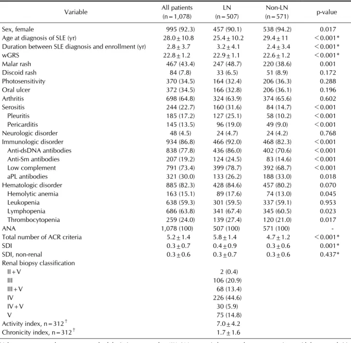
Table 1. Comparison of baseline clinical features and wGRS in patients with lupus nephritis and non-lupus nephritis
Variable All patients
(n=1,078)
LN (n=507)
Non-LN
(n=571) p-value
Sex, female 995 (92.3) 457 (90.1) 538 (94.2) 0.017
Age at diagnosis of SLE (yr) 28.0±10.8 25.4±10.2 29.4±11 <0.001*
Duration between SLE diagnosis and enrollment (yr) 2.8±3.7 3.2±4.1 2.4±3.4 <0.001*
wGRS 22.8±1.2 22.9±1.1 22.6±1.2 <0.001*
Malar rash 467 (43.4) 247 (48.7) 220 (38.6) 0.001
Discoid rash 84 (7.8) 33 (6.5) 51 (8.9) 0.172
Photosensitivity 370 (34.5) 164 (32.4) 206 (36.3) 0.288
Oral ulcer 372 (34.5) 166 (32.8) 206 (36.1) 0.196
Arthritis 698 (64.8) 324 (63.9) 374 (65.6) 0.602
Serositis 244 (22.7) 160 (31.6) 84 (14.7) <0.001
Pleuritis 185 (17.2) 127 (25.1) 58 (10.2) <0.001
Pericarditis 145 (13.5) 96 (19.0) 49 (9.0) <0.001
Neurologic disorder 48 (4.5) 24 (4.7) 24 (4.2) 0.768
Immunologic disorder 934 (86.8) 466 (92.0) 468 (82.3) <0.001
Anti-dsDNA antibodies 838 (77.8) 436 (86.0) 402 (70.6) <0.001
Anti-Sm antibodies 207 (19.2) 124 (24.5) 83 (14.6) <0.001
Low complement 791 (73.4) 399 (78.7) 392 (68.7) <0.001
aPL antibodies 321 (30.0) 133 (26.2) 188 (33.0) 0.018
Hematologic disorder 885 (82.3) 428 (84.6) 457 (80.2) 0.070
Hemolytic anemia 163 (15.1) 89 (17.6) 74 (13.0) 0.045
Leukopenia 638 (59.3) 301 (59.5) 337 (59.1) 0.953
Lymphopenia 686 (63.8) 341 (67.4) 345 (60.5) 0.023
Thrombocytopenia 259 (24.0) 139 (27.4) 120 (21.0) 0.017
ANA 1,078 (100) 507 (100) 571 (100) -
Total number of ACR criteria 5.2±1.4 5.8±1.4 4.7±1.2 <0.001*
SDI 0.3±0.7 0.4±0.9 0.3±0.6 0.001*
SDI, non-renal 0.3±0.6 0.3±0.7 0.3±0.6 0.437*
Renal biopsy classification
II+V 2 (0.4)
III 106 (20.9)
III+V 68 (13.4)
IV 226 (44.6)
IV+V 30 (5.9)
V 75 (14.8)
Activity index, n=312† 7.0±4.2
Chronicity index, n=312† 1.7±1.6
Values are presented as mean±standard deviation, or number (%). LN: systemic lupus erythematosus patients with lupus nephritis, non-LN: systemic lupus erythematosus patients without LN, SLE: systemic lupus erythematosus, wGRS: weighted genetic risk score, dsDNA: double-stranded deoxyribonucleic acid, Sm: Smith, aPL: antiphospholipid, ANA: antinuclear antibodies, ACR:
American College of Rheumatology, SDI: Systemic Lupus International Collaborating Clinics/ACR Damage Index total score at enrollment, SDI, non-renal: total score of SDI at enrollment except for the variables related to LN; such as proteinuria, pyuria, hematuria, and abnormal urinary casts. *p-values are calculated by Student’s t-test and the chi-square test. p-values<0.05.
†Numbers are reduced due to lack of data.
mean follow-up period of 10.4±6.1 years. Table 1 shows the clinical features based on the ACR SLE classification criteria [14] and the wGRS in the biopsy- proven LN and non-LN groups. There were fewer female patients with SLE in the LN group than in the non-LN group (n=457, 90.1% in the LN group vs. n=538, 94.2% in the non-LN
group, p=0.017). Patients in the SLE group were typically diagnosed with SLE at a younger age (25.4±10.2 years vs.
29.4±11.0 years, p<0.001) and had a longer duration be- tween SLE diagnosis and enrollment than those in the non-LN group (3.2±4.1 years vs. 2.4±3.4 years, p<
0.001). Among the clinical features, the presence of a ma-
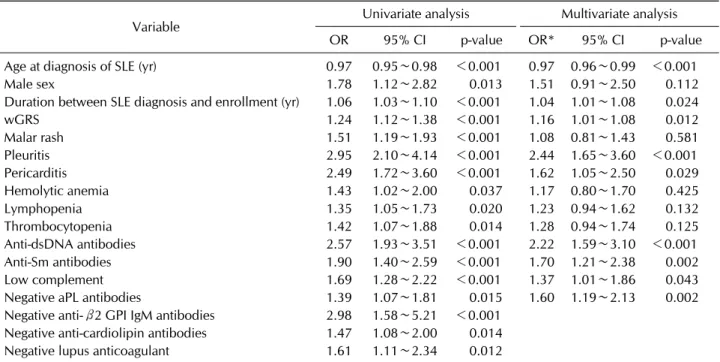
Table 2. Associations between clinical features and the presence of lupus nephritis
Variable Univariate analysis Multivariate analysis
OR 95% CI p-value OR* 95% CI p-value
Age at diagnosis of SLE (yr) 0.97 0.95∼0.98 <0.001 0.97 0.96∼0.99 <0.001
Male sex 1.78 1.12∼2.82 0.013 1.51 0.91∼2.50 0.112
Duration between SLE diagnosis and enrollment (yr) 1.06 1.03∼1.10 <0.001 1.04 1.01∼1.08 0.024
wGRS 1.24 1.12∼1.38 <0.001 1.16 1.01∼1.08 0.012
Malar rash 1.51 1.19∼1.93 <0.001 1.08 0.81∼1.43 0.581
Pleuritis 2.95 2.10∼4.14 <0.001 2.44 1.65∼3.60 <0.001
Pericarditis 2.49 1.72∼3.60 <0.001 1.62 1.05∼2.50 0.029
Hemolytic anemia 1.43 1.02∼2.00 0.037 1.17 0.80∼1.70 0.425
Lymphopenia 1.35 1.05∼1.73 0.020 1.23 0.94∼1.62 0.132
Thrombocytopenia 1.42 1.07∼1.88 0.014 1.28 0.94∼1.74 0.125
Anti-dsDNA antibodies 2.57 1.93∼3.51 <0.001 2.22 1.59∼3.10 <0.001
Anti-Sm antibodies 1.90 1.40∼2.59 <0.001 1.70 1.21∼2.38 0.002
Low complement 1.69 1.28∼2.22 <0.001 1.37 1.01∼1.86 0.043
Negative aPL antibodies 1.39 1.07∼1.81 0.015 1.60 1.19∼2.13 0.002
Negative anti-β2 GPI IgM antibodies 2.98 1.58∼5.21 <0.001 Negative anti-cardiolipin antibodies 1.47 1.08∼2.00 0.014 Negative lupus anticoagulant 1.61 1.11∼2.34 0.012
OR: odds ratio, OR*adjusted for age at diagnosis, sex, and the duration between SLE diagnosis and enrollment, CI: confidence interval, SLE: systemic lupus erythematosus, wGRS: weighted genetic risk score, dsDNA: double-stranded deoxyribonucleic acid, Sm: Smith, aPL: antiphospholipid. p-values and OR are calculated by logistic regression analyses. p-values<0.05.
lar rash (n=247, 48.7% vs. n=220, 38.6%, p=0.001) and serositis (n=160, 31.6% vs. n=84, 14.8%, p<0.001) and immunologic abnormalities including anti-dsDNA (n=436, 86.0% vs. n=402, 70.6%, p<0.001), anti-Sm antibodies (n=124, 24.5% vs. n=83, 14.6%, p<0.001), low complement (n=399, 78.7% vs. n=392, 68.7%, p<0.001), and negative antiphospholipid antibodies (aPL antibodies, n=133, 26.2% vs. n=188, 33%, p=0.018) occurred more frequently in the LN group than in the non-LN group. In addition, with regard to hemato- logic disorders, hemolytic anemia (n=89, 17.6% vs.
n=74, 13%, p=0.045), lymphopenia (n=341, 67.4% vs.
n=345, 60.5%, p=0.023), and thrombocytopenia (n=
139, 27.4% vs. n=120, 21.0%, p=0.017) were more fre- quently reported in the LN group than in the non-LN group. The wGRS was higher in patients in the LN group than in those in the non-LN group (22.9±1.1 vs. 22.6±
1.2, p<0.001). The total number of ACR criteria and SDI score at enrollment were higher in the LN group than in the non-LN group (5.8±1.4 vs. 4.7±1.2, p<0.001 and 0.4±0.9 vs. 0.3±0.6, p=0.001). However, non-renal SDI scores were comparable between the two groups. The re- nal biopsy results are also summarized in Table 1. In pa- tients with LN, the most common histologic LN classi- fication was class IV (n=226, 44.6%), and the number of
patients grew to 256 (50.5%) when classes IV and IV+V were combined. The mean activity index was 7.0±4.2 (range, 0∼18) and the mean chronicity index was 1.7±1.6 (range, 0∼8) in the first renal biopsy.
Factors associated with the presence of LN Factors associated with the presence of LN are summar- ized in Table 2. Among the various clinical features and wGRS in the univariate analysis, younger age at diagnosis (OR=0.97, p<0.001), male sex (OR=1.78, p=0.013), longer duration between SLE diagnosis and enrollment (OR=1.06, p<0.001), presence of a malar rash (OR=
1.51, p<0.001), pleuritis (OR=2.95, p<0.001), peri- carditis (OR=2.49, p<0.001), hemolytic anemia (OR=1.43, p=0.037), lymphopenia (OR=1.35, p=0.020), thrombo- cytopenia (OR=1.42, p=0.014), anti-dsDNA antibodies (OR=2.57, p<0.001), anti-Sm antibodies (OR=1.90, p<0.001), low complement (OR=1.69, p<0.001), ab- sence of aPL (OR=1.39, p=0.015), and higher wGRS (OR=1.24, p<0.001) were significantly associated with the presence of LN. Among aPL antibodies, negative an- ti-beta 2 glycoprotein I IgM antibodies (anti-β2 GPI IgM, OR=2.98, p<0.001), negative anticardiolipin antibodies (OR=1.32, p<0.001), and negative lupus anticoagulant (OR=1.61, p=0.012) test results were associated with
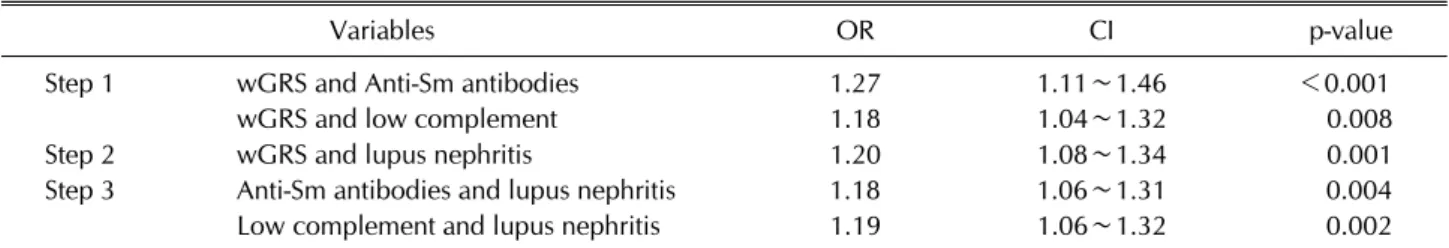
Table 3. Relationships among serologic markers, wGRS, and the presence of lupus nephritis
Variables OR CI p-value
Step 1 wGRS and Anti-Sm antibodies 1.27 1.11∼1.46 <0.001
wGRS and low complement 1.18 1.04∼1.32 0.008
Step 2 wGRS and lupus nephritis 1.20 1.08∼1.34 0.001
Step 3 Anti-Sm antibodies and lupus nephritis 1.18 1.06∼1.31 0.004
Low complement and lupus nephritis 1.19 1.06∼1.32 0.002
OR: odds ratio, CI: confidence interval, wGRS: weighted genetic risk score, Sm: Smith, Step 1: association between the wGRS and serologic markers, Step 2: association between serologic markers and lupus nephritis, Step 3: association between the wGRS and lupus nephritis. p-values and OR are calculated by logistic regression analyses. p-values<0.05.
LN. In the multivariate analysis model, younger age at SLE diagnosis (OR=0.97, p<0.001), longer duration be- tween SLE diagnosis and enrollment (OR=1.04, p=0.024), higher wGRS (OR=1.16, p=0.012), presence of pleuritis (OR=2.44, p<0.001), pericarditis (OR=1.62, p=0.029), anti-dsDNA antibodies (OR=2.22, p<0.001), anti-Sm antibodies (OR=1.70, p=0.002), low comple- ment (OR=1.37, p=0.043), and absence of aPL anti- bodies (OR=1.60, p=0.002) were statistically significant associated factors.
Relationships among the wGRS, serology, and the presence of LN
Since serologic markers, such as anti-dsDNA anti- bodies, anti-Sm antibodies, low complement, absence of aPL antibodies, and the wGRS were independently asso- ciated with the presence of LN and since the wGRS is an inherited trait from birth, we assumed that the wGRS could be the exposure and that serologic markers could be mediators for the presence of LN. All three of the relation- ships in Figure 1 were confirmed to be statistically sig- nificant (Table 3), and the statistical power of the p-values decreased when anti-Sm antibodies and low complement were added in step two (OR before adding anti-Sm anti- bodies in step two: 1.83, OR after adding anti-Sm anti- bodies in step two: 1.16, OR before adding low comple- ment in step two: 1.60, OR after adding low complement in step two: 1.17). Through Sobel’s test, we found an esti- mate of 2.66 for anti-Sm antibodies and an estimate of 2.17 for low complement, which were statistically sig- nificant (Figure 2, standard error [SE]A for anti-Sm anti- bodies: 0.066, SEA for low complement: 0.057, SEB for an- ti-Sm antibodies: 0.157, SEB for low complement: 0.141).
Based on these processes, anti-Sm antibodies and low complement appeared to have indirect effects on the rela- tionship between the wGRS and LN.
DISCUSSION
In this study, we investigated factors associated with the presence of LN in patients with SLE. Younger age at diag- nosis, longer duration between SLE diagnosis to enroll- ment, presence of pleuritis or pericarditis, presence or ab- sence of certain serologic markers, and higher wGRS were independently associated with the presence of LN. In ad- dition, anti-Sm antibodies and low complement mediated an indirect relationship between the wGRS and LN.
Several studies have reported associated clinical charac- teristics in patients with LN, including younger age of SLE onset [3,28], presence of a malar rash [29], hemo- lytic anemia [28], and thrombocytopenia [29], and in- creased number of fulfilled ACR criteria [29], which are in line with the findings of our study. Previous studies have also reported an association between serologic markers including anti-Sm antibodies [3,29] and an- ti-dsDNA antibodies [7], and the presence of LN. Our study revealed that anti-dsDNA antibodies, anti-Sm anti- bodies, low complement, and absent aPL antibodies were factors associated with the presence of LN after adjusting for age, sex, and duration from diagnosis of SLE to enrollment. Regarding the relationship between aPL anti- bodies and LN, inconsistent results have been reported [7,30-32]. One study reported that the anti-β2 GPI IgM antibodies are protective against LN [32], which is in ac- cordance with our results. The authors postulated that a lack of data on anti-β2 GPI antibodies in past research may contribute the negative effect of aPL antibodies against LN. This inconsistent association may be due to different study designs and definitions.
The genetic components of LN have been of recent inter- est in the field of rheumatology [8,33,34]. Previous re- search using transgenic mice produced to express HLA-DR3 and develop antinuclear antibodies, an-
ti-dsDNA antibodies, and glomerulonephritis revealed that HLA-DR3 plays a critical role in generating an auto- immune response to anti-Sm antibodies and progressing to LN [35]. In another study, complement component 4 genes, near the major histocompatibility complex locus, reportedly generated a seven-fold variation in SLE risk [36]. Furthermore, studies have reported that anti-Sm antibodies [5] and low complement [37] are not only risk factors for LN but might also help to predict treatment re- sponse [38,39]. Based on previous research, we could reasonably assume that there is an association between serologic markers, such as anti-Sm antibodies and low complement, and LN. In this study, we revealed that an- ti-Sm antibodies and low complement could be mediators in the relationship between the wGRS and LN through Sobel’s test. The results of our study can be explained by the assumptions of a previous study on possibility of binding of anti-Sm antibodies and complement to the kid- ney structure in LN [40]. It is important to determine the genetic effect of organ involvement to comprehend the pathogenesis of SLE [8]. We expect that the results of our study on the wGRS can be useful for predicting the dis- ease course in patients with SLE in the future.
This study has some crucial limitations including in- sufficient data regarding the treatment regimen and treat- ment adherence in patients with SLE. We were unable to analyze the information on treatment because of a lack of data before enrollment and complexities of the regimen during the long follow-up period. Therefore, we focused on data that were not easily altered by treatment or dis- ease course.
Our study also had some major strengths. First, we in- cluded a large number of patients with SLE with a rela- tively long follow-up period because of good patient compliance. Second, we analyzed high-validity data in- cluding clinical, serologic, histologic, and wGRS that were collected by well-trained medical staff in our center.
Finally, this is the first study to determine the interactions between the wGRS and LN through the mediation of se- rologic markers.
As not all of the pathogenic mechanisms of LN have been elucidated, it is impossible to clearly determine the genetic background and risk factors for the presence of LN in patients with SLE. Further studies are needed to provide precision medicine for patients with LN.
CONCLUSION
Younger age at diagnosis, presence of pleuritis, peri- carditis, anti-dsDNA antibodies, anti-Sm antibodies, low complement, absence of aPL antibodies, and higher wGRS were independently associated with the presence of LN in patients with SLE. Anti-Sm antibodies and low complement seemed to mediate the association between wGRS and the presence of LN.
ACKNOWLEDGMENTS
This work was supported in part by the Bio & Medical Technology Development Program of the National Research Foundation (NRF), funded by the Ministry of Science & ICT (NRF-2017M3A9B4050335) and by Hanyang University Institute for Rheumatology Research, Republic of Korea.
CONFLIC OF INTEREST
No potential conflict of interest relevant to this article was reported.
AUTHOR CONTRIBUTIONS
J.M.S. and D.K. designed this study and drafted the manuscript. G.Y.A. assessed the clinical data and analysis.
Y.C.K. and J.L. collected clinical and genetic data, and per- formed the statistical analysis. Y.P. advised on appropriate statistical techniques, including mediation analysis.
Y.K.L., T.H.L., D.J.P., and Y.J.S. collected clinical data and interviewed patients. E.H. and K.K. analyzed the genetic data. G.Y.A., S.Y.B., C.B.C., and H.S.L. assessed the study design validity. S.C.B. designed this study and critically reviewed the manuscript.
REFERENCES
1. Tsokos GC. Systemic lupus erythematosus. N Engl J Med 2011;365:2110-21.
2. Formiga F, Moga I, Pac M, Mitjavila F, Rivera A, Pujol R.
Mild presentation of systemic lupus erythematosus in eld- erly patients assessed by SLEDAI. SLE Disease Activity Index. Lupus 1999;8:462-5.
3. Hanly JG, O'Keeffe AG, Su L, Urowitz MB, Romero-Diaz J, Gordon C, et al. The frequency and outcome of lupus neph- ritis: results from an international inception cohort study.
Rheumatology (Oxford) 2016;55:252-62.
4. Fiehn C, Hajjar Y, Mueller K, Waldherr R, Ho AD, Andrassy K. Improved clinical outcome of lupus nephritis during the past decade: importance of early diagnosis and treatment.
Ann Rheum Dis 2003;62:435-9.
5. Ntatsaki E, Isenberg D. Risk factors for renal disease in sys- temic lupus erythematosus and their clinical implications.
Expert Rev Clin Immunol 2015;11:837-48.
6. Mavragani CP, Fragoulis GE, Somarakis G, Drosos A, Tzioufas AG, Moutsopoulos HM. Clinical and laboratory predictors of distinct histopathogical features of lupus nephritis. Medicine (Baltimore) 2015;94:e829.
7. Alba P, Bento L, Cuadrado MJ, Karim Y, Tungekar MF, Abbs I, et al. Anti-dsDNA, anti-Sm antibodies, and the lupus anti- coagulant: significant factors associated with lupus nephritis.
Ann Rheum Dis 2003;62:556-60.
8. Reid S, Alexsson A, Frodlund M, Morris D, Sandling JK, Bolin K, et al. High genetic risk score is associated with early disease onset, damage accrual and decreased survival in sys- temic lupus erythematosus. Ann Rheum Dis 2020;79:
363-9.
9. Chen LY, Shi ZR, Tan GZ, Han YF, Tang ZQ, Wang L.
Systemic lupus erythematosus with and without a family history: a meta-analysis. Lupus 2018;27:716-21.
10. Kwon YC, Chun S, Kim K, Mak A. Update on the genetics of systemic lupus erythematosus: genome-wide association studies and beyond. Cells 2019;8:1180.
11. Yin X, Kim K, Suetsugu H, Bang SY, Wen L, Koido M, et al.
Meta-analysis of 208370 East Asians identifies 113 suscept- ibility loci for systemic lupus erythematosus. Ann Rheum Dis 2020;80:632-40.
12. Igo RP Jr, Kinzy TG, Cooke Bailey JN. Genetic risk scores.
Curr Protoc Hum Genet 2019;104:e95.
13. Dudbridge F. Power and predictive accuracy of polygenic risk scores. PLoS Genet 2013;9:e1003348.
14. Tan EM, Cohen AS, Fries JF, Masi AT, McShane DJ, Rothfield NF, et al. The 1982 revised criteria for the classi- fication of systemic lupus erythematosus. Arthritis Rheum 1982;25:1271-7.
15. Joo YB, Park SY, Won S, Bae SC. Differences in clinical fea- tures and mortality between childhood-onset and adult-on- set systemic lupus erythematosus: a prospective single-cen- ter study. J Rheumatol 2016;43:1490-7.
16. Hochberg MC. Updating the American College of Rheumatology revised criteria for the classification of sys- temic lupus erythematosus. Arthritis Rheum 1997;40:
1725.
17. Gladman DD, Ibañez D, Urowitz MB. Systemic lupus eryth- ematosus disease activity index 2000. J Rheumatol 2002;29:288-91.
18. Gladman D, Ginzler E, Goldsmith C, Fortin P, Liang M, Urowitz M, et al. The development and initial validation of the Systemic Lupus International Collaborating Clinics/
American College of Rheumatology damage index for sys- temic lupus erythematosus. Arthritis Rheum 1996;39:363-9.
19. Hill GS, Delahousse M, Nochy D, Rémy P, Mignon F, Méry JP, et al. Predictive power of the second renal biopsy in lupus nephritis: significance of macrophages. Kidney Int 2001;
59:304-16.
20. Weening JJ, D'Agati VD, Schwartz MM, Seshan SV, Alpers CE, Appel GB, et al. The classification of glomerulonephritis in systemic lupus erythematosus revisited. Kidney Int
2004;65:521-30.
21. Austin HA 3rd, Muenz LR, Joyce KM, Antonovych TA, Kullick ME, Klippel JH, et al. Prognostic factors in lupus nephritis. Contribution of renal histologic data. Am J Med 1983;75:382-91.
22. Hill GS, Delahousse M, Nochy D, Tomkiewicz E, Rémy P, Mignon F, et al. A new morphologic index for the evaluation of renal biopsies in lupus nephritis. Kidney Int 2000;58:
1160-73.
23. Kim K, Bang SY, Lee HS, Okada Y, Han B, Saw WY, et al. The HLA-DRβ1 amino acid positions 11-13-26 explain the ma- jority of SLE-MHC associations. Nat Commun 2014;5:5902.
24. MacKinnon DP, Fairchild AJ, Fritz MS. Mediation analysis.
Annu Rev Psychol 2007;58:593-614.
25. Kroon FPB, Veenbrink AI, de Mutsert R, Visser AW, van Dijk KW, le Cessie S, et al. The role of leptin and adiponectin as mediators in the relationship between adiposity and hand and knee osteoarthritis. Osteoarthritis Cartilage 2019;27:
1761-7.
26. He P, Fan SY, Guan JQ, Song WJ, Obore N, Chen WQ, et al.
Mediation analysis for the relationship between dyslipide- mia and coronary artery disease via hypersensitive C-re- active protein in a case-control study. Coron Artery Dis 2020;31:613-9.
27. Sobel ME. Asymptotic confidence intervals for indirect ef- fects in structural equation models. Sociol Methodol 1982;
13:290-312.
28. Mok CC, Kwok RC, Yip PS. Effect of renal disease on the standardized mortality ratio and life expectancy of patients with systemic lupus erythematosus. Arthritis Rheum 2013;
65:2154-60.
29. Reppe Moe SE, Molberg Ø, Strøm EH, Lerang K. Assessing the relative impact of lupus nephritis on mortality in a pop- ulation-based systemic lupus erythematosus cohort. Lupus 2019;28:818-25.
30. Parodis I, Arnaud L, Gerhardsson J, Zickert A, Sundelin B, Malmström V, et al. Antiphospholipid antibodies in lupus nephritis. PLoS One 2016;11:e0158076.
31. Varela DC, Quintana G, Somers EC, Rojas-Villarraga A, Espinosa G, Hincapie ME, et al. Delayed lupus nephritis.
Ann Rheum Dis 2008;67:1044-6.
32. Mehrani T, Petri M. IgM anti-β2 glycoprotein I is protective against lupus nephritis and renal damage in systemic lupus erythematosus. J Rheumatol 2011;38:450-3.
33. Iwamoto T, Niewold TB. Genetics of human lupus nephritis. Clin Immunol 2017;185:32-9.
34. Chen L, Wang YF, Liu L, Bielowka A, Ahmed R, Zhang H, et al. Genome-wide assessment of genetic risk for systemic lu- pus erythematosus and disease severity. Hum Mol Genet 2020;29:1745-56.
35. Chowdhary VR, Dai C, Tilahun AY, Hanson JA, Smart MK, Grande JP, et al. A central role for HLA-DR3 in anti-Smith antibody responses and glomerulonephritis in a transgenic mouse model of spontaneous lupus. J Immunol 2015;195:
4660-7.
36. Kamitaki N, Sekar A, Handsaker RE, de Rivera H, Tooley K, Morris DL, et al. Complement genes contribute sex-biased vulnerability in diverse disorders. Nature 2020;582:577-81.
37. Reátegui-Sokolova C, Ugarte-Gil MF, Harvey GB, Wojdyla D, Pons-Estel GJ, Quintana R, et al. Predictors of renal dam- age in systemic lupus erythematous patients: data from a
multiethnic, multinational Latin American lupus cohort (GLADEL). RMD Open 2020;6:e001299.
38. Ahn SS, Yoo BW, Song JJ, Park YB, Lee SK, Lee SW. Anti-Sm is associated with the early poor outcome of lupus nephritis.
Int J Rheum Dis 2016;19:897-902.
39. Mok CC, Ying KY, Tang S, Leung CY, Lee KW, Ng WL, et al.
Predictors and outcome of renal flares after successful cyclo- phosphamide treatment for diffuse proliferative lupus glomerulonephritis. Arthritis Rheum 2004;50:2559-68.
40. Yung S, Chan TM. Autoantibodies and resident renal cells in the pathogenesis of lupus nephritis: getting to know the unknown. Clin Dev Immunol 2012;2012:139365.