Granulocyte Colony Stimulating Factor Attenuates Hyperoxia-Induced Lung Injury by Down-Modulating
Inflammatory Responses in Neonatal Rats
Ga Won Jeon,
1* Dong Kyung Sung,
3* Yu Jin Jung,
2Soo Hyun Koo,
2Seo Heui Choi,
2Yun Sil Chang,
2Jong Beom Sin,
1and Won Soon Park
21Department of Pediatrics, Pusan Paik Hospital, Inje University College of Medicine, Busan; 2Department of Pediatrics, Samsung Medical Center; 3Samsung Biomedical Research Institute, Sungkyunkwan University School of Medicine, Seoul, Korea.
Received: February 23, 2010 Revised: April 12, 2010 Accepted: April 19, 2010
Corresponding author: Dr. Won Soon Park, Department of Pediatrics, Samsung Medical Center, Sungkyunkwan University School of Medicine, 50 Irwon-dong, Gangnam-gu, Seoul 135-710, Korea.
Tel: 82-2-3410-3523, Fax: Fax: 82-2-3410-0043 E-mail: [email protected]
*These authors contributed equally to this work.
∙ The authors have no financial conflicts of interest.
© Copyright:
Yonsei University College of Medicine 2011 This is an Open Access article distributed under the terms of the Creative Commons Attribution Non- Commercial License (http://creativecommons.org/
licenses/by-nc/3.0) which permits unrestricted non- commercial use, distribution, and reproduction in any medium, provided the original work is properly cited.
Purpose: Granulocyte colony stimulating factor (G-CSF) has been known to in- crease neutrophil production and have anti-inflammatory properties, but the effect of G-CSF on pulmonary system is in controversy. We investigated whether G-CSF treatment could attenuate hyperoxia-induced lung injury, and whether this protective effect is mediated by the down-modulation of inflammatory responses in a neonatal rat model. Materials and Methods: Newborn Sprague-Dawley rats (Orient Co., Seoul, Korea) were subjected to 14 days of hyperoxia (90% oxygen) beginning within 10 h after birth. G-CSF (20 μg/kg) was administered intraperitoneally on the fourth, fifth, and sixth postnatal days. Results: This treatment significantly improved hyperoxia-induced reduction in body weight gain and lung pathology such as in- creased mean linear intercept, mean alveolar volume, terminal deoxynucleotidyl transferase-mediated deoxyuridine triphosphate nick end labeling positive cells. Hy- peroxia-induced activation of nicotinamide adenine dinucleotide phosphate oxidase, which is responsible for superoxide anion production, as evidenced by upregulation and membrane translocation of p67
phoxwas significantly attenuated after G-CSF treatment, as were inflammatory responses such as increased myeloperoxidase activ- ity and mRNA expression of transforming growth factor-β. However, the attenuation of other proinflammatory cytokines such as tumor necrosis factor-α and interleu- kin-6 was not significant. Conclusion: In sum, G-CSF treatment significantly atten- uated hyperoxia-induced lung injury by down-modulating the inflammatory re- sponses in neonatal rats.
Key Words: Granulocyte colony stimulating factor, bronchopulmonary dysplasia, inflammation, animals, infant, newborn
INTRODUCTION
Despite recent improvements in neonatal and perinatal medicine, bronchopulmo-
nary dysplasia (BPD) continues to represent a major cause of mortality and mor-
bidity among premature infants.
1BPD occurs in a growing premature lung, the
mean linear intercept (MLI) and mean alveolar volume, and terminal deoxynucleotidyl transferase-mediated deoxy- uridine triphosphate nick end labeling (TUNEL) staining.
The extent of inflammatory responses was evaluated by measuring myeloperoxidase (MPO) activity and mRNA expression of tumor necrosis factor-α (TNF-α), interleu- kin-6 (IL-6), and transforming growth factor-β (TGF-β).
Nicotinamide adenine dinucleotide phosphate oxidase (NADPH oxidase) activation which was represented by membrane translocation of p67
phoxwas measured to evalu- ate the production of reactive oxygen species (ROS).
MATERIALS AND METHODS
Animal model
The experimental protocols described herein were reviewed and approved by the Animal Care and Use Committee of Samsung Biomedical Research Institute, Seoul, Korea.
This study was also performed in accordance with institu- tional and National Institutes of Health (NIH) guidelines for laboratory Animal Care. Timed pregnant Sprague-Daw- ley rats (Orient Co., Seoul, Korea) were housed in individu- al cages with free access to water and laboratory chow. Rat pups were delivered spontaneously and reared with their dams. The experiment began within 10 h after birth and continued through postnatal day (P) 14.
Rat pups were randomly divided into four groups with six to eight animals in each group: normoxia control group (NC); normoxia with G-CSF treatment group (NG); hyper- oxia control group (HC); and hyperoxia with G-CSF treat- ment group (HG). Rat pups in the NC and NG groups were kept with nursing mothers in standard cages and normal room air throughout the experiment, and pups in the HC and HG groups were maintained with nursing mothers in standard cages within 50 L Plexiglas chambers in which an oxygen concentration of 90% was maintained. The humidi- ty and environmental temperature were maintained at 50%
and 24°C, respectively. Nursing mother rats were rotated daily between litters in the normoxia and hyperxoxia groups to avoid oxygen toxicity.
Rat pups in the NG and HG groups were injected intra- peritoneally with 20 μg/kg of recombinant human G-CSF (Dong-A Pharmaceutical Co. Ltd., Seoul, Korea) in 5%
dextrose solution (2 μg/mL) on P 4, 5 and 6. Pups in the NC and HC groups received the same volume of 5% dex- trose solution injected in the same manner. The survived rat growth of alveoli is arrested, and the interstitium is thick-
ened, resulting in decrease of surface area for gas exchange.
Infants with BPD are prone to respiratory tract infection, asthma, and respiratory decompensation, and outcome of growth impairment and poor neurodevelopment later in life.
2,3There are few effective treatments available for pre- venting or ameliorating this common and serious disorder.
Postnatal dexamethasone has been administered as the sole treatment in patients with BPD. However, it is not recom- mended for routine use to prevent or treat BPD in preterm in- fants because of short-term complications and long-term neurodevelopmental effects.
4Two phase III clinical trials for preventions of BPD with inhaled nitric oxide followed by scheduled doses of late surfactant are presently ongoing.
5,6Therefore, the development of a new therapeutic modality to improve the prognosis of this disease is an urgent subject.
Although the pathogenesis of BPD has not been complete- ly delineated, inflammatory responses mediated by neutro- phils and proinflammatory cytokines are believed to play a key role in the lung injury process, leading to the develop- ment of BPD.
7Therefore, investigation of the effectiveness of new therapeutic modalities with anti-inflammatory capa- bilities in the treatment of BPD is of a great interest.
Granulocyte colony stimulating factor (G-CSF) is a hema- topoietic growth factor that stimulates the production and function of neutrophils.
8Since neutrophils are involved in tis- sue destruction during inflammatory diseases,
9there is a con- cern that G-CSF induced neutrophilia might aggravate in- flammatory tissue damage. However, G-CSF has also been shown to have anti-inflammatory properties.
10This unique combination of improved production and function of neutro- phils and anti-inflammatory effects indicates that G-CSF may be effective for the treatment of various inflammatory condi- tions.
11Furthermore, G-CSF is one of the few growth factors that have been approved for clinical use to treat neutropenia in newborn infants.
12Therefore, any favorable experimental re- sults can readily be translated into clinical practice. In the pul- monary system, G-CSF has been shown to variously aggra- vate,
13exhibit neutral effects on,
14or attenuate
15lung injuries.
Overall, the available data about the role of G-CSF in the de- velopment of lung injury remain largely controversial, and further studies are necessary for clarification.
In the present study, we investigated whether G-CSF treat-
ment could attenuate hyperoxia-induced lung injury, and if
so, whether this protective effect is mediated by down-modu-
lation of the inflammatory responses in neonatal rats. Lung
injuries were evaluated by morphometric analyses such as
buffer for 1 min, and then immersed in a stop/wash buffer and rinsed. The sections tagged with fluorescein isothiocya- nate (FITC)-labeled anti-digoxigenin conjugate were incu- bated at room temperature for 30 min in the dark, and propidium iodide (0.5 μg/mL, Sigma Co.) was used for nu- clear counterstaining. Slides were mounted with a Vecta- shield mounting solution (Vector Laboratories, Burlingame, CA, USA), and visualized by fluorescent microscopy (Nikon E600 fluorescence microscope, Tokyo, Japan). The number of TUNEL positive cells were counted in ten non- overlapping fields with a magnifying power of ×200.
Myeloperoxidase activity
The MPO activity which is an indicator of neutrophil accu- mulation, was made by modification of the method report- ed by Gray, et al.
18The homogenized lung tissues in a phos- phate buffer (pH 7.4) were centrifuged at 30,000 g for 30 min, resuspended in another phosphate buffer with 0.5%
hexadecyltrimethyl ammonium bromide (50 mM, pH 6.0), and measured absorbance changes at 460 nm, using 0.167 mg/mL of O-dianisidine dihydrochloride and 0.0005% hy- drogen peroxide to assay the activity of MPO. One unit of MPO activity equaled the amount of enzyme to degrade 1 μmole of peroxide per minute.
Semiquantitative reverse transcriptase polymerase chain reaction (RT-PCR) of cytokines
The total RNA in the sample was extracted by using a RNA Trizol kit (Invitrogen Corporation, Carlsbad, CA, USA) ac- cording to the manufacturer’s protocol. One microgram of RNA was used to produce cDNA with an Protoscript
®II RT-PCR kit (New England Biolabs, Ipswich, MA, USA).
PCR primers for rat TNF-α, IL-6, TGF-β and glyceralde- hyde-3-phosphate dehydrogenase (GAPDH) were designed with Primer 3 (Whitehead Institute, Cambridge, MA, USA) and were synthesized by Bioneer Inc. (Bioneer, Daejeon, Ko- rea). The sequence of primers used was as follows: (TNF-α) sense 5’-TACTGAACTTCGGGGTGATCGGTCC-3’, anti- sense 5’-CAGCCTTGTCCCTTGAAGAGAACC-3’ (IL- 6) sense 5’-CACCAGGAACGAAAGTCAACTC-3’, anti- sense 5’-GGAAGCATCCATCATTTCTTTG-3’ (TGF-β) sense 5’-CAACTGTGGAGCAACACGTAGA-3’, anti- sense 5’-CAACCCAGGTCCTTCCTAAAGT-3’ (GAP- DH) sense 5’-CTCTACCCACGGCAAGTTCAA-3’, anti- sense 5’-GGGATGACCTTGCCCACAGC-3’.
One μL of cDNA was mixed in 19 μL of reaction mixture of PCR Master Mix (Bioneer, Daejeon, Korea) and then 0.5 pups in each group were weighed daily until sacrifice under
deep pentobarbital anesthesia (60 mg/kg, intraperitoneal) at P 14. Morphometric and biochemical analyses of whole lung tissue was done.
Tissue preparation
For biochemical analyses, transcardiac perfusion using ice- cold phosphate buffered saline (PBS) was done, and then lungs were resected, snap-frozen in liquid nitrogen, and re- frigerated at - 80ºC.
Lungs were inflated by instillation of 10% buffered for- malin with a constant pressure of 20 cm H
2O, and fixed at room temperature through the night for morphometric anal- yses. Then the fixed lung tissue was processed, and embed- ded in paraffin. Four-micrometer-thick sections of paraffin blocks were stained with hematoxylin and eosin. Images of each field were captured using a digital camera through an Olympus BX40 microscope (Olympus Optical Co. Ltd., Tokyo, Japan).
Morphometry
MLI and mean alveolar volume were measured to evaluate the level of alveolarization. The mean inter-alveolar distance was measured as MLI, by dividing the total length of the lines drawn across the lung section by the number of inter- cepts encountered, as described by Thurlbeck.
16The mean alveolar volume was calculated using the method reported by Snyder, et al.
17Briefly, a grid containing equally spaced crosses was placed on a uniformly enlarged photomicro- graph of each lung field. The diameters (ℓ) of the alveoli containing crosses were measured along the horizontal axes of the crosses. The cube of the alveolar diameter times π and divided by 3 (ℓ
3π/3) was used to estimate the mean alveolar volume. A minimum of two sections per rat and six fields per section were selected randomly and examined for each analysis. Analyses were carried out by two independent ob- servers who were blind to treatment conditions.
TUNEL staining
Immunofluorescent TUNEL staining (kit S7110 ApopTag,
Chemicon, Temecula, CA, USA) was done to determine
the extent of apoptosis in the lung. Deparaffinized and re-
hydrated paraffin section slides were digested for 15 min us-
ing proteinase K (20 μg/mL in PBS) (Sigma Co., St. Louis,
MO, USA) at room temperature, washed for 10 min with
PBS. Sections were incubated using working strength TdT
enzyme at 37°C for 1 h after incubated using equilibration
antibody (1 : 1,000, DAKO, Glostrup, Denmark). Developed Western blots using enhanced luminol-based chemilumi- nescent (ECL) detection reagents (Amersham Pharmacia, Uppsala, Sweden) were exposed to X-ray film (Fuji, To- kyo, Japan), and re-probed with antibodies against glyceral- dehyde-3-phosphate dehydrogenase (GAPDH) (1 : 1,000, Santa Cruz Biotechnology Inc., Santa Cruz, CA, USA).
The membrane fraction was presented to immunoblotting for calnexin (1 : 500 Santa Cruz Biotechnology Inc., Santa Cruz, CA, USA), a membrane marker to determine the rel- ative degree of membrane purification.
Statistical analysis
The data are expressed as mean ± SEM. Survival curve comparisons were performed using Kaplan-Meier analysis followed by a log rank test. For continuous variables with a normal distribution, ANOVA with a Bonferroni correction was performed. For variables without normal distribution, we performed Wilcoxon rank sum tests with Bonferroni corrections. p values < 0.05 were considered significant.
RESULTS
Survival rate and body weight gain
Exposure to oxygen (HC) reduced the survival rate of the an- imals to 88% at the end of the experiment (P 14) compared to the 100% survival rate of the normoxia groups (NC and NG), and this reduced survival rate in HC was improved to 92% with G-CSF treatment (HG), however, this improve- μmole of each primer (Bioneer) was added for each reac-
tions which were carried out in a T1 thermocycler (Biome- tra, Goettingen, Niedersachsen, Germany). The cycle profile involved was as follows: 60 s at 95°C, 60 s at X°C, where X is the annealing temperature for each pair of cytokine prim- ers, and 60 s at 68°C. The PCR products were visualized by ethidium bromide after separated using 1.2% agarose gel electrophoresis. Then they were scanned by a Gel Doc 2000 analyzer (Bio-Rad Laboratories, Hercules, CA, USA). The mRNA level of TNF-α, IL-6, and TGF-β were analyzed densitometrically with Quantity One software (Bio-Rad Lab- oratories, Hercules, CA, USA), which were estimated from the density ratio of cytokines to GAPDH (control).
Western blot for p67
phoxmembrane translocation p67
phoxis a subunit of NADPH oxidase, the translocation of which from the cytosol to the plasma membrane generates reactive oxygen species (ROS) and reflects NADPH oxi- dase activation.
19Lung tissues were fractionated into mem- brane and cytosolic components for Western blot analysis.
Homogenized tissues in ice-cold hypertonic solution were centrifuged at 600 g for 10 min, and the supernatant was centrifuged at 100,000 g for 1.5 h. Then the supernatant containing both the cytosolic and the membrane-particulate pellet was resuspended in hypotonic solution containing 1% Triton X-100. Western blot analysis of these products us- ing antibodies against the p67
phox(1 : 500, BD Biosciences, San Diego, CA, USA), the NADPH oxidase cytosolic sub- unit was done, and the bands were recognized by horserad- ish-peroxidase (HRP)-conjugated anti-mouse secondary
Fig. 1. Daily body weight gain in each experimental group. Data; mean ± SEM. NC, normoxia control group (n = 12); NG, normoxia with G-CSF treatment group (n = 12); HC, hyperoxia control group (n = 14); HG, hyperoxia with G-CSF treatment group (n = 14). *p < 0.05 com- pared to NC. †p < 0.05 compared to HC.
35 30 25 20 15 10 5 0
1 2 3 4 5 6 7 8 9 10 11 12 13 14
Postnatal day
Body weight (g)
NC NG HC
HG *,†
*
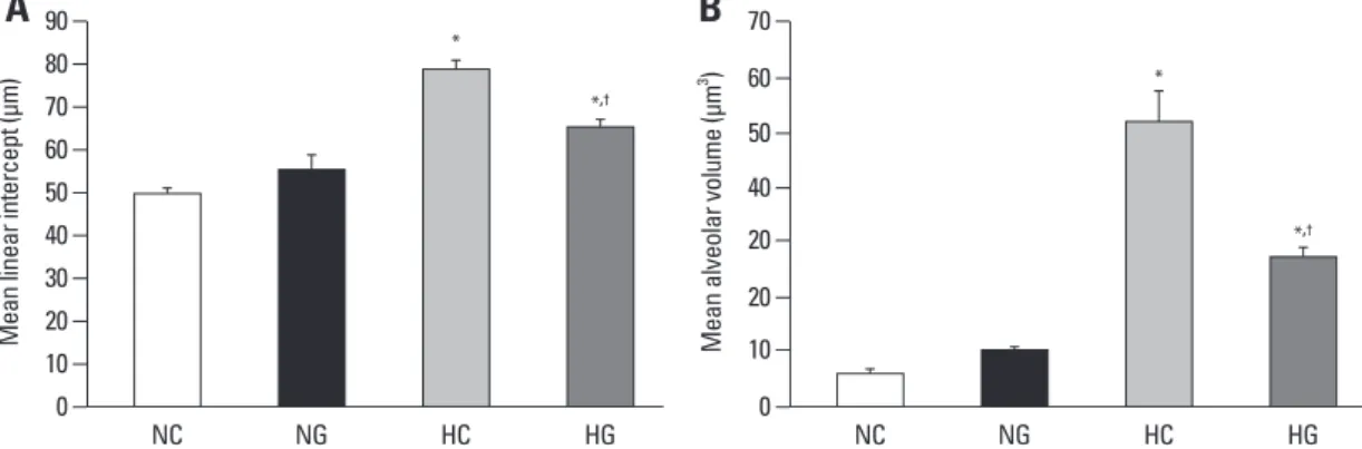
pups in the normoxia groups, indicating hyperoxia-induced impairments in alveolar growth, and these abnormalities seemed to be attenuated with G-CSF treatment (HG). Mor- phometric data demonstrated significantly increased MLI and mean alveolar volume in HC compared to normoxia groups (NC and NG), and significant attenuation of these abnormalities with G-CSF treatment (HG) support these microscopic findings (Fig. 3).
The numbers of TUNEL positive cells in distal lung sam- ples were markedly increased in HC compared to the nor- moxia groups (NC and NP), and G-CSF treatment (HG) ment was not statistically significant.
Although birth weight was not significantly different be- tween the four experimental groups, body weight at P 14 in HC was significantly lower compared to the normoxia groups, and this decreased body weight gain observed in HC was sig- nificantly improved with G-CSF treatment (HG) (Fig. 1).
Lung histopathology
Representative photomicrographs showing differences be- tween the experimental groups are shown in Fig. 2. Fewer and larger alveoli were observed in HC pups compared to
Fig. 2. Representative optical photomicrographs of P 14 rat lungs stained with hematoxylin and eosin. Scale bar = 100 µm, NC, normoxia control group; NG, normoxia with G-CSF treatment group; HC, hyperoxia control group; HG, hyperoxia with G-CSF treatment group.
Fig. 3. Comparisons of alveolarization measured by the mean linear intercept (A) and mean alveolar volume (B). Data; mean ± SEM. NC, normoxia control group (n = 6); NG, normoxia with G-CSF treatment group (n = 6); HC, hyperoxia control group (n = 8); HG, hyperoxia with G-CSF treatment group (n = 8). *p < 0.05 compared to NC. †p < 0.05 compared to HC.
90 80 70 60 50 40 30 20 10
0 NC
Mean linear intercept (µm)
NG HC HG
*
*,†
70 60 50 40 20 20 10
0 NC
Mean alveolar volume (µm3)
NG HC HG
*
*,†
A B
statistically significant.
NADPH oxidase activation
When NADPH oxidase is activated, expression and mem- brane translocation of a cytosolic subunit of NADPH oxidase p67
phoxis increased. Hyperoxia-induced production of ROS was evaluated by NADPH oxidase activation, because NADPH oxidase produces oxygen free radicals both in phagocytic
20and nonphagocytic cells.
21Exposure to oxygen (HC) significantly increased p67
phoxboth in the cytosolic and membrane fractions, indicating activation of NADPH oxi- dase, compared to the normoxia groups (NC and NG) in Western blot analyses, and the G-CSF (HG) significantly at- tenuated this hyperoxia-induced NADPH oxidase activation (Fig. 7).
DISCUSSION
In the present study, prolonged exposure of newborn rat significantly attenuated this hyperoxia-induced increase in
the number of TUNEL positive cells (Fig. 4).
White blood cell counts and lung myeloperoxidase activity
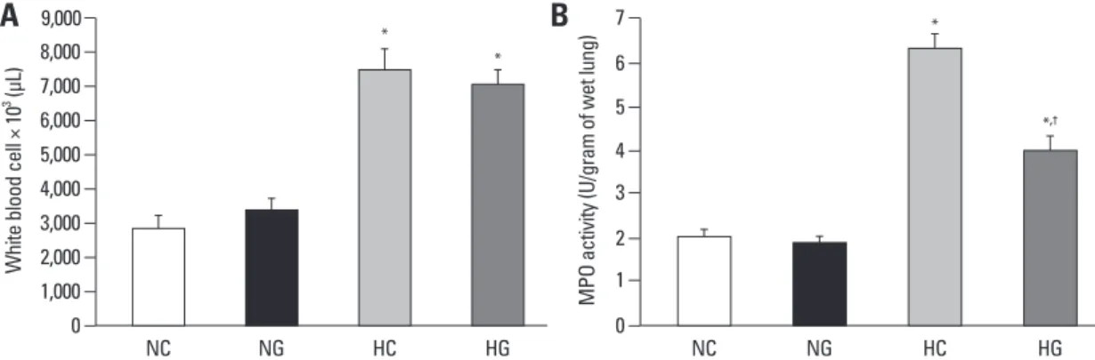
Exposure to oxygen (HC and HG) significantly increased white blood cell counts in the blood in hyperoxia, compared to the normoxia groups (NC and NG), and the G-CSF in- duced granulopoiesis was not observed at P14 (Fig. 5A).
The significantly increased MPO activity observed in HC, compared to the normoxia groups (NC and NG), was signifi- cantly improved with G-CSF treatment (HG) (Fig. 5B).
mRNA expression of TNF-α, IL-6 and TGF-β
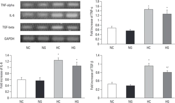
In semi-quantitative RT-PCR, significantly increased mRNA levels of TNF-α, IL-6 and TGF-β were observed in HC, compared to the normoxia groups (NC and NG) (Fig. 6). The hyperoxia-induced increase in TGF-β mRNA expression was significantly attenuated with G-CSF treatment (HG), howev- er, the attenuation of TNF-α and IL-6 mRNA levels was not
Fig. 4. TUNEL positive cells (A) and number of TUNEL positive cells (B) in P14 rat lungs. TUNEL positive cells labeled with FITC (green, ar- row) and the nuclei labeled with DAPI (blue) (Scale bar, 25 µm) (A). Data; mean ± SEM. NC, normoxia control group (n = 6); NG, normoxia with G-CSF treatment group (n = 6); HC, hyperoxia control group (n = 8); HG, hyperoxia with G-CSF treatment group (n = 8). *p < 0.05 com- pared to NC. †p < 0.05 compared to HC. TUNEL, terminal deoxynucleotidyl transferase-mediated deoxyuridine triphosphate nick end label- ing; FITC, fluorescein isothiocyanate; DAPI, 4, 6-diamidino-2-phenylindole; G-CSF, granulocyte colony stimulating factor.
10 9
7 8
6 5 4 3 2 1
0 NC
TUNEL + Cell / High power field
NG HC HG
*
*,†
Fig. 5. White blood cell count in blood (A) and myeolperoxidase activity in P 14 rat lungs (B). Data; mean ± SEM. NC, normoxia control group (n = 6); NG, normoxia with G-CSF treatment group (n = 6); HC, hyperoxia control group (n = 6); HG, hyperoxia with G-CSF treatment group (n = 6). *p < 0.05 compared to NC. †p < 0.05 compared to HC. MPO, myeloperoxidase.
9,000 8,000 7,000 6,000 5,000 4,000 3,000 2,000 1,000
0 NC
White blood cell × 103 (µL)
NG HC HG
*
*
7 6 5 4 3 2 1
0 NC
MPO activity (U/gram of wet lung)
NG HC HG
*
*,†
A
A
B
B COLOR
A
cantly attenuated hyperoxia-induced growth retardation, decreased alveolarization and increased TUNEL positive cells in the newborn rats.
G-CSF is clinically available for use in neonates,
12and its safety and feasibility have been validated in recent human stroke therapy trials.
11Taken together, these findings sug- pups to hyperoxia for two weeks increased mortality, retard-
ed growth, and led to the development of lung injuries simi- lar to those seen in premature human infants with BPD.
22These pups exhibited decreased alveolarization, as shown by increased MLI and alveolar volume,
23and significantly increased TUNEL positive cells.
24G-CSF treatment signifi-
Fig. 6. Representative RT-PCR blots and densitometric histograms for TNF-α, IL-6, and TGF-β. NC, normoxia control group (n = 6); NG, nor- moxia with G-CSF treatment group (n = 6); HC, hyperoxia control group (n = 6); HG, hyperoxia with G-CSF treatment group (n = 6). *p < 0.05 compared to NC. †p < 0.05 compared to HC. RT-PCR, reverse transcriptase polymerase chain reaction; TNF-α, tumor necrosis factor-α; IL- 6, interleukin-6; TGF-β, transforming growth factor-β; GAPDH, glyceraldehyde-3-phosphate dehydrogenase.
Fig. 7. Western blot analysis of p67phox in the cytosol and membrane fractions of P 14 rat lung. NC, normoxia control group (n = 6); NG, nor- moxia with G-CSF treatment group (n = 6); HC, hyperoxia control group (n = 6); HG, hyperoxia with G-CSF treatment group (n = 6). *p < 0.05 compared to NC. †p < 0.05 compared to HC.
GAPDH IL-6
TGF-beta TNF-alpha
NC NG HC HG
1.4 1.6 1.8
1.2 1 0.8 0.6 0.4 0.2
0 NC
Fold increase of TNF-α
NG HC HG
* *
1.4 1.2 1 0.8 0.6 0.4 0.2
0 NC
Fold increase of IL-6
NG HC HG
*
*
1.8 1.6 1.4 1.2 1 0.8 0.6 0.4 0.2
0 NC
% Control of cytosolic p67phox
NG HC HG
*
*
1.4 1 0.8 0.6 0.2
0 NC
Fold increase of TGF-β
NG HC HG
*
*,†
1.8 1.6 1.4 1.2 1 0.8 0.6 0.4 0.2
0 NC
% Control membrane p67phox
NG HC HG
*
*,†
GAPDH 37 kDa
p67phox
NC NG
Cytosol
HC HG
Calnexin 37 kDa p67phox
NC NG
Membrane
HC HG
brane translocation of the p67
phoxcan be served as an in vivo indicator of NADPH oxidase activation as shown in our previous study.
38In the present study, increased expression of the p67
phoxprotein was observed both in the cytosolic and membrane fraction in HC, and G-CSF treatment significantly attenuat- ed hyperoxia-induced NADPH oxidase activation as evi- denced by reduced membrane translocation of the p67
phoxin the lung tissue. As phagocytic NADPH oxidase activation seems to be the main source of ROS production in the lung,
39significant attenuation of hyperoxia-induced increase in lung MPO activity, an indicator of the lung neutrophil accu- mulation, with G-CSF treatment might be responsible for its anti-oxidative effects.
In summary, G-CSF treatment significantly attenuated hyperoxia-induced lung injuries such as decreased alveolar- ization and increased TUNEL positive cells by down-mod- ulating the oxidative stress and inflammatory responses in neonatal rats. These findings suggest the potential use of G- CSF as a new therapeutic agent for BPD. Further studies regarding optimal dosage, timing and safety in multi-spe- cies preterm and term neonates will be necessary for the translation of the benefits of G-CSF treatment observed in this study to clinical trials. In addition, neurological studies have to be done when neonates administered with G-CSF neonatally become adults.
ACKNOWLEDGEMENTS
This work was supported by the IN-SUNG Foundation of Medical Research (C-A7-840-1).
REFERENCES
1. Kinsella JP, Greenough A, Abman SH. Bronchopulmonary dys- plasia. Lancet 2006;367:1421-31.
2. Short EJ, Klein NK, Lewis BA, Fulton S, Eisengart S, Kercsmar C, et al. Cognitive and academic consequences of bronchopulmo- nary dysplasia and very low birth weight: 8-year-old outcomes.
Pediatrics 2003;112:e359.
3. Ehrenkranz RA, Walsh MC, Vohr BR, Jobe AH, Wright LL, Fan- aroff AA, et al. Validation of the National Institutes of Health con- sensus definition of bronchopulmonary dysplasia. Pediatrics 2005;116:1353-60.
4. Committee on Fetus and Newborn. Postnatal corticosteroids to treat or prevent chronic lung disease in preterm infants. Pediatrics 2002;109:330-8.
5. Trial of late surfactant for prevention of bronchopulmonary dys-
gest that G-CSF might be a useful new therapeutic agent for hyperoxia-induced neonatal lung diseases such as clini- cal BPD, for which no effective treatments are currently available. However, better understanding of its mechanism of action must be obtained for translation of this experi- mental result to clinical trials.
We observed significant increases in MPO activity and mRNA of TNF-α, IL-6 and TGF-β in rat pups in the HC group, suggesting that inflammation mediated by neutro- phils
7and proinflammatory cytokines
25plays a central role in the development of BPD.
26G-CSF caused not only a sig- nificant attenuation of hyperoxia-induced increase in MPO activity, but also caused a similar decrease in hyperoxia-in- duced increase in TGF-β. These results suggest that the protective effects of G-CSF against hyperoxic neonatal lung injury are associated with or mediated by anti-inflam- matory effects. The anti-inflammatory action of G-CSF has been reported also in various experimental and clinical con- ditions including murine endotoxemia,
27encephalitis, non- neutropenic surgical intensive care patients,
28pneumonia
29and stroke.
30Although G-CSF suppressed the synthesis of proinflammatroy cytokines such as TNF-α both in vitro and in vivo,
27and upregulated the production of anti-inflamma- tory mediators such as soluble TNF-α receptor type I and II, IL-6, IL-10 and IL-1 receptor antagonist,
15,31the precise mechanisms of its anti-inflammatory action are not yet clear. Further studies are necessary to clarify this action.
Neutrophils have been known to play a role in the exac- erbation of prior lung injury with G-CSF treatment.
13How- ever, in the present study G-CSF treatment significantly at- tenuated lung MPO activity, and did not induce leukocytosis.
The extent of leukocytosis with G-CSF treatment has been known to be dose-dependent.
13The dose of G-CSF used in this study was similar to that prescribed clinically
32,33and relatively low compared to other studies.
34,35Therefore, be- sides differences in animal species and experimental de- sign, different doses of G-CSF might be responsible for these conflicting results. Further studies are needed to de- termine the optimal dosage.
Oxidative stress to the immature lung is a major contrib-
uting factor for the development of BPD.
36NADPH oxi-
dase is responsible for superoxide anion production (O
2-),
which generates other ROS such as hydrogen peroxide, hy-
droxyl radical and hypochlorous acid.
20As the cytoplasmic
subunits p47
phox, p67
phox, p40
phoxand Rac translocate to
membrane-bound cytochrome upon activation of this mul-
ticomponent enzyme complex NADPH oxidase,
37mem-
exposed to hyperoxia. Am J Respir Cell Mol Biol 2001;25:150-5.
25. Chanock SJ, el Benna J, Smith RM, Babior BM. The respiratory burst oxidase. J Biol Chem 1994;269:24519-22.
26. Coalson JJ. Pathology of new bronchopulmonary dysplasia.
Semin Neonatol 2003;8:73-81.
27. Görgen I, Hartung T, Leist M, Niehörster M, Tiegs G, Uhlig S, et al. Granulocyte colony-stimulating factor treatment protects ro- dents against lipopolysaccharide-induced toxicity via suppression of systemic tumor necrosis factor-alpha. J Immunol 1992;149:
918-24.
28. Gross-Weege W, Weiss M, Schneider M, Wenning M, Harms B, Dumon K, et al. Safety of a low-dosage Filgrastim (rhG-CSF) treatment in non-neutropenic surgical intensive care patients with an inflammatory process. Intensive Care Med 1997;23:16-22.
29. Knapp S, Hareng L, Rijneveld AW, Bresser P, van der Zee JS, Florquin S, et al. Activation of neutrophils and inhibition of the proinflammatory cytokine response by endogenous granulocyte colony-stimulating factor in murine pneumococcal pneumonia. J Infect Dis 2004;189:1506-15.
30. Shyu WC, Lin SZ, Lee CC, Liu DD, Li H. Granulocyte colony- stimulating factor for acute ischemic stroke: a randomized con- trolled trial. CMAJ 2006;174:927-33.
31. Hartung T, Döcke WD, Gantner F, Krieger G, Sauer A, Stevens P, et al. Effect of granulocyte colony-stimulating factor treatment on ex vivo blood cytokine response in human volunteers. Blood 1995;85:2482-9.
32. Donini M, Fontana S, Savoldi G, Vermi W, Tassone L, Gentili F, et al. G-CSF treatment of severe congenital neutropenia reverses neutropenia but does not correct the underlying functional defi- ciency of the neutrophil in defending against microorganisms.
Blood 2007;109:4716-23.
33. Carr R, Brocklehurst P, Doré CJ, Modi N. Granulocyte-macro- phage colony stimulating factor administered as prophylaxis for reduction of sepsis in extremely preterm, small for gestational age neonates (the PROGRAMS trial): a single-blind, multicentre, ran- domised controlled trial. Lancet 2009;373:226-33.
34. Minnerup J, Sevimli S, Schäbitz WR. Granulocyte-colony stimu- lating factor for stroke treatment: mechanisms of action and effi- cacy in preclinical studies. Exp Transl Stroke Med 2009;1:2.
35. Kim BR, Shim JW, Sung DK, Kim SS, Jeon GW, Kim MJ, et al.
Granulocyte stimulating factor attenuates hypoxic-ischemic brain injury by inhibiting apoptosis in neonatal rats. Yonsei Med J 2008;49:836-42.
36. Saugstad OD. Bronchopulmonary dysplasia-oxidative stress and antioxidants. Semin Neonatol 2003;8:39-49.
37. Bastian NR, Hibbs JB Jr. Assembly and regulation of NADPH oxidase and nitric oxide synthase. Curr Opin Immunol 1994;6:
131-9.
38. Chang YS, Kim YJ, Yoo HS, Sung DK, Kim SY, Kang S, et al.
Alpha-phenyl-N-tert-butylnitrone attenuates hyperoxia-induced lung injury by down-modulating inflammation in neonatal rats.
Exp Lung Res 2009;35:234-49.
39. Delacourt C, d'Ortho MP, Macquin-Mavier I, Pezet S, Housset B, Lafuma C, et al. Oxidant-antioxidant balance in alveolar macro- phages from newborn rats. Eur Respir J 1996;9:2517-24.
plasia (TOLSIRF) from: URL: http://www.clinicaltrials.gov/ct2/
show/nct01022580.
6. Trial of late surfactant to prevent BPD: A pilot study in ventilated preterm neonates receiving inhaled nitric oxide (TOLSURF Pilot) from: URL: http://www.clinicaltrials.gov/ct2/show/nct00569530.
7. Speer CP. Inflammation and bronchopulmonary dysplasia: a con- tinuing story. Semin Fetal Neonatal Med 2006;11:354-62.
8. Juul S, Felderhoff-Mueser U. Epo and other hematopoietic fac- tors. Semin Fetal Neonatal Med 2007;12:250-8.
9. Weiss SJ. Tissue destruction by neutrophils. N Engl J Med 1989;320:365-76.
10. Boneberg EM, Hartung T. Molecular aspects of anti-inflammatory action of G-CSF. Inflamm Res 2002;51:119-28.
11. Shyu WC, Lin SZ, Yang HI, Tzeng YS, Pang CY, Yen PS, et al.
Functional recovery of stroke rats induced by granulocyte colony- stimulating factor-stimulated stem cells. Circulation 2004;110:
1847-54.
12. Carr R, Modi N, Doré C. G-CSF and GM-CSF for treating or pre- venting neonatal infections. Cochrane Database Syst Rev 2003:CD003066.
13. Adach K, Suzuki M, Sugimoto T, Suzuki S, Niki R, Oyama A, et al. Granulocyte colony-stimulating factor exacerbates the acute lung injury and pulmonary fibrosis induced by intratracheal ad- ministration of bleomycin in rats. Exp Toxicol Pathol 2002;53:
501-10.
14. Miura G, Awaya H, Matsumoto T, Tanaka N, Matsunaga N. Does granulocyte colony-stimulating factor exacerbate radiation-in- duced acute lung injury in rats? Radiat Med 2000;18:227-32.
15. Wiedermann FJ, Mayr AJ, Hobisch-Hagen P, Fuchs D, Schobers- berger W. Association of endogenous G-CSF with anti-inflamma- tory mediators in patients with acute respiratory distress syn- drome. J Interferon Cytokine Res 2003;23:729-36.
16. Cooney TP, Thurlbeck WM. The radial alveolar count method of Emery and Mithal: a reappraisal 1--postnatal lung growth. Thorax 1982;37:572-9.
17. Snyder JM, Jenkins-Moore M, Jackson SK, Goss KL, Dai HH, Bangsund PJ, et al. Alveolarization in retinoic acid receptor-beta- deficient mice. Pediatr Res 2005;57:384-91.
18. Gray KD, Simovic MO, Chapman WC, Blackwell TS, Christman JW, May AK, et al. Endotoxin potentiates lung injury in cerulein- induced pancreatitis. Am J Surg 2003;186:526-30.
19. Babior BM. NADPH oxidase: an update. Blood 1999;93:1464-76.
20. Robinson JM, Badwey JA. The NADPH oxidase complex of phagocytic leukocytes: a biochemical and cytochemical view.
Histochem Cell Biol 1995;103:163-80.
21. Höhler B, Holzapfel B, Kummer W. NADPH oxidase subunits and superoxide production in porcine pulmonary artery endotheli- al cells. Histochem Cell Biol 2000;114:29-37.
22. Bland RD. Neonatal chronic lung disease in the post-surfactant era. Biol Neonate 2005;88:181-91.
23. Kunig AM, Balasubramaniam V, Markham NE, Morgan D, Mont- gomery G, Grover TR, et al. Recombinant human VEGF treatment enhances alveolarization after hyperoxic lung injury in neonatal rats. Am J Physiol Lung Cell Mol Physiol 2005;289:L529-35.
24. McGrath-Morrow SA, Stahl J. Apoptosis in neonatal murine lung