구순구개열 Vol. 7, No. 1 2004
°0
흑성 구순구개열 환자의 치조골 결손부의 재건치료를 위한
bifocal distraction-compression osteosynthesis
이진경1’, 백승학1,, 이종호2)
1서 울대학교 치과대학 치 과교정학교실 2서울대학교 치과대학 구강악안면외과학교실
厂
HBSTRflCT ---
Reconstiuction of alveolar bone defect in bilateral cleft lip and palate using bifocal distraction-compression osteosynthesis
Lee Jin-Kyung1), Seung-Hak Baek1), Jong-Ho Lee2’
v Dept, of Orthodontics, College of Dentistry, Seoul National University 2) Dept, of Oral and Maxillofacial Surgery, College of Dentistry, Seoul National University
The closure of a wide alveolar cleft and fistula in cleft patients and the reconstruction of a maxillary dentoalveolar defect in bilateral cleft lip and palate (BCLP) patients are challenging for both ortliodontists and oromaxillofacial surgeons. It is due to the difficulty in achieving complete closure by 니sing local attached gingiva (palatal flap) and the great volume of bone required for the graft. In this article, the authors used bifocal distraction-compression osteosynthesis(BDCO) to create a segment of new alveolar bone and attached gingiva for the complete approximation of a wide alveolar cleft/fistula and the reconstruction of a maxillary dentoalveolar defect. Since the alveoli and gingivae on both ends of the deft were approximated after BDCO, the need for extensive alveolar bone grafting was eliminated. It also could create new alveolar bone and gingiva for orthodontic tooth movement and implant.
Key word: Bifocal distraction-compression osteosynthesis, bilateral cleft lip and palate
이진경•백승학-이종호
I. 서론
구순구개열 환자에서 치조골 파열부의 골 연속성 을 회복하고, 치조골 결손부를 채우며, 비구강 누공
(oronasal fistula) 을 폐쇄하고, 비익 기저부와 비순 윤곽의 외형을 회복하며, 파열부에 인접한 영구 견치 나 측절치의 맹출 또는 교정치료시 치아의 이동을 위 한골 지지를제공하기 위하여치조골 이식법이 전통 적으로 사용되어져왔다.1시
치조골 이식을 시행하기 전에 협착된 치열궁의 형 태를 정상화하기 위하여 구개 측방확장과 교정치료 에 의한 치아의 배 열이 필요하다.5'6) 그러나측방확장 을 많이 할 경우 협착되었던 치열궁이 측방으로넓어 지고 전상악골(premaxilla) 이 전방으로 이동하면서 치조골 파열부나비구강 누공이 더 넓어지게 된다.
부착치은(attached gingiva) 으로구성된 구개 피판 (palatal flap) 을 이용할경우 사용 가능한 연조직 양 에 제힌-이 있으므로치조골 이식 부위를 덮고 누공을 완전히 폐쇄하기가 어려워지고, 감염 등의 문제점이 발생할수 있다.7’
다른 방법으로 구강점막 피판(oral mucosa flap) 을 이용해 누공을 폐쇄하기도하지만 협점막이나 설 점막은부착 치은을 대체하기에 적당하지 않으며,8’
치아의 맹출이나 교정치료에 의한 이동, 결손부의 보철을 위해서도 부착치은이 필수적이다/이°)따라 서 협착된 치열궁을 치조골 이식 전에 정상에 가깝 게 확장하는 것이 금기시되어 왔으며 누공의 크기를 최소화하거나 유지한 후, 수술 시 폐 쇄할 수 있도록 해 왔다.
최근결손부에 대한치조골 이식이나 치은 이식이 필요 없는bone transport 기법이 제시되었다.7’11’ 생 물학적으로 bone transport 기법은 distraction osteogenesis 오]' transformation osteogenesis의 두 가
지 과정이 기본이 된다. Distraction osteogenesis는 장력을 가하였을 때 서서히 이동되는 골 절편의 표면 사이에서신생골이 형성되는것이다.
Transformation osteogenesis는 병적인 골 조직이 기계적 자극에 의해 정상골로 전환되는 생물학적 과 정이다.12) 병적인 골 조직에 골 생성을 유도하는 기 계적 자극은장력과 압력 모두 가능하다.
Bone transport 기법의 원리는 아래와 같다. 시작 부위 (host bone) 에서 이동 골절편 (transport segment) 을형성한 후 결손부를 가로질러 목표부위 (target bone) 까지 점진적으로 이동시킬 때 시작부 위와이동 골절편사이에장력이 발생하여 신생골이 형성된다. 그리고 이동 골절편이 목표부위에 도달하 게 되면, 접합부에서 입력(compressive force) 이 발 생하게 되고, 그 결과목표부위의 골과 이동 골절편 의 끝이 하나의 신생골로융합된다 (Fig. 1).
Ilizarov에 의하면, 이 방법은 distraction / compression site의 수와골편 사이 에 가해진 힘의 종 류에 따라 분류할 수 있다.7'
첫째, 골 결손부가 작은 경우나 양쪽골편 말단부 의 치유가 비정상적으로 일어나 유합되지 못한경우 monofocal osteosynthesis가이용된다. 전체 골의 길 이를 증가시키지 않아도 되는 경우, 압력을 가하면 병적 조직이 개조되어가골(callus) 이 형성되고 결손 부의 양쪽 말단이 유합된다. 이를 monofocal compression osteosynthesis 라고한다{Fig. 2). 반면, 골의 길이가 신장될필요가 있는 경우, 골말단을분
리시키기 위한 견인력(distraction force) 을 가하게 되면, 골신장(distraction) 이 계속됨에 따라 병적 조 직 이 신생골로전환된다. 이 를monofocal distraction osteogenesis 라고 한다(Fig. 2).
둘째, 골 결손부가 크고골의 길이 신장이 요구되 는 경우라면, 처음에는 골편에 압력을 가하여 가골
=구개열 Vol. 7, No. 1 2004
Fig. 1 Treatment of subtotal tibial defects using Ilizarov’s bone transport technique. Note that the distraction regenerate is formed between the proximal segment and transport disk.
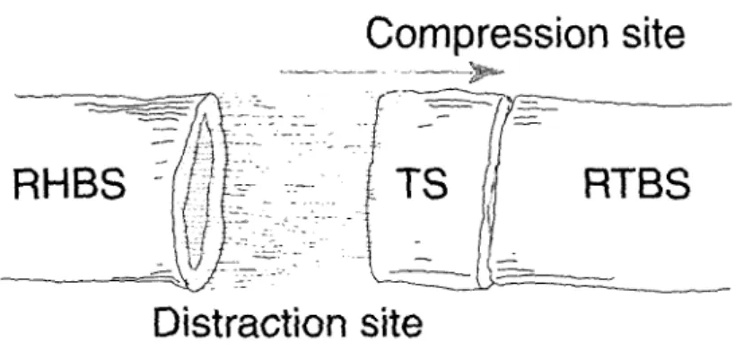
형 성을 자극하고, 골신장(distraction) 을 시행하여 길 이 신장을 유도한다(compression and distraction osteogenesis). 그리고 골 결손부가 더 큰 경우는 한 쪽에서 distraction을 시행하고 다른 쪽에서 압력을 가하는 bifocal osteosynthesis 가 이용된다. 이 경우 는 남아있는 골편의 한쪽을절단하여유리골절편을 형성하고이 절편을점진적으로이동시켜 골결손부 를 통과하여 목표부위 인 반대쪽 골편의 말단에 도달 하게 하는 것이다. 골절편이 이동하는 동안 장력이 발생하고 그 결과 시작부위와 이동 골절편 사이에 신 생골이 형성된다. 그리고 이동골절편이 목표부위에 도달하게 되면, 접합부에서 압력(compressive force) 이 발생하게 되고, 그 결과목표부위의 골과이동 골 절편의끝이 하나의 신생골로 융합된다.
따라서 이 방법을 bifocal distraction-compression
osteosynthesis (BDCO)라고 한다<Fig. 3).
셋째, 골 결손부가 아주큰 경우양 쪽에서 두 개의 골절편을 형성하여 동시에 골 결손부의 중앙으로 이 동시킨 뒤 압력을 가하여 이를 유합시키는 방법인 trifocal distraction-compression osteosynthesis가 있 다<Fig. 4).
구순구개열 환자에서 치조골 파열부와 누공의 크 기를최소화 하면서 상악치조골의 결손부를 재생시 키고치열궁의 길이를신장시키는것이 필요하다.13’
따라서 양측성 구순구개열 환자의 치조골 결손을 BDCO를 이용하여 양쪽의 이동 골절편을 전방으로 이동시켜 전상악골에 근접시켜서 치조골 결손부의 크기를 최소화 하며 골절편의 후방으로는 치조골과 치은조직이 생성되어 치열궁의 전체 길이를 증가시 킨 증례를소개하고자 한다.
이진경-백승학•이종호
Compression site Distraction site
Fig. 2 Monofocal compression osteosynthesis(left side) and monofocal distraction osteosynthesis(right side)
Compression site
Distraction site
Fig. 3 Bifocal distraction-compression osteosynthesis. RHBS means residual host bone segment; TS, transport segment; RTBS, residual target bone segment.
Compression site
Fig. 4 Trifocal distraction-compression osteosynthesis. RHBS means residual host bone segment; TS, transport segment.
구순구개열 Vol. 7, No. 1 2004
II. 중례
1. 의과 및 치과 병력 (Medical and dental history)
13세 10개월의 양측성 구순구개열 환자로 생후 3 개월째 구순열 수술을 받았으며, 1세와6세 때 구개 열 수술을 받았으나 구개부의 누공이 남아있었다. 7 세 때 처음 내원하여 가철식 교정장치(removable appliance) 로 협착된 상악골의 측방확장을시도하였 고 10서] 때 재진 후 bonded RPE(rapid palatal expansion) appliance를 이용하여 계속 상악골 폭경 을 확장하였다.
2. 진단
상악골 열성장에 의한 골격성 III급 부정교합을 보 이는 환자로 전치부 반대교합과 상악궁의 협착으로 인한 양측성 구치부 반대교합을 보이고 있었다<Fig.
5). 좌,우측 상악 측절치와 좌측 상악 중절치가 선천 결손되어 있었으며, 우측상악제2소구치는 치아크 기 이상(microdontia) 을 보였다.
양측성 구순구개열로 인한 치조골의 골결손부가 광범위하였으며 전상악골(premaxilla) 은 비중격에 만연결되어있었고 치조골과 분리되어 있어 심한 유 동성(mobility) 이 관찰되었다. 구개부에는 비강으로 통하는 누공이 크게 남아있는상태였다<Fig. 5). 전상 악골은 누공을 중심으로 협착되어 설측으로 심하게 경사되어 있었으며 우측 상악 중절치도 설측 경사가 심하였다<Fig. 5).
진단은 골격성 III급 부정교합, 전치부 반대교합, 양측성 구치부 반대 교합, 잔존된 비구강 누공과 심 한 치조골결손을 보이는 양측성 구순구개열이다.
3. 치료 목표
치료 목표는 첫째, 심하게 설측경사 되어 있는 전 상악골의 위치를 정상화 하고, 둘째, 치조골 결손부 를 재건하며, 셋째, 우측 상악 중절치와 견치를정상 배 열하여 보철 공간을 확보하는것이다.
4. 치료
Bonded RPE (rapid palatal expansion) appliance를 이용하여 계속 상악골폭경을확장시켰고, 설측 경사 되어있는 전상악골과 상악 중절치를 직립시키기 위 해 고정식 교정장치 (fixed orthodontic appliance) 를 장착하였다<Fig. 5, 6). 교정 력을 이용하여우측 상악 중절치를 포함한 전상악골을 순측 경사시켰고 골신 장술 시 bracket과 archwire 간의 마찰을최소화 하기 위하여 교정장치를 Damon bracket으로교체하였다.
상악의bonded RPE 는 BDCO 전에 제거하고 전치부 설측에 hook이 추가된 설측 호선 (lingual arch) 을 장착하였다(Fig. 6).
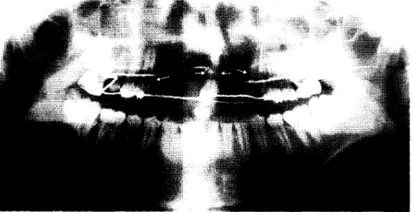
BDCO 시행 전 파노라마 방사선사진에서 우측상 악 중절치 양 쪽으로 넓은골 결손부가관찰되었으며 (Fig. 7), 삼차원 CT상에서 양측성 구순구개열로 인 한 골 결손부의 형태를 입체적으로 관찰할수 있었 다<Fig. 8).
골 절단 수술 시 우측은제1대구치 전방에서 , 좌측 은 제2소구치 전방에서 이동 골절편(transport segment) 을 형성하였고 Liou cleft distractor(KLS Martin LP, Germany) 를 부착하였다{Fig. 9,10).
수술후5일 간의 잠복기를주었고 1개월간 하루에 O.6mm(O.3mm X 2 turns / day) 씩 distractor를 돌려 서 BDCO를 시행하였다. 양쪽의 이동 골절편이 distractor의 방향에 의해 직선으로움직 이는것을막
이진경-백승학-이종호
Fig. 5 Facial, intraoral photos and orthopantomogram when fixed appliance treatment was started, a. Facial photos.
Midface deficiency was seen due to bilateral cleft lip and palate and maxillary hypoplasia, b. Intraoral photos. Note linguoversion of the maxillary central incisor and the premaxilla, remaining oronasal fistula, and severe alve이ar bone defect, c. Orthopantomogram. Right and left alveolar cleft was seen. #12,21,22 were congenitally missing.
구순구개열 Vol. 7, No. 1 2004
Fig. 6 Pre-op. intraoral photos. The premaxilla and the right maxillary central incisor were uprighted using orthodontic force. Therefore, size of the oronasal fistula and alveolar cleft were increased.
Fig. 7 Pre-op. orthopantomogram. Arrows indicate alveolar bone defect in bilateral cleft lip and palate.
이진경•백승학-이종호
Fig. 8 Pre-op. 3-D CT. Note the wide alveolar bone defect due to bilateral cleft lip and palate
Fig. 9 Liou distractor for alveolar cleft transport (KLS Martin LP, Germany)
기 위히 견치에서 lingual arch의 hook 까지 고무줄을 연결하여distraction vector를 조절하였다. 이동골절 편이 전상악골 부위에 도달하게 되었을때 치은골막 성형술(gingiwperiosteophsty) 을 시행하였고, 접합 부에서 압력(compressive force) 이 발생하도록 distraction을 시행하였다. 그후골 경화기를 16주간 부여하였다 (Fig. 10).
5.치료 결과
술전 교정에 의하여우측 상악 중절치의 설측 경사 가 해소되었고, BDCO에 의하여 좌,우측 견치와 소 구치부가 전방이동하여 전상악골과양쪽 견치와 소 구치부의 치조골이 근접하게 되었고 상악우측중절 치와 좌. 우측 견치가인접하게 되었다. 좌,우측 제1
구순구개열 WI.7, No. 1 2004
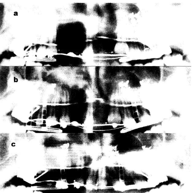
Fig. 10 Orthopantomograms showing the movement of the bony segments and teeth during bifocal distraction
compression osteosynthesis(BDCO). a. Before BDCO, b. During BDCO, c. Consolidation. The alve이ar bone defects(marked by arrows) next to the central incisor were gradually decreasing.
이진경•백승학•이종호
소구치와 제2소구치 사이에는 공간이 발생하였고 이 공간에 치조골이 형성 되었다. 이동중 고무줄을 사 용하여 vector를 조절하여 최종적인 치열궁 형태가 삼각형이 되는 것을 방지하였고 정상적인 전방부만 곡(anterior curvature) 형태를 이루었다<Fig. 11,12).
III.
총괄 및 고안
구순구개열 환자의 치료에 있어서 bone transport 기법의 장점은아래와 같다.
첫째,선천적으로 결손된 치조골과 치은조직을동 시에 형성할 수 있다.14J5) 이 환자의 경우, 전상악골 의 크기가 매우 작고심한 유동성을보이고 있었으며 파열부가 매우 광범위하였으나BDCO를 시행한 결 과치조골 파열부와 구개 누공의 크기가 줄어들었으 며 이동골절편의 후방부에 신생 치조골이 형성되어 치조골길이가 신장되었다. 양 쪽의 골 절편과 전상 악골은 압력을가해 유합시켜서 별도의 골이식이 필 요하지 않았으며, 전상악골의 유동성도 감소하였다 (Fig. 11,12).
Fig. 11 Intraoral photos before and after BDCO. a. Before BDCO, b. After BDCO, c. Consolidation. The right and left canines were approximated to the right central incisor and the maxillary arch form has normal anterior curvature. Note the decrease of oronasal fistula.
Fig. 12 Maxillary occlusogram before and after BDCO. a. Before BDCO. Transporting segments were planned to move along the direction of the arrows, b. After BDCO. The maxillary arch has normal anterior curvature.
구※개열 Vol. 7, No. 1 2004
둘째, 연구개가 전방으로 견인되지 않는다는 점이 다(Fig. 13). 구순구개열 환자에서 악교정수술에 의해 상악골 전체를 전방이동 시킬 경우 연구개가 상악골 과 함께 전방 이동하여 범인두 기능(velopharyngeal function) 에 장애가 올 가능성이 있다.
이러한 환자에서 상악골 양 쪽에서bone transport 를 시행하여 치조골 파열부를 재형성 할 수 있을뿐 아니 라 범 인두 기능에 지장 없이 상악골을 전방으로 이동시킬수있었다<Fig. 14).13)
셋째, 치조골 파열부의 크기를줄이면서 새로운 치 조골을 형성하여 치조골 전체의 길이를증가시킬 수 있다. 치조골 파열부의 크기를 줄이는 것은 악교정 수술시 분절골 수술(segmental osteotomy) 을 이용 해서도 가능하다.16’ 그러나이 방법은 새로운 치조골 을 형성하는 방법이 아니며, 따라서 치조골 전체의 길이를 증가시킬 수도 없다. 그리고 분절골 수술시 소분절(lesser segment) 을 전방으로 위치시켜 치조 골파열부를 폐쇄할 수있다고 하더라도,17" 골 절편
Fig. 13 Lateral cephak아netric radiographs before and after BDCO. a. Before BDCO. The alveolar bone defect to the central incisor was seen. b. After BDCO. The canine and the first prem이ar were moved anteriorly creating space behind them. New alveolar bone was formed in the distraction area.
이진경-백승학’이종호
Fig. 14 Facial profile before and after BDCO. a. Before BDCO. Note the midface deficiency, b. After BDCO. Midface deficiency has been improved more or less.
의 이동량이 많을경우 연조직 피판을 이용해 피개하 기 어려우며 혈액 공급에 지장을 받게 되어 술후 안 정성에도 문제가 야기될 수있다.
넷째, 전치나 구치부총생이 심한경우 새로운 치 조골을형 성하여 치아를 배 열할 공간을 형 성할 수 있 으므로, 발치 치료의 대안으로 사용될 수 있다.13’
Bone transport기법의 단점 중의 하나는 distraction vector 의 조절이 어렵다는 점이다. 일반적으로 distractor는 직선형이기 때문에 신생골의 형태도 직 선형으로 나타나게 되고 치열궁의 정상적인 형태와 만곡을 얻기가 힘들다. 따라서 distraction 양이 많을 경우이를 치열궁 형태대로 재형성하는 것이 중요하 다. 이칸 증례에서는 Liou distractor arm의 유연성 (flexibility) 을 고려하여 elastic force를 부여함으로써 골절편의 이동 방향을 조절하였다. 그 결과 최종적인
악궁형태는 정상적 인 전방부 만곡을가진 치 열궁 형 태가 되었다<Fig. 15).
IV. 결론
BDCO는bone transport 기법 중의 하나이며, 치조 골 파열부가 너무 커서 골 이식 만으로는 파열부를 복원할 수 없고, 부착된 연조직이 결손부를 덮기에 불중분한 경우에 사용이 가능하고 치조골과 부착 치 은을 재생하는데 효과적 인 방법 이 다.
양측성 구순구개열 환자의 본 증례에서 교정력을 이용하여 치조골 파열부와 누공을 최대화시킨 뒤 BDCO를 이용하여 치조골 파열부를 폐쇄하고 신생 치조골과 부착치은을 형성하고 정상적인 치열궁형 태를재생할 수있었다.
58
구순구개열 Vol. 7, No. 1 2004
Fig. 15 Comparison of intraoral photographs, CT, 3-D CT before and after BDCO. a. Before BDCO, b. After BDCO.
Intraoral photos show that the oronasal fistula has been decreased. After BDCO, alveolar continuity can be confirmed in the CT. 3-D CT shows the mesial migration of the transport segment, resulting cleft space closure.
이진경.백승학•이종호
참고문헌
1. Boyne PJ. Autogenous cancellous bone and marrow transplants. Clin Orthop. 1970;73:199-209.
2. Rosenstein SW, Monroe CW, Kernahan DA, Jacobson BN, Griffith BH, Bauer BS. The case of early bone grafting in cleft lip and palate. Plast Reconstr Surg. 1982;70:297-309.
3. Yen SL, Gross J, Wang P, Yaniashita DD. Clos니re of a large alveolar cleft by bony transport of a posterior segment using orthodontic archwires attached to bone: report of a case. J Oral Maxillofac Surg. 2001;59:688-91.
4. Troxell JB, Fonseca KJ, Osbon DB. A retrospective study of alveolar cleft grafting. J Oral Maxillofac Surg. 1982;40:721-5.
5. Witsenb나rg B. The reconstruction of anterior residual bone defects in patients with cleft lip, alveolus and palate. A review. J Maxillofac Surg.
1985;13:197-208.
6. Ross RB. Treatment variables affecting facial growth in complete unilateral cleft lip and palate.
Cleft Palate J. 1987;24:5-77.
7. Samchukov ML, Cope JB, Cherkashin AM. Basic Principles of Bone Transport, in Samchukov ML, Cope JB, Cherkashin AM (eds). Craniofacial Distraction Osteogenesis. St. Louis, MO, Mosby, 2001, pp 349-57.
8. Jackson IT. Closure of secondary palatal fistiilae with intra-oral tissue and bone grafting. Br J Plast Surg. 1972;25:93-105.
9. Abyholm FE, Borchgrevink HC, Eskeland G. Cleft lip and palate in Norway. III. Surgical treatment of
CLP patients in Oslo 1954-75. Scand J Plast Reconstr Surg. 1981;15:15-28.
10. Boyne PJ. Use of marrow-cancellous bone grafts in maxillary alveolar and palatal clefts. Dent Res.
1974;53:821-4.
11. Ilizarov GA. The principles of the Ilizarov method.
Bull Hosp Jt Dis Orthop Inst. 1988;48:1-11.
12. Aronson J, Johnson E, Harp JH. Local bone transportation for treatment of intercalary defects by the Ilizarov technique. Biomechanical and clinical considerations. Clin Orthop. 1989;243:71-9.
13. Liou EJ, Chen PK, Huang CS, Chen YR. Interdental distraction osteogenesis and rapid orthodontic tooth movement: a novel approach to approximate a wide alveolar cleft or bony defect. Plast Reconstr Surg. 2000;105:1262-72.
14. Stucki-McCormick SU, Fox RM, Bmder RB, Mizrahi RD. Cranioacial bone transport in Samchukov ML, Cope JB, Cherkashin AM (eds). Craniofacial Distraction Osteogenesis. St. Louis, MO, Mosby, 2001, pp 358-67.
15. Liou EJ, Polley JW, Figueroa AA. Distraction osteogenesis: the effects of orthodontic tooth movement on distracted mandibular bone. J Craniofac Surg. 1998;9:564-71.
16. Posnick JC, Tompson B. Cleft-orthognathic surgery:
complications and long-term results. Plast Reconstr Surg. 1995;96:255-66.
17. Posnick JC, Witzel MA, Dagys AP. Management of jaw deformities in the cleft patient, in Bardach J, Morris WB (eds): Multidisciplinary Management of Cleft Lip and Palate. Philadelphia, PA, Saunders, 1990, pp 530-542.
구순구개열 Vol. 7, No. 1 2004
18. Wolford LM, Cottrell DA. End-stage reconstruction in the complex cleft lip/palate patient, in Turley TA, Vig KWL, Fonseca RJ (eds): Facial Clefts and
Craniosynostosis. Philadelphia, PA, Saunders, 1996, pp 504-536.
교신 저자
서울대학교 치과대학 치과교정학교실 조교수 백승학
서울시 종로구 연건동 28번지 우편번호) 110-744 / 전화: 02-2072-3952 / E-mail: [email protected]