ORIGINAL ARTICLE
헬리코박터 파일로리 감염 연관 위질환 발생시 형질전환인자-베타 저해신호로서 Smad7의 역할
이호재, 박종민
1, 함기백
1,2가천대학교 의과대학 생화학교실, CHA 의과학대학교 CHA 바이오컴플렉스 CHA 암예방연구센터1, 분당차병원 소화기센터2
Role of Inhibitory Transforming Growth Factor- Signal Smad7 in Helicobacter pylori- associated Gastric Damage
Ho-Jae Lee, Jong Min Park1, and Ki Baik Hahm1,2
Department of Biochemistry, Gachon University School of Medicine, Incheon, CHA Cancer Prevention Research Center, CHA Bio Complex, CHA University1, Digestive Disease Center, CHA Bundang Medical Center2, Seongnam, Korea
Background/Aims: Transforming growth factor-beta (TGF-) is a cytokine implicated in the susceptibility, development, and pro- gression of gastrointestinal cancer and certain other neoplasms. In the later stages of cancer, TGF- not only acts as a bystander of host-immune response, but also contributes to cell growth, invasion, and metastasis. In the current study, we generated gastric mucosal cells that stably express Smad7, and explored the Helicobacter pylori-associated biological changes between mock-transfected and Smad7-transfected RGM1 cells.
Methods: RGM1 cells stably transfected with Smad7 were infected with H. pylori, and molecular changes in apoptotic markers and inflammatory mediators were examined. Several candidate genes were explored in Smad7-overexpressing cells after H.
pylori infection.
Results: Overexpression of Smad7 in RGM1 cells significantly increased the H. pylori-induced cytotoxicity compared to mock-trans- fected cells. Exaggerated increases in inflammatory mediators, cyclooxygenase 2, inducible NO synthase, and augmented apoptosis were noted in Smad7-overexpressing cells, whereas mitigated heme oxygenase 1 was noted in Smad7- overexpressing cells. These phenomena were reversed in cells transfected with Smad7 siRNA.
Conclusions: These data suggest that inhibition of Smad7 is a possible target for mitigating H. pylori-associated inflammation.
(Korean J Gastroenterol 2016;68:186-194)
Key Words: Helicobacter pylori; Transforming growth factor beta; Smad7; Gastritis
Received August 3, 2016. Revised September 2, 2016. Accepted September 27, 2016.
CC This is an open access article distributed under the terms of the Creative Commons Attribution Non-Commercial License (http://creativecommons.org/licenses/
by-nc/4.0) which permits unrestricted non-commercial use, distribution, and reproduction in any medium, provided the original work is properly cited.
Copyright © 2016. Korean Society of Gastroenterology.
교신저자: 함기백, 13496, 성남시 분당구 야탑로 59, 분당차병원 소화기센터
Correspondence to: Ki Baik Hahm, Digestive Disease Center, CHA Bundang Medical Center, 59 Yatap-ro, Bundang-gu, Seongnam 13496, Korea. Tel: +82-31-780- 5005, Fax: +82-31-881-7185, E-mail: [email protected]
Financial support: This work was supported by Korea Institute of Planning and Evaluation for Technology in Food, Agriculture, Forestry and Fisheries (IPET) through High Value-added Food Technology Development Program, funded by Ministry of Agriculture, Food and Rural Affairs (MAFRA) (116015-03-1-CG000).
Conflict of interest: None.
서 론
Transforming growth factor-beta (TGF-)는 항염증, 세 포 항증식 및 암 억제 활성 등 다양한 기능을 가지고 있는 성장인자이다.1,2 이전의 연구에서 TGF- 신호의 제어에 의해
자가면역 췌장염이나 염증성 대장질환과 같은 염증질환의 발 생증가와 Helicobacter pylori 감염에 따른 암의 발생을 촉진 시킬 수 있음을 보고하였다.3-5 이는 이미 규명된 TGF-의 암 억제, 항염증 및 항섬유화작용 등을 설명하는 결과이다. 또한 H. pylori 감염은 bone morphogenic protein (BMP) 신호에
의한 CDX2나 SOX2의 발현 증가를 통해 장형화생을 유발하 며,6 이외에도 sonic hedgehog 신호를 통해 장형화생7 및 점 막증식8을 유발하는 것으로 보고하였다.
최근 H. pylori 감염은 TGF- 또는 BMP 신호전달의 매개 체인 Smad 단백질을 통해 숙주 세포에 영향을 주는 것으로 보고하고 있으나9,10 현재까지 H. pylori 감염에 따른 위염과 위암의 발생에 있어 Smad의 역할에 대해서는 보고가 많지 않다. 이는 TGF-가암 발생에 미치는 영향이 암의 종류와 발생 단계에 따라 차이가 있고,11-16 TGF- 신호 전달도 전형 적(canonical) 및 비전형적 신호 전달 체계(non-canonical pathway), 그리고 다른 신호 전달과의 상호 간섭 현상 (cross-talk)이 나타나기 때문이다.17-19
TGF- 신호 전달의 내인성 억제인자로 작용하는 Smad7 은 TGF-에 의한 항염증 활성에 있어 중요한 매개인자로 작 용할 뿐만 아니라,20 다른 신호 전달계 간의 조절에 있어서도 중요한 상호 간섭 매개체로 작용하고 있다.21 이 연구에서는 H. pylori 감염에 따른 세포사멸과 염증 반응에 있어 Smad7 의 역할을 알아보기 위하여, 정상 위점막 세포주인 RGM1에 서 Smad7를 과발현 또는 발현을 억제시킨 상태에서 세포 독 성과 염증매개물질의 발현에 대하여 분석하였다.
대상 및 방법
1. 세포주 및 세포배양
정상 rat 위점막 세포주인 RGM1 세포는 10%의 fetal bo- vine serum이 포함된 Dulbecco’s modified essential me- dium (DMEM)과 Ham F12 혼합배지에서 37oC 세포배양기 (95% air, 5% CO2)로 배양하였다.
2. H. pylori 균주 및 세포감염
이 실험에 사용된 H. pylori 균주(cytotoxin-associated gene A [CagA]+strain, NCTC 11637)는 American Type Culture Collection (ATCC, Rockville, MD, USA)으로부터 구입하였다. 균주는 5% bovine calf serum과 항생제가 첨가된 Brucella broth에서 1×10⁸ CFU/mL (OD600=1)가 될 때까지 10% CO2 조건에서 진탕 배양하였다. 세포에서의 감염 다중도 (multiplicity of infection, MOI)는 100:1로 사용하였다.
3. Smad7 과발현 세포주 수립
Smad7 단백질을 안정적으로 발현하는 세포주의 제작은 Clontech (Palo Alto, CA, USA)의 pLPCX retroviral vector 를 사용하였다. 바이러스는 Flag epitope이 tagged된 Smad7 DNA construct를 pLPCX vector에 삽입한 pLPCX-Flag-Smad7 vector를 Phoenix A packaging 세포에 형질 주입(transfec-
tion)하여 제조하였다. 이것을 RGM-1 세포주에 감염시킨 후, puromycin (1 g/mL)을 2주간 처리하여 Smad7이 발현되는 세포를 선별하였다. 동일한 방법으로 pLPCX vector를 포함 하는 RGM-1 세포를 control 세포로 사용하였다.
4. MTT assay를 통한 세포 생존측정
세포를 48-well dish에 1×10⁴개/well로 도포한 다음 DMEM 배지에서 배양하였다. 이후 각각의 시약을 일정 시간 처리하여 MTT solution (최종농도 1 mg/mL)을 가하고 2시간 동안 반응한 후 dimethyl sulfoxide로 용해시킨 후 540 nm에서 optical density를 microplate reader로 측정하였다.
5. Flow cytometry를 통한 apoptosis 측정
각각의 실험군에 대한 Annexin V (PharMingen, San Diego, CA, USA) 염색은 flow cytometer (FACScan; Becton Dickinson, Mountain View, CA, USA)와 CellQuest pro- gram (Becton Dickinson)을 사용하여 1-2×10⁴개의 세포들 에서 측정하였다. 중복 세포들은 FL3A (PI) vs. FL3W (PI) 도수 분포도에서 제외하였다. 자료의 분석은 WinList 4.0과 ModiFit LT 3.0 software program (Verity Software House, Topsham, ME, USA)을 이용하였다.
6. Western blot 분석
배양된 세포를 phosphate buffered saline 용액으로 세척 한 후, cell lysis buffer (150 mM NaCl, 0.5% triton×100, 50 mM tris-HCl, pH 7.4, 25 mM NaF, 20 mM ethyl- eneglycol-bis (-aminoethylether)-N, N’-tetraacetic acid, 1 mM dithiothreitol, 1 mM Na3VO4, Protease Inhibitor Cocktail tablet [Boehringer, Manneim, Germany])로 용해 시켜 protein lysate를 만들었다. 이것을 sodium dodecyl sul- fate-polyacrylamide gel electrophoresis로 전기 영동한 후 전개시킨 단백질을 PVDF membrane (Gelman Sciences, Ann Arbor, MI, USA)에 옮겨 각각의 primary antibody와 secondary antibody로 반응시킨 후, chemoluminescence system을 이용하여 분석하였다.
7. Reverse transcription PCR 분석
RNA는 TRIzol (Gibco BRL, Rockville, MD, USA)을 이용 하여 추출하였으며, 추출된 RNA는 moloney murine leuke- mia virus 역전사효소(Perkin Elmer, Morrisville, NC, USA) 를 이용하여 cDNA로 합성하였다. PCR 분석에 사용된 각각 의 primer 서열은 Table 1에 나타내었으며, internal standard 로서 glyceraldehyde 3-phosphate dehydrogenase (GAPDH) 을 사용하였다.
Fig. 1. Cytotoxicity against Helicobacter pylori in Smad7 overexpressed cells. (A) Stable expression of Smad7 in RGM1 cells was detected with Flag-tag (upper) and Smad7 (lower) antibodies. (B) MTT assay in mock- or Smad7-transfected RGM1 cells after treatment with 100 multiplicity of infection (MOI) H. pylori. (C) mock- or Smad7-transfected RGM1 cells were treated with H. pylori (100 MOI) for 24 hours, followed by Annexin V staining. Cells were stained with Annexin V-FITC and propidium iodide, and analyzed by flow cytometry. Bars represent mean±SD (n=3). (D) Western blot analysis of PARP and Bax after treatment with 100 MOI H. pylori in mock-transfected or Smad7-transfected RGM1 cells. (E) Expression of XBP-1, GADD34, CHOP, and Smad7 mRNA after treatment with 100 MOI H. pylori in mock-transfected or Smad7-transfected RGM1 cells.
Table 1. List of Primer Sequences Used for RT-PCR Analysis
Gene Forward Reverse
Xbp-1 5'-AAACAGAGTAGCAGCGCAGACTGC-3' 5'-GGATCTCTAAAACTAGAGGCTTGGTG-3'
Gadd34 5'-CCCTCCAACTCTCCTTCTTCAG-3' 5'-CAGCCTCAGCATTCCGACAA-3'
Ddit3 5'-GAAAGCAGAAACCGGTCCAAT-3' 5'-GGATGAGATATAGGTGCCCCC-3'
Smad7 5'-GGGCCTTTTTGGGGAGAGGCG-3' 5'-GTCCTGAACATGCGGGGCGAG-3'
Cox2 5'-GAAATGGCTGCAGAGTTGAA-3' 5'-TCATCTAGTCTGGAGTGGGA-3'
Nox2 5'-TTTTCCCAGGCAACCAGACG-3' 5'-GTAGCGGGGCTTCAGAATGG-3'
TNF- 5'-CCCTCACACTCAGATCATCTTCTCAA-3' 5'-TCTAAGGTACTTGGGCAGGTTGACCTC-3'
Ho1 5'-GACAGCATGTCCCAGGATTT-3' 5'-GGTTCTGCTTGTTTCGCTCT-3'
Il6 5'-CTTCCAGCCAGTTGCCTTCT-3' 5'-GAGAGCATTGGAAGTTGGGG-3'
Gapdh 5'-GGTGCTGAGTATGTCGTGGA-3' 5'-TTCAGCTCTGGGATGACCTT-3'
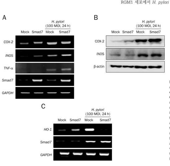
Fig. 2. Inflammation mediators against Helicobacter pylori in Smad7 over- expressed cells. (A) Expression of Cox-2, iNOS, TNF-, and Smad7 mRNA after treatment with 100 multiplicity of infection (MOI) H. pylori in mock-transfected or Smad7-trans- fected RGM1 cells. (B) Western blot analysis of COX-2 and iNOS after treatment with 100 MOI H. pylori in mock-transfected or Smad7-transfected RGM1 cells. (C) Expression of HO-1 and Smad7 mRNA after treatment with 100 MOI H. pylori in mock- transfected or Smad7-transfected RGM1 cells.
8. Microarray for genes implicated in Smad7-regulation after H. pylori infection
Microarray 분석을 위한 RNA는 TRIzol을 이용하여 추출하 였으며, total RNA로부터 cDNA를 만든 후 Agilent Whole Genome 8x60K arraykit (GenoCheck, www.genocheck.
com)를 이용하여 수행하였다. 통계적으로 의미 있는 유전자에 대해서 Panther Classification System (www.pantherdb.
org)을 이용하여 수행하였다.
9. 통계 분석
모든 결과값은 독립적으로 세 번 반복 실험 후 통계 처리하 였으며, 실험에 얻어진 값들의 평균±표준편차로 기술하였다.
대조군과 실험군 사이의 통계적 유의성 검증은 SPSS Statistics ver. 20.0 (IBM Co., Seoul, Korea) 프로그램을 이 용하여 ANOVA 분석한 후 Duncan’s multiple range test와 Scheffe’s multiple range test를 사용하였으며, p<0.05 수 준에서 처리군 간의 유의성을 검증하였다.
결 과
1. 세포 손상
RGM1에서 Smad7 retrovirus 감염을 통해 Smad7이 과발 현됨을 확인하였다(Fig. 1A). Mock-transfected RGM1 세포 와 Smad7-transfected RGM1 세포에서 H. pylori를 24시간 동안 감염시킨 후 세포 독성을 확인한 결과, RGM1 세포에서 H. pylori의 감염은 세포 독성을 증가시키는 것으로 나타났으 며, 특히 Smad7 과발현 세포의 경우 mock-transfected 대조 군 세포보다 더욱 통계적으로 유의한 세포 독성의 증가(p
<0.01)를 보였다(Fig. 1B). Smad7 과발현 세포에서의 세포 독성 증가가 apoptosis와 관련이 있는지를 알아보기 위하여 PI와 Annexin V를 이용하여 flow cytometry 분석을 시행하 였고, Smad7 과발현 세포주에서 통계적으로 유의한(p
<0.01) apoptosis의 증가가 발생하였다(Fig. 1C). H. pylori 의 감염에 의해 apopotosis가 일어남을 apoptosis executor 인 poly (ADP-ribose) polymerase (PARP)의 cleavage와 proapoptotic marker 단백질인 Bax의 발현을 조사한 결과, control RGM1 세포주(mock)보다 Smad7 과발현 세포주에 서 cleaved PARP와 Bax의 발현이 증가하였다(Fig. 1D).
RGM1 세포에서 H. pylori 감염에 의해 endoplasmic retic-
Fig. 3. Inflammation mediators against Helicobacter pylori in Smad7 knock- down cells. (A) Target-specific siRNA for Smad7 (Smad7 siRNA) or a nega- tive control RNA (mock) was trans- fected into RGM1 cells for 24 hours.
Expression of Cox-2 and Smad7 mRNA was checked by RT-PCR. (B) Expression of COX-2 and Smad7 were compared between mock-transfected and Smad7 siRNA-transfected cells.
(C) Expression of IL-6, TNF-, and iNOS mRNA were compared between mock-transfected and Smad7 siRNA- transfected cells.
Table 2. Genes Down-regulated in Smad7-overexpressing RGM-1 Cells after Helicobacter pylori Infection
H. pylori Gene symbol Description H. pylori/control intensity
0.24766 Mpzl3 Myelin protein zero-like 3 0.78
0.24227 Tmem209 Transmembrane protein 209 0.83
0.23147 Pde2a Phosphodiesterase 2A, cGMP-stimulated 0.69
0.22563 Stc2 Stanniocalcin 2 1.09
0.21645 Tnfrsf21 Tumor necrosis factor receptor superfamily, member 21 0.69
0.21413 Cdc42se2 CDC42 small effector 2 0.44
0.21286 CD44 CD44 molecule 0.87
0.20468 Dnajc3 DnaJ (Hsp40) homolog, subfamily C, member 3 0.88
0.19036 Cenpk Centromere protein K 0.72
0.18423 Ptgs2 Prostaglandin-endoperoxide synthase 2 2.31
0.17253 Riok3 RIO kinase 3 (yeast) 0.88
0.12621 Atf6 Activating transcription factor 6 0.85
0.06935 Abcb1a ATP-binding cassette, sub-family B (MDR/TAP), member 1A 1.28
0.06903 Ccl4 Chemokine (C-C motif) ligand 4 1.27
0.04475 Il23a Interleukin 23, alpha subunit p19 1.27
0.04155 Cox6a2 Cytochrome c oxidase, subunit VIa, polypeptide 2 0.73
0.03243 Derl3 Der1-like domain family, member 3 0.56
ulum (ER) stress가 유발되는지를 확인하기 위하여 XBP-1, GADD34, CHOP의 mRNA 발현을 RT-PCR로 분석한 결과, Smad7 과발현 세포에서 H. pylori 감염에 의해 ER stress가 증가되었다(Fig. 1E).
2. 염증 반응
Mock-transfected 세포와 Smad7-transfected RGM1 세 포에 H. pylori 감염에 따른 염증유발 인자의 mRNA 발현을 조사한 결과, control RGM-1 세포에서의 H. pylori 감염은 Cox-2, iNOS, TNF-
mRNA의 발현이 증가하였으며(pTable 3. Genes Up-regulated in Smad7-overexpressing RGM-1 Cells after Helicobacter pylori Infection
H. pylori Gene symbol Description H. pylori/control intensity
12.90167 Expi Extracellular proteinase inhibitor 2.35
9.19571 Stc1 Stanniocalcin 1 2.87
8.39839 Cxcl11 Chemokine (C-X-C motif) ligand 11 1.36
7.40992 Krt14 Keratin 14 1.64
6.48686 Rab40b Rab40b, member RAS oncogene family 1.25
6.42740 Terc Telomerase RNA component 1.26
6.37652 Ubd Ubiquitin D 1.11
6.10055 Cfb Complement factor B 1.67
5.95001 Pla2g2a Phospholipase A2, group IIA (platelets, synovial fluid) 1.32
5.29546 Hsd11b1 Hydroxysteroid 11-beta dehydrogenase 1 2.10
5.23783 Ppp1r3c Protein phosphatase 1, regulatory (inhibitor) subunit 3C 3.07
4.84217 Aldoc Aldolase C, fructose-bisphosphate 1.62
4.70297 Myh11 Myosin, heavy chain 11, smooth muscle 1.75
4.66639 Mxi1 MAX interactor 1 1.41
4.49582 Tnk2 Tyrosine kinase, non-receptor, 2 6.09
4.43258 Smad6 SMAD family member 6 1.00
4.40368 Mapt Microtubule-associated protein tau 1.36
4.36186 Pitpnc1 Phosphatidylinositol transfer protein, cytoplasmic 1 5.34 4.19300 Rasgrp2 RAS guanyl releasing protein 2 (calcium and DAG-regulated) 1.21
4.15559 Rragd Ras-related GTP binding D 1.15
4.08929 Ndrg1 N-myc downstream regulated gene 1 2.10
4.06843 Stat1 Signal transducer and activator of transcription 1 1.11
4.06837 Tmem140 Transmembrane protein 140 1.16
<0.01), Smad7이 과발현된 세포주에서는 이러한 염증유발 인자의 발현이 더욱 증가하였다(Fig. 2A). Smad7이 과발현된 세포주에서 H. pylori 감염에 의한 Cox-2와 iNOS mRNA의 발현 증가는 COX-2와 iNOS의 단백질 발현으로 재확인하였 다(Fig. 2B). 한편, Smad7 과발현 세포주에서는 H. pylori 감 염에 따라 항산화 및 항염증 인자인 heme oxygenase-1 (HO-1) mRNA의 발현 취소를 관찰하였다(Fig. 2C).
3. Smad7 발현 억제에 따른 염증 반응
Smad7의 발현 억제는 H. pylori 감염에 의한 염증 반응이 증가하는지를 확인하기 위해 control siRNA (mock)와 Smad7 specific siRNA를 형질 주입하여 염증인자를 분석하 였다. RGM1 세포에서 Smad7 siRNA를 통한 발현 억제는 Cox-2 mRNA의 발현이 감소하였고(Fig. 3A), 이는 Western blot을 통해서 확인하였다(Fig. 3B). 이외의 H. pylori 감염과 연관된 다른 염증 매개인자인 IL-6, TNF-
, 그리고 iNOS mRNA의 변화를 살펴본 결과, Smad7의 발현 억제로 인하여 염증매개 인자의 mRNA 발현이 증가하지 않았다(Fig. 3C).4. 유전자 분석
Smad7의 과발현 세포주에 H. pylori를 100 MOI로 24시 간 감염시킨 후 발현이 변화되는 유전자를 microarray로 분 석한 결과, 통계적으로 유의하게 감소하는 유전자로는 mye- lin protein zero-like3, transmembrane protein 209, phos-
phodiesterase 2A, stanniocalcin 2, TNFSF21, Cdc42, CD44 등이 있었으며(Table 2), 반대로 통계적으로 유의하게 증가하는 유전자로는 extracellular proteinase inhibitor, stanniocalcin 1, CXCL11, keratin 14, Rab40b 등을 확인하 였다(Table 3).
고 찰
저자들은 이전의 연구에서 H. pylori 감염에 의한 위염의 전파 및 악화, 그리고 위염 연관 위암 발생의 기전으로, 암 억제인자로 알려진 TGF- 신호의억제는신호 전달계의 변화 와 함께 염증 및 암화의 촉진과 연관됨을 보고한 바가 있다.
이번 연구에서는 이러한 신호전달에 있어 H. pylori 감염이 TGF-의 억제 신호인 Smad7의 증가와 연관되어 있음을 증 명하였다.
H. pylori의 감염은 위점막 조직에서 직접적인 접촉을 통해 다양한 염증유발인자의 발현을 유도하여 염증 반응을 촉진하 며, H. pylori가 가지고 있는 cytotoxcin-associated gene A (CagA), vacuolating toxin (VacA) 등과 같은 독성인자는 염 증 반응 뿐만 아니라, 위 상피세포에서 ER stress를 유발하여 세포 독성을 일으키는 것으로 최근 보고되었다.22 특히 Smad7 과발현 세포에서의 H. pylori 감염은 ER stress를 증 가시켰으며, 이러한 결과로 Smad7 과발현 세포주에서 apop- tosis에 의한 세포 독성이 증가됨을 알 수 있었다. 한편,
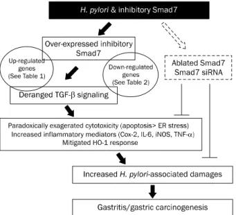
Fig. 4. Schematic summary. The current study raises hope that inhibiting Smad7 can be a strategy to alleviate Helicobacter pylori-induced gastric damage.
ER stress, endoplasmic reticulum stress.
Smad7 과발현주에서 H. pylori 감염 후 HO-1의 발현 감소는 Smad7의 과발현에 의해 항염증 사이토카인인 TGF-의 작용 을 억제시킨 것으로 해석된다. 또한 Smad7의 과발현 세포에 서 H. pylori의 감염에 따라 변화하는 유전자의 분석으로 위 점막 세포주에서 TGF-신호전달의억제인자로작용하는
Smad7의 과발현은 H. pylori 감염에 따른 세포 독성 및 염증 매개인자의 증가와 관련이 있었다(Fig. 4)
이번 연구는 이미 염증성 대장질환에서 Smad7의 anti- sense oligonucleotide를 통한 Smad7 발현 억제를 통해 항 염증 약제로 매우 효과적인 임상결과를 보이는 Morgensen 약물 연구23-25에서와 유사한 연구결과를 보였다. H. pylori에 감염된 환자의 조직에서 TGF- 신호 전달의 중요한 매개단 백질인 Smad3 활성화가 억제되고 이러한 결과로 Smad7의 발현이 증가하는 것으로 나타난다는 보고26에서와 같이, H.
pylori 감염 환자에서 억제된 TGF-의 신호전달을 정상적인 상태로 조절할 수 있는 약물은 TGF- 고유의 항염증 활성을 유도할 수 있을 것이다.
Smad7을 과발현시킨 세포에서 H. pylori에 대한 염증 반 응과 세포사멸이 현저히 증가되어 위점막 세포 손상이 유발될 수 있으나, 반대로 Smad7 siRNA를 이용한 Smad7의 발현 억제는 H. pylori 감염에 따른 염증 반응을 약화시킬 수 있음 을 증명하였다. 이번 연구에서는 H. pylori 감염에 따른 위 상피세포에서의 세포 독성과 염증 반응을 중심으로 Smad7의 역할을 규명하였으나, Monteleone 등27은 크론병과 같은 만 성염증성 대장질환에서 상피세포나 침윤 T 림프구 모두에서
Smad7이 과발현되어 있으며, 이를 antisense로 억제시켰을 때 염증이 약화되었다고 보고하였는데, 염증성 장질환 환자의 장조직에서도 같은 현상28-31이 관찰되었다. 또한 Smad7의 과 발현은 염증성 대장질환의 주요 전사인자인 NF-B의 활성화 와도 연관됨을 보고하였고, 위나 장과 같은 장기 이외에도 신 장 섬유화 및 신장염에도 Smad7이 주요하게 작동함을 보고 한 바 있다.32 이러한 연구결과는 Smad7 발현을 억제할 수 있는 anti-sense oligonucleotides에 의한 대장염 동물모델 에서의 효능33의 단계를 넘어서 염증성 대장질환 환자에서도 성공적인 임상효능을 발표한 바 있다.
저자들은 TGF- 신호매개의 Smad2 또는 Smad3가 아닌 dominant negative TGF- receptor를 과발현시킨 동물 모 델을 통해 TGF- 작용의 결손으로 인한 염증성 대장질환의 악화, 자가면역성 췌장염의 발생과 바렛 식도나 H. pylori 감 염에 대한 감수성의 증가에 따른 위식도암 및 염증성 대장암 의 발생이 증가된다는 연구결과를 보고한 바 있다.1-5 이러한 비이상적인 TGF- 신호 전달은TGF- receptor의 haplo-in- suficiency state에 의해 염증의 악화 및 암 발생으로 이어질 수 있는데, 다른 연구자들도 구강염,34 H. pylori 연관 위염 및 위암 발생,35,36 probiotics의 작용,37 gastrin 발현38 등에 Smad7이 미치는 영향에 대하여 보고한 바가 있다.
이번 연구에서는 H. pylori 감염에 있어 Smad7의 역할에 대하여 조사하였으나, Nguyen 등10의 연구에 의하면 H. py- lori 독성인자 CagA는 Smad3와 특이적으로 결합하여 TGF-에의해유도되는IL-8, CXCL1, CXCL3 등과 같은 염 증 매개인자들의 발현 억제를 못하게 함으로써 H. pylori에 의한 염증 반응을 증대시킬 수 있고, 더불어 이러한 항염증 사이토카인인 TGF-나 그 수용체 활성을 억제시켜 H. pylori 감염에 따른 면역 감시체계를 교란시킨다는 결과를 얻을 수 있었다. 또한 Jo 등39의 연구에 의하면 H. pylori 감염 환자에 서 TGF-수용체의 발현이 감소되어 있으며, in vitro 연구에 서 H. pylori 감염 24시간 이내에 유의한 TGF- 발현이 감소 되고 이러한 현상은 24시간 이후에 회복이 되는데, 이는 H.
pylori균이 숙주에 유의한 염증전파를 위해 숙주를 속이는
“stupid host defense response”일 수 있다고 보고하였다.
한편, 세포 보호 기능을 하는 Nrf2가 결손된 생쥐에서는 H.
pylori 감염이 악화된다는 결과(저자 등의 미발표 자료)와 H.
pylori 감염 후 활성산소 유도에 의한 자가탐식 저해가 주로 암 줄기세포에서 유발되어 H. pylori 증식에 유리한 반응을 초래한다는 보고40 등을 비추어 볼 때, H. pylori 감염에 의해 위염이나 위암으로 진전되는 비율이 1% 이내로 낮지만 심각 한 위염이나 위암이 발생하는 경우에는 H. pylori에 의한 숙 주 간의 반응이 매우 중요하다는 사실을 알 수 있다.
결론으로 H. pylori가 class I carcinogen이기 때문에 위암
발생을 예방하기 위해서는 제균이 일차적인 예방 및 치료 방 안이 될 수 있으나, 염증성 대장질환에서와 같이 Smad7 an- tisense oligonucleotide가 유망한 치료제로 임상시험 중인 점에 비추어 볼 때 TGF- 신호의 조절인자로 작용하는 Smad7을 제어할 수 있는 약물의 개발은 H. pylori에 의한 세포 손상이나 염증을 제어할 수 있는 새로운 전략으로 제시 될 수 있을 것이다.
요 약
목적: Helicobacter pylori 감염에 따른 세포 독성과 염증 반 응에 있어 형질전환인자-베타에 의한 항염증 활성의 중요한 매개인자로 작용하는 Smad7의 역할을 알아보고자 하였다.
대상 및 방법: 정상 위점막 세포주 RGM1에서 Smad7을 과발 현 또는 발현을 억제시킨 상태에서 H. pylori를 감염시킨 후 세포사멸 인자의 변화와 염증 유발인자의 발현을 세포생물학 적 방법으로 분석하였다.
결과: 위점막 세포주에서 Smad7의 과발현은 H. pylori 감염 에 의한 세포사멸을 증가시켰다. 감염에 의한 COX-2, iNOS, TNF- 등의 염증 유발인자의 발현 또한 대조 세포주에 비해 Smad7 과발현 세포주에서 증가하였으며, 이러한 염증 유발 인자의 발현은 Smad7 발현 억제에 의해 감소되었다.
결론: 형질전환인자-베타 신호의 저해인자로 작용하는 Smad7 이 H. pylori 감염에 의한 세포손상 및 위염 억제를 위한 새로 운 치료표적으로 제시될 수 있을 것으로 생각된다.
색인단어: Helicobacter pylori; 형질전환인자-베타; Smad7;
위염
REFERENCES
1. Im YH, Kim HT, Kim IY, et al. Heterozygous mice for the trans- forming growth factor-beta type II receptor gene have increased susceptibility to hepatocellular carcinogenesis. Cancer Res 2001;61:6665-6668.
2. Hahm KB, Cho K, Lee C, et al. Repression of the gene encoding the TGF-beta type II receptor is a major target of the EWS-FLI1 oncoprotein. Nat Genet 1999;23:222-227.
3. Hahm KB, Lee KM, Kim YB, et al. Conditional loss of TGF-beta sig- nalling leads to increased susceptibility to gastrointestinal carci- nogenesis in mice. Aliment Pharmacol Ther 2002;16 Suppl 2:115-127.
4. Hahm KB, Im YH, Parks TW, et al. Loss of transforming growth fac- tor beta signalling in the intestine contributes to tissue injury in inflammatory bowel disease. Gut 2001;49:190-198.
5. Hahm KB, Im YH, Lee C, et al. Loss of TGF-beta signaling contrib- utes to autoimmune pancreatitis. J Clin Invest 2000;105:1057- 1065.
6. Camilo V, Barros R, Sousa S, et al. Helicobacter pylori and the
BMP pathway regulate CDX2 and SOX2 expression in gastric cells. Carcinogenesis 2012;33:1985-1992.
7. Shiotani A, Kamada T, Yamanaka Y, et al. Sonic hedgehog and CDX2 expression in the stomach. J Gastroenterol Hepatol 2008;23 Suppl 2:S161-S166.
8. Lee KM, Lee JS, Jung HS, Park DK, Park HS, Hahm KB. Late re- activation of sonic hedgehog by Helicobacter pylori results in population of gastric epithelial cells that are resistant to apopto- sis: implication for gastric carcinogenesis. Cancer Lett 2010;
287:44-53.
9. Barros R, Pereira B, Duluc I, et al. Key elements of the BMP/
SMAD pathway co-localize with CDX2 in intestinal metaplasia and regulate CDX2 expression in human gastric cell lines. J Pathol 2008;215:411-420.
10. Nguyen TT, Kim SJ, Park JM, Hahm KB, Lee HJ. Repressed TGF-
signaling through CagA-Smad3 interaction as pathogenic mech- anisms of Helicobacter pylori-associated gastritis. J Clin Biochem Nutr 2015;57:113-120.
11. Pickup M, Novitskiy S, Moses HL. The roles of TGF in the tumour microenvironment. Nat Rev Cancer 2013;13:788-799.
12. Wakefield LM, Hill CS. Beyond TGF: roles of other TGF super- family members in cancer. Nat Rev Cancer 2013;13:328-341.
13. Akhurst RJ, Hata A. Targeting the TGF signalling pathway in disease. Nat Rev Drug Discov 2012;11:790-811.
14. Ikushima H, Miyazono K. TGFbeta signalling: a complex web in cancer progression. Nat Rev Cancer 2010;10:415-424.
15. Park S, Kim WS, Choi UJ, et al. Amelioration of oxidative stress with ensuing inflammation contributes to chemoprevention of H. pylori-associated gastric carcinogenesis. Antioxid Redox Signal 2004;6:549-560.
16. Han SU, Kim YB, Joo HJ, et al. Helicobacter pylori infection pro- motes gastric carcinogenesis in a mice model. J Gastroenterol Hepatol 2002;17:253-261.
17. Schmierer B, Hill CS. TGFbeta-SMAD signal transduction: molec- ular specificity and functional flexibility. Nat Rev Mol Cell Biol 2007;8:970-982.
18. Yingling JM, Blanchard KL, Sawyer JS. Development of TGF-beta signalling inhibitors for cancer therapy. Nat Rev Drug Discov 2004;3:1011-1022.
19. Siegel PM, Massagué J. Cytostatic and apoptotic actions of TGF-beta in homeostasis and cancer. Nat Rev Cancer 2003;3:
807-821.
20. Hong S, Lim S, Li AG, et al. Smad7 binds to the adaptors TAB2 and TAB3 to block recruitment of the kinase TAK1 to the adaptor TRAF2. Nat Immunol 2007;8:504-513.
21. Yan X, Chen YG. Smad7: not only a regulator, but also a cross-talk mediator of TGF- signalling. Biochem J 2011;434:1-10.
22. Akazawa Y, Isomoto H, Matsushima K, et al. Endoplasmic retic- ulum stress contributes to Helicobacter pylori VacA-induced apoptosis. PLoS One 2013;8:e82322.
23. Monteleone G, Neurath MF, Ardizzone S, et al. Mongersen, an or- al SMAD7 antisense oligonucleotide, and Crohn's disease. N Engl J Med 2015;372:1104-1113.
24. Monteleone G, Di Sabatino A, Ardizzone S, et al. Impact of patient characteristics on the clinical efficacy of mongersen (GED-0301), an oral Smad7 antisense oligonucleotide, in active
Crohn's disease. Aliment Pharmacol Ther 2016;43:717-724.
25. Ardizzone S, Bevivino G, Monteleone G. Mongersen, an oral Smad7 antisense oligonucleotide, in patients with active Crohn's disease. Therap Adv Gastroenterol 2016;9:527-532.
26. Monteleone G, Del Vecchio Blanco G, Palmieri G, et al. Induction and regulation of Smad7 in the gastric mucosa of patients with Helicobacter pylori infection. Gastroenterology 2004;126:674- 682.
27. Monteleone G, Kumberova A, Croft NM, McKenzie C, Steer HW, MacDonald TT. Blocking Smad7 restores TGF-beta1 signaling in chronic inflammatory bowel disease. J Clin Invest 2001;108:
601-609.
28. Monteleone G, Mann J, Monteleone I, et al. A failure of trans- forming growth factor-beta1 negative regulation maintains sus- tained NF-kappaB activation in gut inflammation. J Biol Chem 2004;279:3925-3932.
29. Monteleone G, Pallone F, MacDonald TT. Smad7 in TGF-be- ta-mediated negative regulation of gut inflammation. Trends Immunol 2004;25:513-517.
30. Monteleone G, Boirivant M, Pallone F, MacDonald TT. TGF-beta1 and Smad7 in the regulation of IBD. Mucosal Immunol 2008;1 Suppl 1:S50-S53.
31. Monteleone G, Caruso R, Pallone F. Role of Smad7 in in- flammatory bowel diseases. World J Gastroenterol 2012;18:
5664-5668.
32. Wang W, Huang XR, Li AG, et al. Signaling mechanism of TGF-be- ta1 in prevention of renal inflammation: role of Smad7. J Am Soc Nephrol 2005;16:1371-1383.
33. Boirivant M, Pallone F, Di Giacinto C, et al. Inhibition of Smad7
with a specific antisense oligonucleotide facilitates TGF-be- ta1-mediated suppression of colitis. Gastroenterology 2006;
131:1786-1798.
34. Han G, Bian L, Li F, et al. Preventive and therapeutic effects of Smad7 on radiation-induced oral mucositis. Nat Med 2013;19:
421-428.
35. Yuasa Y. Control of gut differentiation and intestinal-type gastric carcinogenesis. Nat Rev Cancer 2003;3:592-600.
36. Beswick EJ, Pinchuk IV, Earley RB, Schmitt DA, Reyes VE. Role of gastric epithelial cell-derived transforming growth factor beta in reduced CD4+ T cell proliferation and development of regulatory T cells during Helicobacter pylori infection. Infect Immun 2011;79:2737-2745.
37. Yang YJ, Chuang CC, Yang HB, Lu CC, Sheu BS. Lactobacillus acid- ophilus ameliorates H. pylori-induced gastric inflammation by in- activating the Smad7 and NFκB pathways. BMC Microbiol 2012;12:38.
38. Datta De D, Bhattacharjya S, Maitra M, et al. IL1B induced Smad 7 negatively regulates gastrin expression. PLoS One 2011;
6:e14775.
39. Jo Y, Han SU, Kim YJ, et al. Suppressed gastric mucosal TGF-be- ta1 increases susceptibility to H. pylori-Induced gastric in- flammation and ulceration: a stupid host defense response. Gut Liver 2010;4:43-53.
40. Tsugawa H, Suzuki H, Saya H, et al. Reactive oxygen species-in- duced autophagic degradation of Helicobacter pylori CagA is specifically suppressed in cancer stem-like cells. Cell Host Microbe 2012;12:764-777.