pISSN 2288-3886 / eISSN 2288-3959
자외선 조사와 병행된 녹차추출물 식이공급이 무모생쥐의 표피보습 개선 및 유리아미노산 생성 관련 대사에 미치는 영향*
최수민
·
신지혜·
이보민·
조윤희†경희대학교동서의학대학원의학영양학과
Dietary effect of green tea extract on hydration improvement and metabolism of free amino acid generation in epidermis of UV-irradiated hairless mice *
Choi, Sumin · Shin, Jihye · Lee, Bomin · Cho, Yunhi
†Department of Medical Nutrition, Graduate School of East-West Medical Science, Kyung Hee University, Yongin 17104, Korea
서 론
피부의 최외곽층인 표피는 인체 내 수분의 손실을 막고 보습을 유지하여 피부에 유연성을 부여하고, 피부장벽 역 할을 수행한다.1,2표피의 각질층은 세포 사이의 다중층상 구조를 이루어 체액손실을 억제하는 피지막과 각질세포 내의 친수성 성분으로 수분과 결합하여 흡습성에 직접적 인 영향을 미치는 자연보습인자 (natural moisturizing factor, NMF)에 의해 피부의 보습을 유지한다. 자연보습인 자에는 유리아미노산, pyrrolidone carboxylic acid (PCA), lactate, urea 등이 있으며 그 중 유리아미노산은 약 40%의 높은 함량을 차지한다.1,3유리아미노산은 filaggrin의 대사 를 거쳐 생성되는데 과립층의 keratohyalin granule 안에
있는 profilaggrin은 불활성 및 불용성인 상태로 저장되어 있다가 탈인산화 및 단백분해과정을 거쳐 filaggrin으로 분 해되어 각질층으로 방출된다.4 Filaggrin은 세포골격을 형 성하는 keratin filament와 결합하여 matrix protein으로 피 부장벽을 구성하며 peptidylarginine-deiminase3 (PAD3) 등의 효소에 의해 유리아미노산으로 최종 분해되어 자연 보습인자의 역할을 수행한다.5
외부적인 스트레스 중 하나인 자외선은 피부건조 및 과 증식, 주름형성과 같은 광노화뿐만 아니라 면역억제와 피 부세포의 DNA 손상을 일으키고6-8표피 filaggrin의 발현 및 유리아미노산으로 분해되는 과정의 변화를 야기하는 것으 로 알려져 있다.1,9광노화에 따른 피부건조, 주름형성 등을 완화시키기 위해 여러 기능성 식이소재 개발이 이루어지고 ABSTRACT
Purpose: Ultraviolet (UV) irradiation decreases epidermal hydration, which is maintained by reduction of natural moisturizing factors (NMFs). Among various NMFs, free amino acids (AA) are major constituents generated by filaggrin degradation. This experiment was conducted to determine whether or not dietary supplementation of green tea extract (GTE) in UV-irradiated mice can improve epidermal levels of hydration, filaggrin, free AAs, and peptidylarginine deiminase-3 (PAD3) expression (an enzyme involved in filaggrin degradation). Methods: Hairless mice were fed a diet of 1% GTE for 10 weeks in parallel with UV irradiation (group UV+1%GTE). As controls, hairless mice were fed a control diet in parallel with (group UV+) or without (group UV-) UV irradiation. Results: In group UV+, epidermal levels of hydration and filaggrin were lower than those in group UV-; these levels increased in group UV+1% GTE to levels similar to group UV-. Epidermal levels of PAD3 and major AAs of NMF, alanine, glycine and serine were similar in groups UV- and UV+, whereas these levels highly increased in group UV+1% GTE. Conclusion: Dietary GTE improves epidermal hydration by filaggrin generation and degradation into AAs.
KEY WORDS: green tea extract (GTE), filaggrin, peptidylarginine deiminase-3 (PAD3), natural moisturizing factor (NMF)
Received: June 10, 2016 / Revised: July 6, 2016 / Accepted: September 5, 2016
*This study was supported by a grant of the Korean Health Technology R & D Project, Ministry of Health & Welfare, Republic of Korea (Grant No. HN13C0076).
†To whom correspondence should be addressed.
tel: +82-31-201-3817, e-mail: [email protected]
© 2016 The Korean Nutrition Society
This is an Open Access article distributed under the terms of the Creative Commons Attribution Non-Commercial License (http://creative- commons.org/licenses/by-nc/3.0/) which permits unrestricted non-commercial use, distribution, and reproduction in any medium, provided the original work is properly cited.
있는데10,11이 중 녹차의 카테킨은 피부의 항염증, 항암 및 항산화 효과를 입증한 연구가 많이 보여졌으며,12,13 자외 선에 의해 유도되는 종양형성과 광노화를 저해하는 물질
로써14,15치료 및 화장품으로 상용되고 있다. 선행연구를
통해 녹차의 카테킨 중 주요 활성물질인 epigallocatechin- 3-gallate (EGCG)로 처리한 각질형성세포에서 filaggrin의 발현을 유도함이 보고되었다.16그러나 녹차 추출물 식이 공급을 통한 피부 보습 관련 filaggrin 대사 기전 및 유리아 미노산에 대한 연구는 미흡한 실정이다. 이에 본 연구에서 는 무모생쥐에 자외선 조사와 함께 녹차추출물을 식이공 급하고 피부 보습의 변화를 비롯하여, 표피 filaggrin과 유 리아미노산의 함량 및 filaggrin 분해 관련 효소의 단백질 발현에 미치는 영향을 살펴보았다.
연구방법
실험 동물생후 6주된 암컷 무모생쥐 [Albino hairless mouse (SKH- 1)]를 (주)오리엔트바이오에서 공급받아 실험식이로 사육하 기 전 1주간 고형배합 사료로 적응시켰다. 체중에 따른 난괴 법을 이용하여 자외선을 조사하지 않은 정상 대조군 (UV−
군, n = 5), 자외선 조사군 (UV+군, n = 5) 및 자외선 조사와 함께 녹차추출물 (green tea extract, GTE)을 전체식이의 1%
수준에서 공급한 실험군 (UV+1%GTE군, n = 5)으로 나누 었다. 사육기간 동안 사료와 물을 마음껏 섭취하도록 하였 으며 온도는 24 ± 2oC, 습도는 55 ± 10%를 유지하고 광주기 와 암주기가 매일 12시간이 되도록 조절하였다. 본 동물실 험에 대한 프로토콜은 경기과학기술 진흥원의 동물실험 윤리위원회에 의해 승인되었다 (승인번호 : 2013-12-0006).
자외선 조사
사료 투여 개시일로부터 1주일에 3회씩 UVB lamp (30%
의 UVA를 함유함: TL 20W/12RS SLV, wave length 290~
390 nm, Phillips, Netherlands)로 자외선 조사를 하였다.
자외선 조사량의 측정은 UV meter (VARIOCONTROL, Waldmann ver. 2.03, Germany)를 사용하였다.13 1주차에 는 1MED (minimal erythema dose; 75 mJ/cm2, 15 min)로 시작해서 2주차에 2MED (150 mJ/cm2, 30 min), 3주차에 3MED (225 mJ/cm2, 45 min), 4주차부터 10주차까지 4MED (250 mJ/cm2, 50 min)를 동물의 등 부위 (등 중앙기 준, 2 cm × 3 cm)에 조사 하였다.17
실험 식이
UV-군과 UV+군은 GTE가 첨가되지 않은 일반 식이
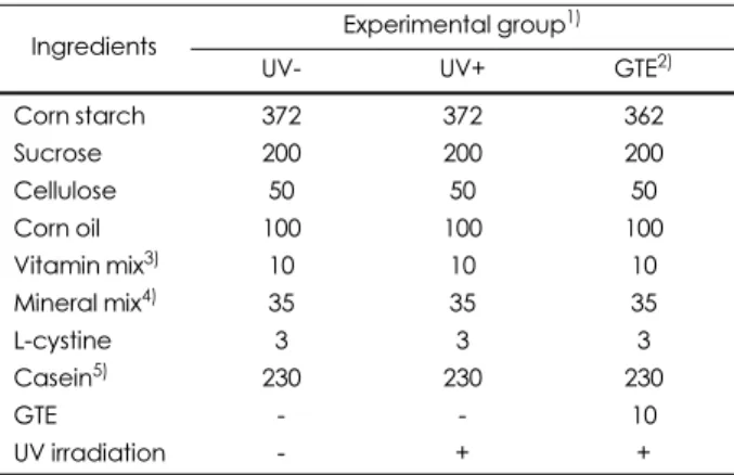
(AIN-93G)를 공급하였다.13 UV+1%GTE군은 전체 식이 무게의 1.0%에 상응하는 GTE [Catechin 50%, EGCG 28% 함유]를 식이에 첨가하여 공급하였다. 식이 1%의 GTE 첨가 수준은 독성을 유발하지 않으며18 피부산도개 선의 효과가 나타난 예비실험을 바탕으로 결정하였다.19 GTE 시료 첨가에 따른 건분 식이 무게 차이는 corn starch 를 감하여 조정하였으며, GTE 시료는 (주)아모레퍼시픽 (South Korea)에서 공급받았다. 각 실험군의 식이 조성은 Table 1과 같다.
피부 보습 측정
피부의 보습측정은 Corneometer CM825 (Courage &
Khazaka, Cologne, Germany)를 사용하여 10주차에 측정 하였다. 측정 시 각 개체 별 측정부위는 동일하게 등 중앙 부분으로 하였으며 측정면에 대한 압력을 일정하게 눌러 총 3회 측정하여 수치의 평균값을 기록하였다.
동물의 희생과 표피 분리
10주간의 실험식이 및 자외선 조사 후 경추 탈골로 희생 시키고 등 부위의 피부조직을 얻었다. 피부조직은 Dispase II (2.4 unit/mL, Roche, Germany)와 Hank solution (Hank’s balanced salt solution)이 혼합된 용액 (1:1 v/v)에 담가 4oC 에서 16시간 동안 처리한 후 표피를 분리하였다.11
표피 filaggrin 추출 및 분석
분리한 표피는 2 mM EDTA, 9 M urea, 2% SDS 및 protease inhibitor cocktail가 포함된 50 mM Tris buffer (P-
Table 1. Diet composition and UV irradiation of experimental groups (g/kg)
Ingredients Experimental group1)
UV- UV+ GTE2)
Corn starch 372 372 362
Sucrose 200 200 200
Cellulose 50 50 50
Corn oil 100 100 100
Vitamin mix3) 10 10 10
Mineral mix4) 35 35 35
L-cystine 3 3 3
Casein5) 230 230 230
GTE - - 10
UV irradiation - + +
1) Groups UV- and UV+, hairless mice fed a control diet without (group UV-) or with (group UV+) UV irradiation for 10 weeks; Group GTE, hairless mice fed a diet containing 1.0% green tea extract (GTE) in parallel with UV irradiation for 10 weeks. 2) GTE: green tea extract 3) AIN-93 vitamin mix #310025 (Dyets Inc., Bethle- hem, PA) 4) AIN-93G salt mix #210025 (Dyets Inc., Bethlehem, PA) 5) Casein (nitrogen × 6.25), 870 g/kg
8340, Sigma Chemical Co., St. Louis, MO, USA)를 처리 하여20 polytron으로 분쇄하였다. 분쇄한 조직을 12,000 g 에서 4oC로 20분동안 원심분리 후 상층액을 얻었고, 단백 질 정량을 하였다. 동량의 단백질 (20 µg/well)을 4%
(profilaggrin 발현) 또는 8% (filaggrin 발현) SDS/PAGE gel에 전기영동 후 nitrocellulose membrane에 흡착시켰다.
Membrane을 1차항체인 anti-rabbit polyclonal filaggrin antibody (Enzo Biochem Inc., NY, USA, cat no. p20930) 를 1/1,000의 농도로 희석한 PBS solution과 함께 4oC에서 17시간 동안 반응시키고 PBST solution으로 3회 세척하였 다. 이어서 anti-rabbit IgG-HRP (Bethly Laboratories Inc., Mongomery, TX, USA, cat no. A120-101P)로 2시간동안 실온에서 반응시킨 후 PBST solution으로 3회 세척하고 Super Signal West Pico Chemiluminescent Substrate (Thermo Scientific, Rockford, IL, USA)를 통해 detection 하였다. 각 band는 imaging densitometer (Lab work version 4.6, UVP, upland, CA, USA)를 이용하여 측정한 후 actin의 발현을 기준으로 보정하였다.
표피 PAD3 발현 분석
분리된 표피에 0.5% Nonidet P-40 (TE-NP40 buffer extract), 10 mM EDTA, 40 mM Tris-HCl (pH 7.5), protease inhibitor 및 Tris-EDTA buffer을 첨가하여 polytron을 사 용하여 분쇄하였다.5 분쇄한 조직은 원심 분리 (1,2000 g, 4oC, 20 min)하여 상층액을 얻었고, 단백질 정량 후 8%
SDS/PAGE gel에 시료 20 µg/well을 전기영동 하여 nitro- cellulose membrane에 흡착시켰다. Membrane은 1차항체 anti- goat polyclonal PAD3 antibody (cat no. sc-55693, Santa Cruz Biotechnology Inc., Santa Cruz, California, USA) 및 2차 항체 anti-goat IgG-HRP (bethly Laboratories Inc., Montgomery, TX, USA)를 이용하여 동일한 방법으 로 반응시킨 뒤 Super Signal West Pico Chemiluminescent substrate를 통해 detection하여 발색하였다. 각 band의 intensity는 imaging densitometer를 사용하여 측정 후 actin의 발현을 기준으로 정량되었다.
표피 유리 아미노산 함량 분석
표피 조직에 15% trichloroacetic acid (TCA) 용액을 첨 가하여 polytron을 이용해 분쇄하고 4oC에서 15시간 동안 반응시켜 단백질을 침전시켰다. 12,000 g에서 10분간 원심 분리를 하여5상층액을 분리한 후 0.45 µm filter (Millipore, USA)를 사용해 상층액을 여과하여 high performance liquid chromatography (HPLC)를 통해 각 유리아미노산 의 농도를 분석하였다. Acetonitrile : methanol : water (45
: 45 : 10, volume)의 용액을 Model 510 solvent delivery system Pico Tag column (3.9 × 300 nm, 4 µm)에서 유속 1.0 mL/min의 속도로 아미노산을 분리하고 각 2487 UV detector (254 nm, Waters)를 사용하여 함량분석 후 사용 표피 무게의 정량 값으로 보정하였다.
통계분석
실험 결과는 SPSS statistics 22 program (statistical package for social science)을 사용하여 통계 처리하였다.
각 실험군 별 결과는 평균 (mean)과 표준편차 (standard deviation, SD)로 나타냈으며, 각 군간 비교는 one-way ANOVA로 분석한 후 Tukey’s multiple range test로 p <
0.05 수준에서 검증 하였다.
결 과
피부 보습 측정자외선 조사 및 녹차추출물 식이공급 후 10주차에 피부 보습 측정 결과는 Fig. 1에 나타내었다. 자외선 조사 10주 에 측정한 보습함량은 자외선 조사군인 UV+군이 정상대 조군인 UV-군보다 유의적으로 낮았는데 이 결과는 지속 적인 자외선 조사에 의해 피부 보습함량이 감소하였음을 의미한다. 반면에 자외선 조사와 병행하여 녹차추출물을 10주간 공급한 실험군인 UV+1%GTE 군의 보습은 UV+
군에 비해 유의적으로 높았으나 UV-군의 함량에는 미치 지 못하였다. 이는 전체식이 1.0%에 달하는 녹차추출물의 식이공급이 자외선 조사에 의해 유도된 피부 건조를 완화 시켰음을 의미한다.
Fig. 1. Altered epidermal hydration of groups. Hairless mice fed a control diet without UV irradiation for 10 weeks (group UV-); UV- irradiated hairless mice fed a control diet (group UV+) or a diet supplemented with 1.0% green tea extract (group UV+1%GTE) for 10 weeks. Values are mean ± SD (n = 5). Values with different alphabetical letters are significantly different (p < 0.05) using one- way ANOVA and Tukey's multiple range test.
표피 profilaggrin 및 filaggrin의 함량 변화
표피의 profilaggrin 및 filaggrin 함량 변화를 western blot assay로 분석 하였고 그 결과를 Fig. 2에 나타내었다.
자연보습인자의 전구체인 filaggrin은 각질형성세포가 분 화단계에서 발현시키는 단백질 중 하나로 피부의 보습 및 피부 막 기능의 지표로 사용된다.4,21 UV+군의 profilaggrin 함량은 정상 대조군 인 UV-군과 유의적인 차이가 없었으 나, filaggrin 함량은 UV-군에 비해 유의적으로 낮았다. 반 면 UV+1%GTE 군의 profilaggrin 함량은 UV-군 및 UV+
군보다 현저히 높았다. Filaggrin의 함량은 UV+군에 비해 유의적으로 높았으며 UV-군의 함량과 유사하였다. 이는 녹차추출물의 식이공급이 pro-filaggrin의 합성을 촉진시 키고 자외선에 의한 표피 filaggrin 함량의 감소를 정상화 시켰음을 의미한다.
PAD3 단백질 발현 변화
PAD3는 filaggrin의 분해과정에서 filaggrin의 deimination 을 통해 유리아미노산의 분해를 유도한다.22표피 PAD3의
단백질 발현을 western blot assay로 분석한 결과 (Fig. 3) UV+군의 PAD3 단백질 발현은 UV-군에 비해 유의적으로 낮았다. 이는 자외선 조사가 PAD3의 발현을 억제시켰음을 의미한다. UV+1%GTE군에서는 UV+군에 비해 유의적으 로 높았으며 UV-군의 함량보다 유의적으로 높았다. 따라 서, 녹차추출물의 식이공급이 PAD3 발현을 현저하게 활 성화함을 의미한다.
표피 유리아미노산 함량 변화
Filaggrin은 serine, glycine, glutamate, histidine, alanine 등의 아미노산을 다량 함유한 단백질로23 분해과정에서 유리아미노산으로 전환되어 아미노산의 친수성 성질에 의해 자연보습인자로서 표피의 보습 유지를 담당한다.3,24 유리아미노산 분석결과 serine과 glycine, alanine은 UV- 군과 UV+군에서 유의적인 차이를 보이지 않은 반면, UV+1%GTE군에서는 두 군에 비해 유의적으로 높았다.
Histidine, arginine, asparagine 또한 UV-군과 UV+군에서 유 의적인 차이는 나타나지 않았으나, UV+1%GTE군에서는 UV-군에 비해 유의적으로 높았다. UV+군에서 glutamine 및 tryptophan은 UV+1%GTE군과 유사하였다. 그 외
Fig. 3 Altered protein level of peptidylarginine deiminase-3 in the epidermis of groups. Hairless mice fed a control diet without UV irradiation for 10 weeks (group UV-); UV-irradiated hairless mice fed a control diet (group UV+) or a diet supplemented with 1.0%
green tea extract (group UV+1%GTE) for 10 weeks (A) Represen- tative expression of peptidylarginine deiminase-3 (PAD3) protein in the epidermis of groups (B) Signal intensities from multiple exper- iments of (A) were quantified and the integrated areas were nor- malized, first to the corresponding value of actin and then to the signal observed in the normal control group (group UV-). Values are mean ± SD (n = 5). Values with different alphabetical letters in PAD3 is significantly different (p < 0.05) using one-way ANOVA and Tukey's multiple range test.
Fig. 2. Altered protein levels of profilaggrin and filaggrin in the epi- dermis of groups. Hairless mice fed a control diet without UV irradi- ation for 10 weeks (group UV-): UV-irradiated hairless mice fed a control diet (group UV+) or a diet supplemented with 1.0% green tea extract (group UV+1%GTE) for 10 weeks (A) Representative expression of profilaggrin and filaggrin proteins in the epidermis of groups (B) Signal intensities from multiple experiments of (A) were quantified and the integrated areas were normalized, first to the corresponding value of actin and then to the signal observed in the normal control group (group UV-). Values are mean ± SD (n = 5). Values with different alphabetical letters in profilaggrin and fil- aggrin are significantly different (p < 0.05) using one-way ANOVA and Tukey's multiple range test.
isoleucine, leucine, lysine, methionine, phenylalanine, threonine, valine, aspartate, glutamate, proline 및 tyrosine 의 함량은 모든 군에서 군간 유의성이 없었다. 이는 녹차추 출물의 식이공급이 자연보습인자인 serine, glycine, arginine 등 보습인자역할을 하는 주요 유리아미노산 함량 증가시 켜 피부 보습을 증가시켰음을 의미한다.
고 찰
지속적인 자외선 조사에 의해 초래되는 광노화는 내인 성 노화와 달리 산화 스트레스 증가와 함께 피부 보습 및 탄력 감소에 의한 주름을 야기하고, 색소침착, 피부암 등을 유발하는 것으로 보고되어진다.7,8주 3회 15~50분간 매우 높음 수준의 자외선 지수 3.3배에 해당하는 양을 10주동안 조사 하였을 때, 피부 보습함량이 낮았는데 이 결과는 사전 연구에서 자외선 비조사군에 비해 자외선 조사군의 피부 의 보습함량이 낮고 경피수분손실 (transepidermal water loss, TEWL)이 높은 결과와 일치하였다.25반면 자외선 조 사를 병행한 녹차추출물의 식이공급은 UV+군에 비해 피 부 보습함량이 높았다. 즉, 녹차추출물의 식이공급은 지속 적인 자외선 조사로 야기된 피부 건조를 완화시켰음을 의 미한다.
사전연구에서 filaggrin을 구성하는 아미노산과 표피 내 자연보습인자로 작용하는 유리아미노산의 구성이 유사하 게 나타남에 의해 자연보습인자로 작용하는 표피 유리아 미노산의 유일한 전구체는 filaggrin임이 입증되었다.23,26 과립층의 keratohyalin granule 내에 존재하는 profilaggrin 은 표피의 칼슘 기울기가 증가함에 따라 phosphatase
(PPase),27 furin28등의 효소에 의해 탈인산화와 단백분해 과정을 거쳐 histidine이 풍부한 filaggrin으로 분해 된다.4,26 자외선을 조사한 UV+군에서 profilaggrin의 함량은 UV-군 과의 차이가 나타나지 않았으나, filaggrin의 함량은 UV-군 에 비해 낮게 발현되었다. 이는 자외선 조사에 의해 피부장 벽이 손상되면 표피의 칼슘 기울기가 감소하고 경피수분 손실이 증가한다는 기존 연구와 함께29지속적인 자외선 조 사가 profilaggrin이 filaggrin으로 분해되는 과정에 장애를 일으킨 것을 의미한다. 반면, UV+1%GTE군의 profilaggrin 함량은 UV+군에 비해 높았고, filaggrin의 함량은 UV-군과 유사한 수준으로 높았다. 사전연구에서 녹차의 주요 카테 킨인 EGCG가 profilaggrin발현의 regulator4,30로 작용하는 AP-1 (activator protein-1)을 활성화하며,31 EGCG에 대한 in vitro 실험을 통해 정상각화세포에서 filaggrin 발현의 증가가 보고되었다.16 즉, 녹차추출물의 식이공급이 표피 profilaggrin 함량을 높이고, 자외선에 의한 대사 장애를 정 상화시켰음을 나타낸다.
Profilaggrin은 filaggrin으로 분해 후 과립층과 각질층 경 계부에서 keratin intermediate filaments와 결합하여 세포의 골격을 이루는 거대원섬유를 형성한다.4,21 Keratin과 결합 된 filaggrin은 PADs에 의해 아미노산의 잔기가 arginine에 서 citrulline으로 치환되는 deimination 과정을 거쳐 keratin 과 분리되고 caspase-14,32 bleomycin hydrolase4 등의 효 소에 의한 단백분해 과정을 거쳐 유리아미노산으로 분해 된다. Filaggrin의 deimination 에는 칼슘 의존형 효소인 PAD1과 PAD3가 관여하는데, 그 중 PAD3의 발현장소는 모발과 표피로 제한되며, 사전연구에서 PAD3의 발현은 과 립층 및 각질층에서 (pro)filaggrin과 함께 발현되었다.5,22
Fig. 4. Free amino acid contents in the epidermis of groups. Hairless mice fed a control diet without UV irradiation for 10 weeks (group UV-); UV-irradiated hairless mice fed a control diet (group UV+) or a diet supplemented with 1.0% green tea extract (group UV+1%GTE) for 10 weeks. Data are mean ± SD (n = 5). Values with different alphabetical letters in each amino acids are significantly different (p <
0.05) using one-way ANOVA and Tukey’s multiple range test.
PAD3는 Profilaggrin이 filaggrin으로 분해된 후 유리아미 노산으로 분해되는 과정의 도입부로써 중요한 역할을 하 며, filaggrin의 citrullination에 PAD3가 관여되지 못하면 표피의 항상성을 손상시키고 피부의 장벽기능을 잃게 한 다.33 PAD3의 발현은 UV+1%GTE군이 UV-군 및 UV+군 보다 현저히 높았다. EGCG를 처리한 각질형성세포에서 filaggrin을 유리아미노산으로 분해하는 caspase-14효소의 발현이 증가되었다는 선행연구도 보고되었는데,34 이를 종합하여 녹차추출물의 식이공급이 PAD3 및 filaggrin의 분해에 관여하는 효소의 발현을 촉진하여 궁극적으로 유 리아미노산의 생성이 높게 나타난 것으로 사료되며, 추후 PAD3이외 caspase-14 등 아미노산으로 분해되는 과정에 관여하는 효소에 대해서도 추가적인 연구가 필요할 것으 로 여겨진다.
자연보습인자인 유리아미노산 중 serine과 glycine은 총 유리아미노산의 40%을 차지하며 alanine 또한 약 10% 가 량 함유되어 있다고 보고되었다. 또한, 표피의 건조화 및 과증식에 의해 serine과 glycine의 급격한 농도감소가 나타 남에 따라35 추가적인 대사과정을 거치는 다른 아미노산 과 달리 serine과 glycine은 이후의 대사과정을 거치지 않 으며 그 자체로 표피의 보습유지에 주요한 역할을 담당한 다.24,36,37 UV+군에서 serine과 glycine 및 alanine의 함량 은 UV-군과 차이가 없는 반면, 피부 보습은 UV-군에 비해 유의적으로 낮았다. 이 결과는 자외선이 투과성 피지막을 구성하는 주요 지질성분의 함량을 감소시킨다는 사전연 구와 함께38,39자외선에 의한 피부 보습함량이 낮은 이유 는 자연보습인자인 유리아미노산 보다는 피부 장벽을 통 해 보습을 유지하는 지질 메커니즘에 의한 것으로 여겨진 다. 추후 자외선에 따른 피지막의 주요 구성성분인 지질, 그 중에서 특히 ceramide 함량 감소 확인 및 녹차추출물의 효 능에 대한 연구가 필요할 것이라고 사료된다. UV+1%GTE 군에서 serine과 glycine, alanine의 함량은 UV+군 및 UV- 군보다 현저히 높게 나타났는데, 이는 serine과 glycine, alanine이 피부 보습에 대한 효과를 직접적으로 나타내는 자연보습 인자로써 녹차섭취가 자외선에 의해 야기되었 던 피부건조를 완화시켰음을 의미한다. Filaggrin으로부터 분해된 유리아미노산 중 glutamate와 histidine은 각각 PCA와 urocanic acid (UCA)로 빠르게 대사되어 자연보습 인자의 역할을 수행 하며,40,41 arginine은 urea cycle의 중 간 물질로 작용한다.36 따라서, glutamate, histidine 및 arginine으로는 자외선 조사 및 GTE 의 섭취 효과를 절대 적으로 반영하는 데에는 적절하지 않을 것으로 사료된다.
asparagine과 tryptophan은 선행연구를 통해 isoleucine, leucine, phenylalanine, threonine, tryptophan 등과 같이
미량 아미노산으로 존재한다고 보고되어36 피부 보습에 있어서는 큰 영향을 미치지 않을 것으로 여겨진다.
위 결과를 종합해보면, 장기간의 자외선 조사는 pro- filaggrin 및 filaggrin, 유리 아미노산의 대사보다는 ceramide 를 포함한 피지막 구성성분의 변화와 함께 피부 보습을 감 소시키는 반면, 녹차추출물의 식이공급은 자외선 대조군 인 UV+군에 비해 표피 (pro)filaggrin, PAD3, 주요 유리아 미노산의 발현변화를 유의적으로 증가시켜 자외선 조사 로 인한 피부 건조화를 개선하였다.
요 약
본 연구에서는 자외선 조사를 병행하여 녹차 추출물의 식이공급을 10주간 공급한 무모생쥐 (UV+1%GTE군)의 표피 보습과 filaggrin 의 대사물질 및 관련 효소인 PAD3 의 발현 및 함량 변화를 정상대조군 (UV-군) 및 자외선 조 사군 (UV+군)과 비교 분석하였으며 결과는 다음과 같다.
UV+군의 피부 보습은 UV-군보다 유의적으로 낮은 반면 UV+1%GTE군 의 피부 보습은 UV+군에 비해 유의적으로 높았고, UV-군에는 미치지 못하였다.
UV+군의 profilaggrin 함량은 UV-군과 유의적인 차이가 없었으나, filaggrin의 함량은 UV-군에 비해 유의적으로 낮 았다. UV+1%GTE군 에서 profilaggrin은 UV- 및 UV+에 비해 현저히 높았고, filaggrin은 UV-군과 유사한 수준으로 높았다. UV+군의 PAD3 효소 발현은 UV-군에 비해 유의 적으로 낮았다. UV+1%GTE군은 UV+군에 비해 유의적으 로 높았으며, UV-군에 비해 높았다. 자연보습인자의 주요 아미노산인 serine 및 glycine, alanine의 함량은 UV-군과 UV+군간 유사하였으나 UV+1%GTE군에서는 UV- 및 UV+군에 비해 현저히 높았다.
UV-군과 비교하여 UV+군의 피부 보습함량이 낮게 측정 된 것에 비해 유리아미노산 함량에는 변화가 없어 자외선 에 따른 피부 보습에 관여되는 또 다른 메커니즘인 표피 지 질함량 분석과 함께 녹차추출물의 효과에 대한 연구가 추 후 이루어질 필요성이 있다. 그러나, 결론적으로 자외선 조 사와 병행된 녹차 추출물 식이 공급은 (pro)filaggrin의 함 량 및 PAD3의 발현이 높게 나타남에 의한 표피 유리아미 노산의 함량이 높아 궁극적으로 자외선 조사에 의해 야기 된 피부건조를 완화시켰다.
References
1. Rawlings AV, Harding CR. Moisturization and skin barrier func- tion. Dermatol Ther 2004; 17 Suppl 1: 43-48.
2. Baroni A, Buommino E, De Gregorio V, Ruocco E, Ruocco V, Wolf R. Structure and function of the epidermis related to barrier properties. Clin Dermatol 2012; 30(3): 257-262.
3. Verdier-Sévrain S, Bont F. Skin hydration: a review on its molecu- lar mechanisms. J Cosmet Dermatol 2007; 6(2): 75-82.
4. Sandilands A, Sutherland C, Irvine AD, McLean WH. Filaggrin in the frontline: role in skin barrier function and disease. J Cell Sci 2009; 122(Pt 9): 1285-1294.
5. Nachat R, Méchin MC, Takahara H, Chavanas S, Charveron M, Serre G, Simon M. Peptidylarginine deiminase isoforms 1-3 are expressed in the epidermis and involved in the deimination of K1 and filaggrin. J Invest Dermatol 2005; 124(2): 384-393.
6. Gilchrest BA. Skin aging and photoaging: an overview. J Am Acad Dermatol 1989; 21(3 Pt 2): 610-613.
7. Chung JH. Photoaging in Asians. Photodermatol Photoimmunol Photomed 2003; 19(3): 109-121.
8. Tagami H. Functional characteristics of the stratum corneum in photoaged skin in comparison with those found in intrinsic aging.
Arch Dermatol Res 2008; 300 Suppl 1: S1-S6.
9. Gruber JV, Holtz R. Examining communication between ultravio- let (UV)-damaged cutaneous nerve cells and epidermal keratino- cytes in vitro. Toxicol Ind Health 2009; 25(4-5): 225-230.
10. Hwang E, Sun ZW, Lee TH, Shin HS, Park SY, Lee DG, Cho BG, Sohn H, Kwon OW, Kim SY, Yi TH. Enzyme-processed Korean Red Ginseng extracts protects against skin damage induced by UVB irradiation in hairless mice. J Ginseng Res 2013; 37(4): 425- 434.
11. Kim J, Lee Y, Cho Y. Effects of dietary royal jelly on epidermal generation of ceramides from acidic sphingomyelin and glucosyl- ceramides in UV-irradiated hairless mice. Curr Top Nutraceutical Res 2012; 10(3/4): 151-164.
12. Khan SG, Katiyar SK, Agarwal R, Mukhtar H. Enhancement of antioxidant and phase II enzymes by oral feeding of green tea poly- phenols in drinking water to SKH-1 hairless mice: possible role in cancer chemoprevention. Cancer Res 1992; 52(14): 4050-4052.
13. Min J, Lee Y, Han SM, Choi Y. Dietary effect of royal jelly supple- mentation on epidermal levels of hydration, filaggrins, free amino acids and the related enzyme expression in UV irradiated hairless mice. Korean J Nutr 2013; 46(2): 109-118.
14. Katiyar SK, Elmets CA. Green tea polyphenolic antioxidants and skin photoprotection (Review). Int J Oncol 2001; 18(6): 1307- 1313.
15. Jackson JK, Zhao J, Wong W, Burt HM. The inhibition of collage- nase induced degradation of collagen by the galloyl-containing polyphenols tannic acid, epigallocatechin gallate and epicatechin gallate. J Mater Sci Mater Med 2010; 21(5): 1435-1443.
16. Hsu S, Bollag WB, Lewis J, Huang Q, Singh B, Sharawy M, Yamamoto T, Schuster G. Green tea polyphenols induce differenti- ation and proliferation in epidermal keratinocytes. J Pharmacol Exp Ther 2003; 306(1): 29-34.
17. Kiss I, Chen S, Tramposch KM. The effect of high and low ultravi- olet-B dose exposure on the degree of hairless mouse skin wrin- kling. Photochem Photobiol 1991; 53(1): 109-112.
18. Takami S, Imai T, Hasumura M, Cho YM, Onose J, Hirose M.
Evaluation of toxicity of green tea catechins with 90-day dietary administration to F344 rats. Food Chem Toxicol 2008; 46(6):
2224-2229.
19. Lee B, Kim J, Hwang J, Cho Y. Dietary effect of green tea extract on epidermal levels of skin pH related factors, lactate dehydroge- nase protein expression and activity in UV-irradiated hairless mice.
J Nutr Health 2016; 49(2): 63-71.
20. Ginger RS, Blachford S, Rowland J, Rowson M, Harding CR. Fil- aggrin repeat number polymorphism is associated with a dry skin phenotype. Arch Dermatol Res 2005; 297(6): 235-241.
21. Dale BA, Resing KA, Lonsdale-Eccles JD. Filaggrin: a keratin fil- ament associated protein. Ann N Y Acad Sci 1985; 455: 330-342.
22. Kanno T, Kawada A, Yamanouchi J, Yosida-Noro C, Yoshiki A, Shiraiwa M, Kusakabe M, Manabe M, Tezuka T, Takahara H.
Human peptidylarginine deiminase type III: molecular cloning and nucleotide sequence of the cDNA, properties of the recombinant enzyme, and immunohistochemical localization in human skin. J Invest Dermatol 2000; 115(5): 813-823.
23. Steinert PM, Cantieri JS, Teller DC, Lonsdale-Eccles JD, Dale BA.
Characterization of a class of cationic proteins that specifically interact with intermediate filaments. Proc Natl Acad Sci U S A 1981; 78(7): 4097-4101.
24. Visscher M, Robinson M, Wickett R. Stratum corneum free amino acids following barrier perturbation and repair. Int J Cosmet Sci 2011; 33(1): 80-89.
25. Yamada Y, Obayashi M, Ishikawa T, Kiso Y, Ono Y, Yamashita K.
Dietary tocotrienol reduces UVB-induced skin damage and sesa- min enhances tocotrienol effects in hairless mice. J Nutr Sci Vita- minol (Tokyo) 2008; 54(2): 117-123.
26. Scott IR, Harding CR, Barrett JG. Histidine-rich protein of the ker- atohyalin granules. Source of the free amino acids, urocanic acid and pyrrolidone carboxylic acid in the stratum corneum. Biochim Biophys Acta 1982; 719(1): 110-117.
27. Kam E, Resing KA, Lim SK, Dale BA. Identification of rat epider- mal profilaggrin phosphatase as a member of the protein phospha- tase 2A family. J Cell Sci 1993; 106(Pt 1): 219-226.
28. Pearton DJ, Nirunsuksiri W, Rehemtulla A, Lewis SP, Presland RB, Dale BA. Proprotein convertase expression and localization in epidermis: evidence for multiple roles and substrates. Exp Derma- tol 2001; 10(3): 193-203.
29. Jiang SJ, Chu AW, Lu ZF, Pan MH, Che DF, Zhou XJ. Ultraviolet B-induced alterations of the skin barrier and epidermal calcium gradient. Exp Dermatol 2007; 16(12): 985-992.
30. Jang SI, Steinert PM, Markova NG. Activator protein 1 activity is involved in the regulation of the cell type-specific expression from the proximal promoter of the human profilaggrin gene. J Biol Chem 1996; 271(39): 24105-24114.
31. Balasubramanian S, Efimova T, Eckert RL. Green tea polyphenol stimulates a Ras, MEKK1, MEK3, and p38 cascade to increase activator protein 1 factor-dependent involucrin gene expression in normal human keratinocytes. J Biol Chem 2002; 277(3): 1828- 1836.
32. Hoste E, Kemperman P, Devos M, Denecker G, Kezic S, Yau N, Gilbert B, Lippens S, De Groote P, Roelandt R, Van Damme P, Gevaert K, Presland RB, Takahara H, Puppels G, Caspers P, Van- denabeele P, Declercq W. Caspase-14 is required for filaggrin deg- radation to natural moisturizing factors in the skin. J Invest Dermatol 2011; 131(11): 2233-2241.
33. Kim Y, Han SM, Cho Y. The dietary effect of royal jelly supple- mentation on epidermal levels of filaggrin and free amino acids
during menopause in rats. J Korean Soc Food Sci Nutr 2013; 42(3):
389-396.
34. Hsu S, Yamamoto T, Borke J, Walsh DS, Singh B, Rao S, Takaaki K, Nah-Do N, Lapp C, Lapp D, Foster E, Bollag WB, Lewis J, Wataha J, Osaki T, Schuster G. Green tea polyphenol-induced epi- dermal keratinocyte differentiation is associated with coordinated expression of p57/KIP2 and caspase 14. J Pharmacol Exp Ther 2005; 312(3): 884-890.
35. Koyama J, Horii I, Kawasaki K, Nakayama Y, Morikawa Y, Mitsui T, Kumagai H. Free amino acids of stratum corneum as a biochem- ical marker to evaluate dry skin. J Soc Cosmet Chem 1984; 35(4):
183-195.
36. Tabachnick J, LaBadie JH. Studies on the biochemistry of epider- mis. IV. The free amino acids, ammonia, urea, and pyrrolidone car- boxylic acid content of conventional and germ-free albino guina pig epidermia. J Invest Dermatol 1970; 54(1): 24-31.
37. Joo KM, Han JY, Son ED, Nam GW, Jeong HJ, Lim KM, Cho JC.
Study on the relationship between skin dryness and amino acids in stratum corneum. J Soc Cosmet Sci Korea 2012; 38(1): 75-82.
38. Bak H, Hong SP, Jeong SK, Choi EH, Lee SE, Lee SH, Ahn SK.
Altered epidermal lipid layers induced by long-term exposure to suberythemal-dose ultraviolet. Int J Dermatol 2011; 50(7): 832- 837.
39. Kim H, Oh I, Park KH, Kim NM, Do JH, Cho Y. Stimulatory effect of dietary red ginseng on epidermal hydration and ceramide levels in ultraviolet-irradiated hairless mice. J Med Food 2009; 12(4):
746-754.
40. Kezic S, Kammeyer A, Calkoen F, Fluhr JW, Bos JD. Natural moisturizing factor components in the stratum corneum as bio- markers of filaggrin genotype: evaluation of minimally invasive methods. Br J Dermatol 2009; 161(5): 1098-1104.
41. Mark H, Harding CR. Amino acid composition, including key derivatives of eccrine sweat: potential biomarkers of certain atopic skin conditions. Int J Cosmet Sci 2013; 35(2): 163-168.