I. Introduction
Mucogingival surgery is a plastic surgical procedure designed to correct defects in the morphology, position, and dimensions of the gingiva surrounding the teeth. Many surgi- cal techniques have been reported in muco- gingival surgery. Since these procedures al- so include the soft tissue esthetic approach, the term “periodontal plastic surgery” has been proposed to be more appropriate.1 Root coverage is a procedure that falls with this definition, and it has attracted more interest than others. Root cov- erage has been performed through many surgical techniques. The autogenous masticatory graft(free gingival autograft) was proposed by Miller2 as the first predictable technique to obtain root coverage.
However, the procedure didn't show ideal esthetic results and offered a low degree of predictability in
the correction of gingival recessions. Pedicle grafts have been reported to offer good re- sults in terms of root coverage3.1)Pedicle grafts provide the most esthetic result when adequate amount of keratinized gingiva exists. However, adequate gingiva does not always exist in adjacent locations.4 The guided tissue regeneration and the bilaminar techniques (BTs) consisting of connective tissue grafts with a pedicle graft have also been proposed as a possible therapeutic al- ternative in the management of gingival recessions.5-8 Comparative studies have dem- onstrated that bilaminar techniques show a significantly greater degree of predictability when the aim of the clinical trials is to ob- tain complete root coverage.9,10 The rationale of bilaminar techniques is to enhance the degree of predictability of therapeutic result
* Corresponding author:Seoung-Ho Lee, Department of Periodontology, School of Medicine / Graduate School of Clinical Dentistry, Ewha Womans University, 911-1 Mok-6-dong, Yangch대ngu, Seoul, Korea.
Tel:+82-2-2650-5680, Fax:+82-2-2650-5764, E-mail:perio772@ewha.ac.kr
대한치주과학회지 : Vol. 36, No. 1, 2006
Experimental Study on the Acellular Dermal Matrix Graft for the Root Coverage in Dog
Min-Young Cho1, Seoung-Ho Lee2,3, Keum-Ah Han3, Jun-Young Lee3, Hye-Ran Jeon1,3, Na-Ra Kang1, Myung-Rae Kim1
Department of Implantology, Graduate School of Clinical Dentistry, Ewha Womans University1, Department of Periodontology, Graduate School of Clinical Dentistry, Ewha Womans
University2, Department of Periodontology, School of Medicine, Ewha Womans University3
by increasing the blood supply to the grafted tissue in comparison to the free gingival flap. Currently, bilaminar techniques are considered to be the surgical procedures of- fering the most predictable results when the maxium percentage of root coverage(%RC) represents the main goal.14 Bilaminar tech- niques make use of a autogenous connective tissue graft from the palate to increase the gingival dimensions, and therefore require a second surgical procedure to harvest the tis- sue from the donor area. This causes dis- comfort to the patient because two surgical sites must be prepared: The palatal donor site and the recipient site. As a result, postsurgical pain and the risk of bleeding from the donor site can be increased. The use of a barrier membrane underneath a co- ronally sliding flap in a GTR procedure has been proposed in order to avoid the need for tissue drawing from the palate. However, it has been observed that although the degree of root coverage yielded by GTR is similar to bilaminar techniques, the GTR procedure produces a lower increase in gingival thickness.15 And, the ability of guided tissue regeneration to produce a stable long term result has been questioned.16
Recently, acellular dermal graft(ADM) has been used as a substitute for the palatal donor sites for the root coverage, the treatment of alveolar ridge deformities, and the increasing the width of kerati- nized tissue around teeth.19 In addition, an acellular dermal matrix has been reported to have a favor- able clinical outcome in root coverage at gingival recession sites.20-23 Originally, ADM was introduced in plastic surgery for the treatment of full-thick- ness burn wounds.24 It was subsequently in-
troduced to periodontal surgery in 1994, as an alternative to the autogenous free gin- gival graft to increase the amount of kerati- nized attached gingiva around natural teeth or implants.25 A periodontal plastic surgery procedure using ADM offers the advantage of avoiding the need for a palatal donor site, while offering to the clinician a tissue with a thickness similar to an autogenous con- nective graft. This allograft is obtained from a human donor skin tissue. Its cell compo- nents have been removed while the remain- ing bioactive components and the ex- tracellular matrix preserved. Then the ma- terial is subsequently freeze-dried. The ADM exhibits undamaged collagen and elas- tin matrices that function as a scaffold to allow ingrowth by host tissues.26 Due to its non-vital structure, it depends on cells and blood vessels from the recipient site to ach- ieve reorganization.26 The healing and re- vascularization of an autograft are based on the anastomoses between blood vessels of the gingival corium and the pre-existing blood vessels in the graft.27,28 When the ADM is associated with a coronally posi- tioned flap, the overlying flap provides an extrasource of blood supply. Finally, the fa- vorable healing and the incorporation of the allograft are enhanced as well.29
Previous studies compared the clinical results obtained with ADM(test group) and the sub- epithelial connective tissue graft(SCTG;control group) for the treatment of gingival recessions.20,21,30 None of them showed any sig- nificant differences in recession reduction between the procedures. However, there is no information regarding the histologic evaluation of the ADM
graft or connective tissue graft.
The aim of the present study was to eval- uate the effectiveness and histological heal- ing appearance of an acellular dermal ma- trix allograft for the root coverage and to compare it with connective tissue grafts, when used with a coronally positioned flap.
II. Materials and Methods
1. Materials
Three male mongrels were used for this study. The left and right maxillary canines were prepared in each animal. At the ini- tiation of the study, a mild chronic gingivi- tis with moderate amounts of calculus was present in all animals. All canines were scaled and polished prior to surgery. The acellular dermal matrix graft (SureDermTM ; Hans Biomed, Seoul, Korea) and the autogenous con- nective tissue graft was used as the graft material.
2. Experimental methods
Prior to surgery, each dog was selected with an intramuscular injection of 50mg/ml
Ketamine HCL (Ketarlar; Yuhan-Kimberly, Seoul, Korea) and 1.5mg/10kg Xylazine (Rompun; Bayer-Korea, Seoul, Korea). In addition, the surgical area was locally anes- thetized with 2% lidocaine solution contain- ing epinephrine.
Before creating artifical gingival defect, a notch was placed at the gingival margin level with fis- sure bur. It served reference point at the coverage surgery and observations.
1) Surgical procedures
Surgical preparation of the artificial de- fect was previously described in detail by Guiha et al.30 Artificial gingival recessions were created surgically by removing all ker- atinized gingiva and raising a mucoper- iosteal flap and removing buccal alveolar bone. Finally, the buccal alveolar bone around canine was removed at level of 12mm apical direction from CEJ. The flaps were coronally positioned and sutured back leaving the some part of root surface exposed. The gingival wounds were left un- treated for 35 days. Clinical data were re- corded three times; beginning of the experi- ment, before surgical coverage, 4 weeks af- ter the surgery. All measurements were per- formed at the buccal surface of the canines and were made at the experimental sites by a Williams probe (Vivacare TPS probe;
Ivoclar Vivadent, Schaan, Liechtenstein) accu- rate to the nearest 0.5mm.
a. Recession height (RH):the distance between the cementoenamel junc- tion(CEJ) and the most apical point of the gingival margin.
b. Probing pocket depth (PPD)
c. Clinical attachment levels (CALs):the distance from the bottom of the pocket to the CEJ. (RH +PPD)
d. Height of the keratinized tissue (HKT):
between the most apical portion of the gingival margin to the mucogingival junction.
All surgical procedures were done by one operator. The same surgical procedures were used for both groups, except that one group received the acellular dermal matrix graft (test group), while
the other group received connective tissue graft (control group). Following intra- muscular injection for sedation and local an- esthesia, root planning of the exposed root surfaces was performed in order to remove the contaminated root surfaces. The exposed root surfaces were gently planed with sharp curettes and root surfaces were conditioned with a solution of citric acid (pH=1.0) for about 2 minutes 30 seconds. The cotton pel- lets were soaked in the solution and bur- nished on the surfaces as a chemical preparation. That was changed approx- imately every 30 seconds. The tooth surfaces were then thoroughly flushed with sterile saline. The coronally positioned flap used in both groups was based on a design described by Bernimoulin et al.31A partial thickness flap was reflected by sharp dissection as close to the periosteum as possible, beyond the mucogingival junction, and was extended until the partial thickness flap could be pas- sively positioned over the defects without tension. Following flap elevation, the ex- posed root surface gently planed with sharp curettes.
The surgical protocol used for the ADM group(
Fig 1) was described in detail by Dodge et al32. The exposed root surface of the test group was treated with acellular dermal matrix allograft that was aseptically rehydrated in sterile saline, accord- ing to the manufacturer's instructions. The graft was trimmed to the shape and size designed to cover the root surface and the surrounding bone.
The acellular dermal matrix was then positioned basement membrane (white side) of the ADM ad- jacent to the pedicle and the connective tissue side (red side) adjacent to the tooth and periosteum.
In the case of treating with a connective tissue graft, a connective tissue graft was removed from the middle palatal area (Figure 2). The graft was taken as full thickness including epithelium, connective tissue. The epithelium was removed with No. 15 blade on the surgical table. Finally, the harvested CTG was trimmed to same di- mensions of the ADM. The palatal donor site was sutured with silk.
The graft material was placed against the root surface and was sutured using a sling suture technique with a Vicryl (Ethicon;
Somerville, NJ, USA) 4-0 suture material after placement of the graft material. The flap was coronally positioned and sutured using a sling suture technique with a silk.
The vertical releasing incisions were closed with interrupted sutures. All dogs under- went the same postsurgical management, consisting of intramuscular injection of Antibiotic (Ubactam; Hanmi Pharm, Seoul, Korea) and the prescription of 0.12%
Chlorhexidine gluconate (Hexamedin sol- ution; Bu Kwang pharm, Seoul, Korea) rinse weekly for 4 weeks following the surgery.
The sutures were removed after 7 days healing.
2) Histologic examination
At 28 days after surgery, the specimens were taken in block with a scalpel and fissure burs under deep sedation. Blocks including the canines and the surrounding tissues were fixed in 10%
formalin and decalcified with a solution which con- tained 5% nitric acid. The specimens were proc- essed and embedded in paraffin. They were seri- ally sectioned longitudinally at 4㎛ using
LEICA RM 2145 (Leica Microsystems Nussloch Gmbh. Heidelberger, Germany) and stained with hematoxylin and eosin (H&E).
Each stained specimen was evaluated un- der a light microscope at varying magnifica- tions to determine orientation. After initial evaluation, each stained section was magni- fied and photographed using the KAPPA Image Base (KAPPA opto-electronics, Gottingen, Germany). Analysis of each sec- tion was carried out under the light micro- scope at varying magnifications to record in- formation pertaining to the overall structure and composition of the tissues.
3) Statistical analyses
Descriptive statistics are expressed as means ± standard deviations(SD). while all the hypothesis testings were by non-para- metric methods;
The percentage of root coverage was cal- cualted as :
gingival recession baseline-gingival recession at 4 weeks ×100 gingival recession baseline
Quantitative data were recorded as mean
± standard deviation. The Wilcoxon signed rank test was used to analyze whether clin- ical measurements differed before and after
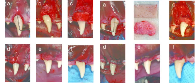
Figure 1. ADM group (Acellular dermal matrix graft).
a) Preoperative recession on a maxillary right canine with a narrow zone of keratinized tissue.
b) Reflection of partial thickness flap and recipient bed preparation.
c) Acellular dermal matrix graft sutured in place with a bioabsorbable suture.
d) Flap coronally sutured to completely cover the acellular dermal matrix graft.
e) Acellular dermal matrix graft at 2 weeks.
f) Acellular dermal matrix graft at 4 weeks.
Figure 2. CT group (Connective tissue graft).
a) Preoperative recession on a maxillary left canine with a narrow zone of keratinized tissue.
b) Comparison of two graft material. (upper;
acellular dermal matrix graft, lower; con- nective tissue graft)
c) Connective tissue graft sutured in place with a bioabsorbable suture material.
d) Coronally positioned flap sutured to com- pletely cover the connective tissue graft.
e) Connective tissue graft at 2 weeks.
f) Connective tissue graft at 4 weeks.
a b c
f e
d
a b c
d e f
treatment. The Mann-Whitney U test was used to determine if one surgical procedure produced better clinical result after 4 weeks. For all statistical analyses, a sig- nificance level of 5% was used.
Ⅲ. Results
1. Clinical evaluation
Uneventful healing was observed through the experimental period in all dogs. Three dogs with six gingival recessions were treat- ed with acellular dermal matrix graft (3 re- cessions) or connective tissue graft (3 re- cessions) associated with coronally posi- tioned flap.
As comparison with the preoperative measurements, small amounts of keratinized tissue and moderate degree of reduction in recession height were observed at 4 weeks after graft surgery in all groups. But there were little change in pocket depth after healing. No significant differences were ob-
served between two groups in the clinical parameters evaluated.
The average initial recession height for the six sites was 6.78±0.71mm. After 4 weeks, the average residual recession height for all the sites was 2.50±0.22mm. (coverage percentage of root coverage = 58.50%±3.80%).
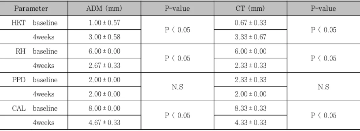
The results of the intragroup analysis are summarized in Table 1. Both treatments produced significant changes from baseline in all the parameters evaluated (p < 0.05) with the exception of PPD.
2. Histological evaluation
In all groups, the overlying epithelium of the gingiva and mucosa as well as the sulcu- lar epithelium and junctional epithelium were similar. No gross inflammatory
reaction was seen, and the inflammatory infiltrate was predominately located within the connective tissue adjacent to the sulcu- lar epithelium in ADM-grafted sites. (Fig 3)
With in the gingiva, the collagen fibers of
Table 1. Gingival changes after ADM graft compared to CT graft.
Parameter ADM (mm) P-value CT (mm) P-value
HKT baseline 1.00 ± 0.57
P < 0.05 0.67 ± 0.33
P < 0.05
4weeks 3.00 ± 0.58 3.33 ± 0.67
RH baseline 6.00 ± 0.00
P < 0.05
6.00 ± 0.00
P < 0.05
4weeks 2.67 ± 0.33 2.33 ± 0.33
PPD baseline 2.00 ± 0.00
N.S 2.33 ± 0.33
N.S
4weeks 2.00 ± 0.00 2.00 ± 0.00
CAL baseline 8.00 ± 0.00
P < 0.05 8.33 ± 0.33
P < 0.05
4weeks 4.67 ± 0.33 4.33 ± 0.33
*Satistically significant at p < 0.05
#Wilcoxon signed rank test before and after treatment.
the host's overlying connective tissue and those of the underlying area corresponding to the grafted area were similarly dense and incorporated such that it was difficult to distinguish one from the other with stand- ard H&E. The attachments to the root sur- faces were similar.(Figure 3 and Figure 4) Adjacent to the tooth and coronal to the osseous crest, beneath the graft, dense col- lagen was generally arranged parallel to the root surface in two groups. But, the colla- gen fiber density and quantity in CT-grafted
sites was considered mostly as 'sparse' while as 'dense' in the ADM-grafted site. As comparison with the autogenous connective tissue samples, a significantly increased bucco-lingual thickness of collagen fibers in the ADM grafted site was noted in the areas of previ-ous graft placement. The osseous crest was present at the apical from the root notch in all groups. And alveolar bone remodeling appearance was observed in all sites. But, the remodeling process appeared to be more active in CT grafted sites
Figure 3. Acellular dermal matrix graft specimen.
a) root notch( ),connective tissue at- tachment( CT ),collagen fiber( CF ), osseous crest( O ).(original magnifica- tion 12.5; H&E)
b) root( R ), cementum( C ). (original magnification 40; H&E)
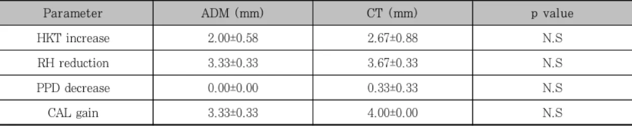
Table 2. Clinical difference between CT group and ADM group after 4 weeks.
Parameter ADM (mm) CT (mm) p value
HKT increase 2.00±0.58 2.67±0.88 N.S
RH reduction 3.33±0.33 3.67±0.33 N.S
PPD decrease 0.00±0.00 0.33±0.33 N.S
CAL gain 3.33±0.33 4.00±0.00 N.S
*Satistically significant at p < 0.05
#Mann-Whitney U test between treatments
Figure 4. Connective tissue graft specimen.
a) root notch( ),connective tissue at- tachment( CT ),collagen fiber( CF ), osseous crest( O ).(original magnifica- tion 12.5; H&E)
b) root ( R ), cementum( C ).blood ves- sel( V ) (original magnification 40)
In one specimen of CT grafted sites, blood vessels were observed between alveolar bone and periodontal ligament. The additional deposition or resorption of cementum was not observed in all specimens of each group.
Ⅳ. Discussion
The main goal of this study was to com- pare the effectiveness of two bilaminar tech- niques; the ADM graft and the autogenous CT graft. The results of present study showed that both periodontal plastic surgical procedures were able to significantly im- prove the clinical parameters evaluated from the preoperative baseline to the 4 weeks postoperative inspection. The only exception was seen for PPD, which did not show sig- nificant changes at the 4 weeks examination in either group. This result was expected, because the baseline PPD was compatible with a condition of gingival health in these experimental animals.
Similar amounts of root coverage were ob- tained with both procedures. When the acel- lular dermal matrix was used, the mean root coverage was 61.33%±5.67%, and when a connective tissue graft was used, the mean root coverage was 55.67%±5.67%.
Those rates of mean root coverage imply that acellular dermal matrix may be a suit- able substitute for autogenous connective tissue grafts in clinical practice.
The reported root coverage in this study is lower than that from other studies, in a comparative study between CT and ADM grafts used in a bilaminar techni- ques16,20,21,33-37
, but well within the range of
reported root coverage.8,10,20,21,38-44. Harris20 reported comparable percentage root cover- ages from both treatments(CT:96.2%, ADM:
95.8%). The connective tissue graft pro- duced a great mean probing reduction and mean keratinized tissue increase than the acellular dermal matrix. However, this did not appear to be clinically significant.
Similarly, no significant differences were re- ported between CT and ADM grafts by Aichelmann-Reidy et al.21 These authors21 observed mean root coverages of 74.1%(CT) and 65.9%(ADM). Paolantonio16 reported mean percentage of root coverages were 88.80% and 83.33 % in the CT and ADM groups. No significant differences were ob- served between the two techniques for gin- gival recession, clinical attachment level.
Relatively lower percentage of root cover- age was due to the lack of postsurgical care with possible graft mobility and inadequate oral hygiene. In order to obtain more pre- dictable results, careful surgical technique and meticulous postsurgical care might be required. During the postsurgical main- tenance period, all patients were routinely instructed not to touch or brush surgical areas and daily chlorhexidine rinse several times a day is critical in human. However, it is impossible to put a routine oral hygiene regimen for the experimental animals. As a result, the graft material could not be main- tained intact over the surgical sites. Some part of the graft material was necrotized and gone away. This event might have con- tributed relatively low percentage of root coverage.
Another issue to mention is how much
time is needed to heal the root coverage surgery in these experimental animals. Rami Guiha et al30 who studied histological heal- ing of subepithelial connective tissue grafts in dogs, reported that demarcation zones were not at all visible and could not be de- tected at 28 days. This is in agreement with the findings of Garguilo and Arrocha et al45 who studied the histological healing of free gingival grafts in human. In addition, the microvasculature of the flap and the graft was almost normal. In the present study, biopsy and clinical measurement were per- formed after 28 days according to the pre- vious study30. In the present study, light microscopical observations show the epi- thelium area contain more chronic in- flammatory cell infiltrate in the specimens of ADM groups than that of CT groups.
These suggest that healing of the ADM have been generally retarded compared to autograft. There are some reports, which have described the retarded healing of the ADM like Wei et al's studies26, 27. In their studies26, 27, the clinical healing of the ADM was generally retarded by approximately 2 weeks when compared to free gingival tissue graft. At the present, there is no report to describe about the complete healing time when ADM graft is applied for root coverage in dogs. Therefore, further study will be fol- lowed to find out the exact time necessary to obtain a mature tissue structure using acellular dermal matrix graft placed under- neath a pedicle flap.
V. Conclusion
The experimental studies on the healing of the ADM disclosed that:
1. As comparison with the preoperative measurements, reduction in recession height and increase in keratinized tissue, improvement in clinical attachment level were observed in all groups.(p≤0.05) 2. The mean root coverage was 61.33±5.67%
(n=3) for ADM group and 55.67±5.67%
(n=3) for CT group.
3. No statistically significant differences were observed between ADM group and CT group in the parameters evaluated.(p
>0.05)
4. In histologic evaluation, all graft materi- als were well incorporated with recipient sites. Also similar healing appearances were observed in two groups.
5. The acellular dermal matrix can be a suitable for autogenous connective tissue grafts for root coverage in the treatment of gingival recession.
References
1. Miller PD Jr. Root coverage grafting for regeneration and aesthetics. Periodontol 2000. 1993;1:118-27.
2. Miller PD. Root coverage using a free soft tissue autograft following citric acid application. part 1: technique. Int J Periodontics Restorative Dent. 1982;2(1) :65-70
3. Wennstrom JL, Lang NP. Proceedings of the 1st European Workshop on Periodontology. London, Quintessence
Publishing Co.Ltd; 1994. p. 193-209.
4. Cummings LC, Kaldahl WB, Allen EP.
Histologic evaluation of autogenous con- nective tissue and acellular dermal ma- trix grafts in humans. J Periodontol.
2005;76(2):178-86.
5. Gottlow J, Karring T, Nyman S. Guided tissue regeneration following treatment of recession-type defects in the monkey.
J Periodontol. 1990;61(11):680-5.
6. Cortellini P, DeSanctis M, Pini Prato G, Baldi C, Clauser C. Guided tissue re- generation procedure using a fibrin-fi- bronectin system in surgically induced recession in dogs. Int J Periodontics Restorative Dent. 1991;11(2):150-63.
7. Cortellini P, Clauser C, Prato GP.
Histologic assessment of new attachment following the treatment of a human buc- cal recession by means of a guided tis- sue regeneration procedure. J Periodontol.
1993;64(5):387-91.
8. Parma-Benfenati S, Tinti C. Histologic evaluation of new attachment utilizing a titanium-reinforced barrier membrane in a mucogingival recession defect. a case report. J Periodontol. 1998;69(7):834-9.
9. Sbordone L, Ramaglia L, Spagnuolo G, De Luca M. A comparative study of free gingival and subepithelial connective tis- sue grafts. Periodontal Case Rep. 1988;
10(1):8-12.
10. Paolantonio M, Di Murro C, Cattabriga A, Cattabriga M. Subpedicle connective tissue graft versus free gingival graft in the coverage of exposed root surfaces. a 5-year clinical study. J Clin Periodontol.
1997 ;24(1):51-6.
11. Langer B, Langer L. Subepithelial con-
nective tissue graft technique for root coverage. J Periodontol. 1985;56(12):715-20.
12. Raetzke PB. Covering localized areas of root exposure employing the "envelope"
technique. J Periodontol. 1985;56(7):397-402.
13. Nelson SW. The subpedicle connective tissue graft. a bilaminar reconstructive procedure for the coverage of denuded root surfaces. J Periodontol. 1987;58(2):95-102.
14. Harris RJ. A comparative study of root coverage obtained with guided tissue re- generation utilizing a bioabsorbable membrane versus the connective tissue with partial-thickness double pedicle graft. J Periodontol. 1997;68(8):779-90.
15. Rateitschak KH, Egli U, Fringeli G.
Recession: a 4-year longitudinal study after free gingival grafts. J Clin Periodontol.
1979;6(3):158-64.
16. Paolantonio M, Dolci M, Esposito P, Archivio D, Lisanti L, Di Luccio e.
Subpedicle acellular dermal matrix graft and autogenous connective tissue graft in the treatment of gingival recessions:
a comparative 1-year clinical study. J Periodontol. 2002; 73(11):1299-307.
17. Muller HP, Eger T. Gingival phenotypes in young male adults. J Clin Periodontol.
1997;24(1):65-71.
18. Muller HP, Eger T, Schorb A. Gingival dimensions after root coverage with free connective tissue grafts. J Clin Periodontol. 1998;25(5):424-30.
19. Cortes Ade Q, Martins AG, Nociti FH Jr, Sallum AW, Casati MZ, Sallum EA.
Coronally positioned flap with or with- out acellular dermal matrix graft in the treatment of Class I gingival recessions:
a randomized controlled clinical study. J
Periodontol. 2004;75(8):1137-44.
20. Harris RJ. A comparative study of root coverage obtained with an acellular der- mal matrix versus a connective tissue graft: results of 107 recession defects in 50 consecutively treated patients. Int J Periodontics Restorative Dent. 2000;20(1) :51-9.
21. Aichelmann-Reidy ME, Yukna RA, Evans GH, Nasr HF, Mayer ET. Clinical evaluation of acellular allograft dermis for the treatment of human gingival recession. J Periodontol. 2001;72(8):998-1005.
22. Tal H. Subgingival acellular dermal ma- trix allograft for the treatment of gin- gival recession: a case report. J Periodontol.
1999;70(9):1118-24.
23. Henderson RD, Greenwell H, Drisko C, Regennitter FJ, Lamb JW. Predictable multiple site root coverage using an acellular dermal matrix allograft. J Periodontol. 2001;72(5):571-82.
24. Wainwright DJ. Use of the AlloDerm in the management of full- . 1995;21(4):243-8.
25. Wei PC, Laurell L, Geivelis M, Lingen MW, Maddalozzo D. Acellular dermal matrix allografts to achieve increased attached gingiva. part 1. a clinical study. J Periodontol 2000;71(8):1297-1305.
26. Batista EL Jr, Batista FC, Novaes AB Jr. Management of soft tissue ridge de- formities with acellular dermal matrix.
clinical approach and outcome after 6 months of treatment. J Periodontol.
2001;72(2):265-73.
27. Oliver RC, Loe H, Karring T. Microscopic evaluation of the healing and revascula- rization of free gingival grafts. J Periodontal Res. 1968;3(2):84-95.
28. Janson WA, Ruben MP, Kramer GM, Bloom AA. Development of the blood supply to split-thickness free ginival autografts.J Periodontol. 1969;40(12):707-16.
29. Barros RR, Novaes AB, Grisi MF, Souza SL, Taba MJ, Palioto DB. A 6-month comparative clinical study of a conven- tional and a new surgical approach for root coverage with acellular dermal matrix. J Periodontol. 2004; 75(10):1350-6.
30. Guiha R, Khodeiry S, Mota L, Caffesse R. Histological evaluation of healing and revascularization of the subepithelial connective tissue graft. J Periodontol.
2001; 72(4):470-8.
31. Bernimoulin JP, Luscher B. Coronally repositioned periodontal flap. clinical evaluation after one year. J Clin Periodontol. 1975; 2(1):1-13.
32. Dodge JR, Henderson RD. Root coverage without a palatal donor site, using an acellular dermal graft. Periodontal Insights 1998; 5(4):5-9.
33. Novaes AB, Grisi DC, Molina GO, Souza SL, Taba M, Comparative 6-month clin- ical study of a subepithelial connective tissue graft and acellular dermal matrix graft for the treatment of gingival recession. J Periodontol 72(11):1477-84.
34. Harris RJ. The connective tissue and partial thickness double pedicle graft: a predictable method of obtaining root coverage. J Periodontol. 1992; 63(5):
477-86.
35. Harris RJ. The connective tissue with partial thickness double pedicle graft:
the results of 100 consecutively-treated defects. J Periodontol. 1994; 65(5):448-61.
36. Levine RA. Covering denuded maxillary
root surfaces with the subepithelial con- nective tissue graft. Compendium. 1991;
2(8):568 -572.
37. Wennstrom JL. Increased gingival dimensions. A significant factor for suc- cessful outcome of root coverage proce- dures? a 2-year prospective clinical study. J Clin Periodontol. 1996; 23(8):
770-7.
38. Baldi C, Pini-Prato G, Pagliaro U, Nieri M, Saletta D, Muzzi L et al. Coronally advanced flap procedure for root coverage. Is flap thickness a relevant predictor to achieve root coverage? a 19-case series. J Periodontol. 1999; 70(9):
1077-84.
39. Borghetti A, Louise F. Controlled clin- ical evaluation of the subpedicle con- nective tissue graft for the coverage of gingival recession. J Periodontol. 1994;
65(12):1107-12.
40. Borghetti A, Glise JM, Monnet-Corti V, Dejou J. Comparative clinical study of a bioabsorbable membrane and sub- epithelial connective tissue graft in the treatment of human gingival recession. J
Periodontol. 1999; 70(2):123-30.
41. Bouchard P, Etienne D, Ouhayoun JP.
Subepithelial connective tissue grafts in the treatment of gingival recessions. a comparative study of 2 procedures. J Periodontol. 1994; 65(10):929-36.
42. Bouchard P, Nilveus R, Etienne D.
Clinical evaluation of tetracycline HCl conditioning in the treatment of gingival recessions. a comparative study. J Periodontol. 1997; 68(3):262-9.
43. Jahnke PV, Sandifer JB, Gher ME, Gray JL, Richardson AC. Thick free gin- gival and connective tissue autografts for root coverage. J Periodontol. 1993;
64(4):315-22.
44. Ricci G, Silvestri M, Tinti C, Rasperini G. A clinical/statistical comparison be- tween the subpedicle connective tissue graft method and the guided tissue re- generation technique in root coverage.
Int J Periodontics Restorative Dent.
1996; 16(6):538-45.
45. Gargiulo AW, Arrocha R. Histo-clinical evaluation of free gingival grafts.
Periodontics. 1967; 5(6):285-91.
-Abstract-
성견에서 ADM(acellular dermal matrix)의 치근피개 효과에 관한 실험적 연구
조민영1, 이승호2,3, 한금아3, 이준영3, 전혜란1,3, 강나라1, 김명래1
이화여자대학교 임상치의학대학원 임플란트학과1,
이화여자대학교 임상치의학대학원 치주과학교실2,
이화여자대학교 의과대학/이화의료원 목동병원 치주과3
결합조직이식을 이용한 치근피개 술식에 많은 관심이 집중되고 있다. 최근에는 acellular dermal matrix 가 자가 결합조직 이식편의 대체물로써 소개되었다. 본 실험의 목적은 치근피개를 위해 acellular dermal matrix을 사용시, 그 임상 효과 및 조직 치유 양상을 평가하고, 이를 자가 결합조직 이식시의 결과와 비교하 기 위함이다.
3마리의 성견에 인위적인 치은 퇴축부 형성을 위해서, 상악 좌우견치의 협측에서 각화치은을 모두 제거하고, 법랑백악경계부로부터 12mm 정도 치조골을 삭제한 후에 판막을 봉합하였다. 그 후 35일을 치유 기간으로 부 여하였다. 총 6 부위의 결손부가 실험에 포함되었고, 각각 3 부위씩이 대조군과 실험군으로 분류되었다. 실험 군에서는 acellular dermal matrix 이식과 치관측 변위 판막을 시행하였고, 대조군에서는 치은 퇴축부위에 자가 결합조직 이식과 치관측 변위 판막을 시행하였다.
치주낭 깊이, 임상적 부착 수준, 치은 퇴축 높이, 각화조직 높이 등을 인위적 결손부 형성전, 치은 피개술 시행 직전, 피개술 시행후 4주 경과시에 각각 측정하였다. 술후 4주시에, 상악 좌우 견치 부위에서 시편을 얻 어 조직학적으로 관찰하고 Wilcoxon signed rank test 와 Mann-Whitney U test으로 통계처리하였다.
임상결과 관찰시, 대조군과 실험군 모두에서, 술전과 비교시 치은 퇴축 감소와 각화조직의 증가, 임상 부착 수준의 개선이 나타났다.(p ( 0.05) 평균 치근 피개율은 실험군에서 61.33(5.67%(n=3), 대조군에서 55.67(5.67%(n=3) 이었고, 대조군과 실험군에서 임상결과에서는 통계학적으로 유의성 있는 차이는 없었 다.(p ( 0.05). 조직학상으로는, 두 군 모두에서 이식편이 수여부에 잘 융화되어 있었고 비숫한 치유 양상을 보였다.
이상의 실험결과에 의하면, Acellular dermal matrix은 치근피개술 시행시에 결합조직 이식편의 대용품 으로 사용할 수 있고 비숫한 피개 결과를 얻을수 있었다.2)
주요어:acellular dermal matrix, 치근피개술식, 치관변위판막술, 결합조직이식