ISSN 0378-6471 (Print)⋅ISSN 2092-9374 (Online)
https://doi.org/10.3341/jkos.2018.59.7.697
Case Report
매독에 동반된 양안 긴장동공의 치료
Treatment of Bilateral Tonic Pupil Associated with Syphilis
문용석⋅양희경⋅황정민
Yong Seok Mun, MD, Hee Kyung Yang, MD, Jeong-Min Hwang, MD, PhD
서울대학교 의과대학 분당서울대학교병원 안과학교실
Department of Ophthalmology, Seoul National University Bundang Hospital, Seoul National University College of Medicine, Seongnam, Korea
Purpose: We report a case of a male with bilateral tonic pupils associated with syphilis, that partially improved after syphilis treatment.
Case summary: A 27-year-old male presented with a 2-month history of near vision impairment. The right and left pupils were 5.5 mm and 6.5 mm in diameter, respectively, in the dark and 5.3 mm and 6.1 mm, respectively, in the light. Both pupils demon- strated light-near dissociation, slow constriction and redilation when looking at near, and constriction after instillation of 0.0625%
pilocarpine. Serological tests were positive for syphilis, while cerebrospinal fluid testing was negative. Two months after treat- ment with intramuscular injection of benzathine penicillin G, his near vision subjectively improved and the right and left pupils were 5.9 mm and 6.4 mm, respectively, in the dark and 4.8 mm and 5.3 mm, respectively, in the light. The size of both pupils de- creased and the pupillary light responses partially improved in both eyes.
Conclusions: Patients with bilateral tonic pupils should have serological tests for syphilis. Recovery of tonic pupils can be ex- pected after early treatment with effective antibiotics.
J Korean Ophthalmol Soc 2018;59(7):697-701 Keywords: Syphilis, Tonic pupil
■Received: 2018. 3. 23. ■ Revised: 2018. 4. 30.
■Accepted: 2018. 6. 21.
■Address reprint requests to Hee Kyung Yang, MD Department of Ophthalmology, Seoul National University Bundang Hospital, #82 Gumi-ro 173beon-gil, Bundang-gu, Seongnam 13620, Korea
Tel: 82-31-787-7379, Fax: 82-31-787-4057 E-mail: [email protected]
* Conflicts of Interest: The authors have no conflicts to disclose.
ⓒ2018 The Korean Ophthalmological Society
This is an Open Access article distributed under the terms of the Creative Commons Attribution Non-Commercial License (http://creativecommons.org/licenses/by-nc/3.0/) which permits unrestricted non-commercial use, distribution, and reproduction in any medium, provided the original work is properly cited.
매독에 동반되는 대표적 동공운동장애인 아르길 로버트 슨(Argyll-Robertson) 동공은 대개 양안의 축동과 함께 대광 반사 소실, 대광-근접반사해리를 특징으로 한다.1,2 긴장동 공은 아르길 로버트슨 동공과 달리 매독에 동반되는 경우 는 적다고 알려져 있다.2-10 기존에 보고된 증례들은 대부분 뇌척수액 venereal diseases research laboratory (VDRL) 검사
를 통해 신경매독을 진단받았으며, 외국의 보고에서 신경매 독 치료 후 단안을 침범한 긴장동공이 호전된 예는 있으나 동공반응과 그 호전 양상에 대해 자세히 기술한 경우는 없 었다.3-12 한편, 중추신경계 침범이 없는 매독 환자에서 발생 한 양안의 긴장동공이 매독 치료 후 호전을 보인 경우는 국 내외에 보고된 적이 없다. 이에 저자들은 양안의 긴장동공 으로 내원하여 중추신경계 침범이 없는 매독으로 진단된 환자에서, 매독 치료 후 긴장동공의 호전을 보인 1예를 경험하였기에 이를 보고하고자 한다.
증례보고
27세 남자 환자가 2개월 전부터 서서히 시작된 근거리 시 력저하를 주소로 내원하였다. 눈부심이 함께 동반되었고 두 통, 안통은 동반하지 않았다. 4년 전 뇌진탕 및 좌측안면마
A
B
C
D
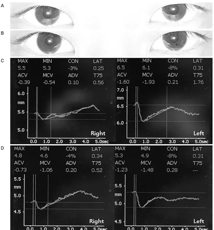
Figure 1. Pupil sizes before and after instillation of 0.0625% pilocarpine. (A) On initial examination, the minimal pupil sizes were
5.3 mm on the right and 6.1 mm on the left. (B) After instillation of 0.0625% pilocarpine, both pupils showed significant con- striction, suggesting cholinergic denervation supersensitivity. (C) Results of digital pupillometry on initial examination showing de- creased constriction (CON) to light. (D) Results of digital pupillometry after instillation of 0.0625% pilocarpine showing decreased pupil sizes in both eyes. MAX = maximum pupil diameter; MIN = minimum pupil diameter; CON = (MAX-MIN)/MAX; LAT= latency; ACV = average constriction velocity; MCV = maximum constriction velocity; ADV = average dilation velocity; T75
= time to reach 75% recovery.
비의 과거력이 있었으며, 같은 해 양안 레이저각막상피절삭 성형술(laser epithelial keratomileusis, LASEK)을 시행받았 다. 그 외 당뇨병, 고혈압과 같은 전신 질환이나 안외상, 안
과 수술의 과거력은 없었다.
원거리 나안시력은 우안 1.2, 좌안 1.2였으며, 근거리 나 안시력은 우안 0.5, 좌안 1.0이었다. 자동굴절검사상 우안
A
B
Figure 2. Pupil sizes after treatment with benzathine penicillin G. (A) Two months after treatment with benzathine penicillin G, mini-
mal pupil sizes decreased to 4.8 mm on the right and 5.3 mm on the left. (B) Results of digital pupillometer two months after treatment show partially improved pupillary light responses (CON, MCV, ACV) in both eyes. MAX = maximum pupil diameter; MIN = mini- mum pupil diameter; CON = (MAX-MIN)/MAX; LAT = latency; ACV = average constriction velocity; MCV = maximum con- striction velocity; ADV = average dilation velocity; T75 = time to reach 75% recovery.+0.50 Dsph. -0.50 Dcyl. × 180˚, 좌안 0.00 Dsph. -0.25 Dcyl.
× 180˚였고, 안압은 우안 8 mmHg, 좌안 9 mmHg였다. 반대 눈을 가리고 시표를 원거리에서 근거리로 옮길 때 시표가 흐려보이기 시작하는 지점을 조절근점으로 정의하였으며, 우안의 조절근점은 35 cm로 조절력이 저하되었고, 근거리 시력은 +2.50 D의 볼록 렌즈를 대면 1.0으로 교정되었다. 교 대가림 검사상 원거리와 근거리 모두 정위였고, 눈운동은 정상이며 눈을 움직일 때 동반되는 통증은 없었다. 세극등 현미경검사상 각결막 및 수정체에 이상 소견이 없었고, 전 방에 염증세포 및 홍채의 부분마비는 관찰되지 않았다. 자 동동공측정계(PLR-200, NeurOptics Inc., Irvine, CA, USA)를 이용하여 측정한 양안의 동공크기는 원거리를 주시한 상태 에서 실내등을 껐을 때(dark) 우안 5.5 mm, 좌안 6.5 mm, 실 내등을 켰을 때(light) 우안 5.3 mm, 좌안 6.1 mm로 동공수 축비율이 우안 3%, 좌안 8%로 양안 대광반사가 저하되었 고, 근접반사에서는 느린 동공의 수축과 확대를 보이는 대 광-근접반사해리를 보였다. 0.0625% 필로카르핀을 점안했 을 때 동공크기는 양안 모두 감소하였다(Fig. 1).
양안의 긴장동공 진단하에 전신질환을 감별하기 위해 시 행한 혈액검사에서 rapid plasma reagin (RPR) 양성, fluo- rescent treponemal antibody absorption (FTA-ABS) 양성으로
매독이 확진되었다. 피부 병변이나 기타 중추신경계 이상은 동반되지 않았고, 뇌척수액 검사에서 VDRL 음성, 뇌척수액 백혈구 증가증은 보이지 않았으며 동반된 신경학적 징후는 없었다. 후천성면역결핍증후군 동반을 감별하기 위해 실시 한 항체 검사는 음성이었다.
Benzathine penicillin 240만 단위를 1주 간격으로 3회 근 주하였으며, 치료 2개월 후 환자의 주관적인 근거리 시력저 하 증상은 호전되었다. 동공크기는 원거리를 주시한 상태에 서 실내등을 껐을 때(dark) 우안 5.9 mm, 좌안 6.4 mm, 실내 등을 켰을 때(light) 우안 4.8 mm, 좌안 5.3 mm로 동공수축 비율이 우안 18%, 좌안 16%로 호전되었고, 치료 전에 비해 대광 반사와 평균 동공수축속도가 호전되었다(Fig. 2).13
고 찰
신경매독에서 나타나는 전형적인 동공운동 장애는 양안 의 동공 축동과 대광-근접반사해리를 보이는 아르길-로버트 슨 동공이다.1 한편, 긴장동공은 대광-근접반사해리를 보이 는 점에서 아르길-로버트슨 동공과 유사하나, 연령, 성별, 동 공 크기, 양측 이환율 등에서 아르길-로버트슨 동공과 다르 며, 80% 이상이 단안에 발생하고, 근접반사 시 느리고 과도
한 축동, 홍채의 부분마비로 인한 벌레모양운동(vermiform movement), 저농도의 부교감신경 자극제에 대한 과민반응 등을 특징적으로 보인다.2,4
Sakai et al4은 신경매독에 동반된 양안 긴장동공 총 4예 를 보고하였다. 이 중 2예는 양안 시신경병증, 1예는 보행장 애와 건반사소실, 1예는 눈운동장애를 동반하고 있었으며, 4예 모두 매독 치료 후에도 동공운동장애는 호전되지 않았 다. Fletcher and Sharpe3도 신경매독에 동반된 양안 긴장동 공 4예와 잠복매독에 동반된 양안 긴장동공 1예를 보고하 였으며, 5예 모두 매독 치료 후에도 긴장동공은 호전되지 않았다. Englestein et al5이 보고한 신경매독에 동반된 양안 긴장동공 1예 또한 치료 후에도 호전되지 않았다. Gu et al11 은 신경매독에 동반된 좌안의 긴장동공을 보고하였며, 매독 치료 9개월 후 조명하에 좌안 동공 크기는 치료 전보다 5 mm 감소하였으나 대광반사는 회복되지 않았다. Takata et al12은 신경매독에 동반된 좌안의 긴장동공 1예를 보고하였 으며, 매독 치료 6개월 후 대광반사가 회복되고 좌안 동공 크기는 치료 전보다 2 mm 감소하였다. 이처럼 대부분의 보 고에서 매독 치료 후 동공반응의 호전을 보이지 않았으며, 치료 후 호전을 보인 기존 보고 2예는 신경매독에 동반되어 단안에 발생하였으며, 동공반응의 회복 양상에 대한 자세한 기술이 없다.11,12
긴장동공은 섬모체신경절(ciliary ganglion) 또는 신경절후 부교감신경(postganglionic parasympathetic fibers)의 손상에 의해 유발되는 것으로 알려져 있다.6,14,15 긴장동공의 발생 원인은 대부분 불명인 경우가 많으나 외상, 종양, 수술에 의 한 신경 손상, 안와 연조직염, 바이러스 감염, 매독감염, 자 율신경병증 등에 의해 발생할 수 있으며, 바이러스 감염의 경우 혈청검사에서 역가가 상승되어 있지 않은 경우가 많 다.3,16 특히 양안에 동시에 발생한 긴장동공의 경우 매독 외 에 라임병, 편두통, 쇼그렌증후군, 밀러-피셔증후군, 신경사 르코이드증, 신경모세포종, 히르슈슈프룽병 등의 가능성이 있으므로 관련된 검사를 해야 한다.17-23
매독 감염에 의한 긴장동공은 명확한 병태생리가 알려져 있지 않으며, 일부 문헌에서 매독이 섬모체신경절 또는 신 경절후 부교감신경에 염증이나 허혈을 유발하여 손상시켰 을 것으로 추측하고 있다.3,4 대부분의 보고에서 매독치료 후 동공반응의 호전을 보이지 않았으나,3-10 증상 발생 후 1 개월 만에 시행한 매독 치료 후 긴장동공이 회복되었다고 보고한 외국의 두 증례와 2개월 만에 치료하여 부분적인 호 전을 보인 이 증례를 통해, 매독에 의해 직접 또는 간접적인 섬모체신경절 또는 신경절후 부교감신경에 가역적인 손상 이 발생하여, 이를 조기에 치료하면 회복될 수 있음을 시사 한다.11,12
매독에 의한 긴장동공의 경우, 80% 이상이 단안에 발생 하는 특발성 긴장동공과 달리 대부분 양안에 발생하여, 이 증례와 앞서 보고된 10예 모두 양안을 침범하였고 단안 침 범은 2예 뿐이었다.3-5,11,12 일반적인 긴장동공의 경우 반대 안 이환율이 매년 4% 정도로 낮은 것을 감안하면, 매독에 의한 긴장동공은 양안의 섬모체신경절 또는 신경절후 부교 감신경을 침범하는 전신적인 병태생리학적 기전을 추측해 볼 수 있다.24 다만 이 증례는 앞선 보고들과 달리 뇌척수액 검사를 통해 신경매독이 음성이고 다른 신경학적 증상이나 징후를 동반하지 않았기에, 긴장동공이 매독에 의한 직접적 인 중추신경계 침범 뿐 아니라 간접적인 말초신경병증의 가능성이 있음을 시사한다.
결론적으로 양안에 동시에 발생한 긴장동공이 관찰되었 을 때, 매독을 포함한 전신질환의 가능성을 염두에 두고 혈 액검사를 시행해야 하고, 뇌척수액 검사를 통해 신경매독을 감별해야 한다. 또한 매독의 진단과 동시에 조기에 적절한 항생제를 투여하면 긴장동공의 부분적인 회복을 기대할 수 있다.
REFERENCES
1) Loewenfeld IE. The Argyll Robertson pupil 1869-1969. A critical survey of the literature. Surv Ophthalmol 1969;14:199-299.
2) Thompson HS, Kardon RH. The argyll robertson pupil. J Neuroophthalmol 2006;26:134-8.
3) Fletcher WA, Sharpe JA. Tonic pupils in neurosyphilis. Neurology 1986;36:188-92.
4) Sakai T, Shikishima K, Mizobuchi T, et al. Bilateral tonic pupils as- sociated with neurosyphilis. Jpn J Ophthalmol 2003;47:368-71.
5) Englestein ES, Ruderman MI, Troiano RA, Digiovanni VJ. Dilated tonic pupils in neurosyphilis. J Neurol Neurosurg Psychiatry 1986;49:1455-7.
6) Thompson HS. Adie's syndrome: some new observations. Trans Am Ophthalmol Soc 1977;75:587-626.
7) Bowsher D, Lahuerta J. A case of tabes dorsalis with tonic pupils and lightning pains relieved by sodium valproate. J Neurol Neurosurg Psychiatry 1987;50:239-41.
8) Yasaki S, Ohshima J, Yonekura J, et al. A case of early syphilis pre- senting general paresis-like symptoms and bilateral tonic pupils.
Rinsho shinkeigaku 1992;32:994-9.
9) Ohya Y, Matsumura T, Kojima S, et al. Bilateral internal carotid ar- tery stenoses in a patient with meningovascular neurosyphilis.
Rinsho shinkeigaku 1993;33:875-9.
10) Kim HB, Kwon OW, Chun GH. Bilateral tonic pupil in syphilis. J Korean Ophthalmol Soc 1983;24:877-81.
11) Gu X, Guan Z, Chai Z, Zhou P. Unilateral mydriasis as the primary sign of neurosyphilis. Infection 2014;42:215-7.
12) Takata T, Kamada M, Ikeda K, et al. Unilateral mydriatic tonic pu- pil as an early isolated symptom of neurosyphilis. J Neurol Sci 2014;344:219-20.
13) Bak E, Yoo YJ, Yang HK, Hwang JM. Quantitative pupillometry of
= 국문초록 =
매독에 동반된 양안 긴장동공의 치료
목적: 매독 환자에서 발현된 양안의 긴장동공이 매독 치료 후 부분적인 호전을 보인 1예를 경험하였기에 이를 보고하고자 한다.
증례요약: 27세 남자가 2개월 전부터 시작된 근거리 시력저하를 주소로 내원하였다. 양안의 동공크기는 실내등을 껐을 때 우안 5.5 mm, 좌안 6.5 mm, 실내등을 켰을 때 우안 5.3 mm, 좌안 6.1 mm였으며 동공반응은 대광-근접반사해리를 보이며 근거리 주시 때 느린 동공의 수축과 확대, 그리고 0.0625% 필로카르핀 점안 후 양안의 동공이 축동되었다. 혈청검사상 매독이 확진되었다. Benzathine penicillin G를 근주하고 2개월 경과 후 주관적인 근거리 시력저하 증상이 호전되었으며, 동공크기는 실내등을 껐을 때 우안 5.9 mm, 좌안 6.4 mm, 실내등을 켰을 때 우안 4.8 mm, 좌안 5.3 mm로 일부 호전되었고, 양안 대광반사도 부분적으로 호전되었다.
결론: 양안 긴장동공 관찰 시 혈청검사를 통해 매독 여부를 확인해야 하며, 조기에 적절한 항생제를 투여하면 긴장동공의 상대적으로 빠른 회복을 기대할 수 있다.
<대한안과학회지 2018;59(7):697-701>
the pupillary light reflex in Koreans. J Korean Ophthalmol Soc 2017;58:712-7.
14) Loewenfeld IE, Thompson HS. The tonic pupil: a re-evaluation.
Am J Ophthalmol 1967;63:46-87.
15) Miller NR, Walsh FB, Hoyt WF. Walsh and Hoyt's clinical neu- ro-ophthalmology, 6th ed. Vol. 1. Philadelphia: Lippincott Williams
& Wilkins, 2005; 761-4.
16) Wabbels BK, Elflein H, Lorenz B, Kolling G. Bilateral tonic pupils with evidence of anti-hu antibodies as a paraneoplastic manifes- tation of small cell lung cancer. Ophthalmologica 2004;218:141-3.
17) Fugimoto F, Ghanem RC, Monteiro ML. Bilateral tonic pupil as the only remaining ophthalmic sign of Lyme disease: case report.
Arq Bras Oftalmol 2005;68:381-4.
18) Millar E, Habib M, Gnanaraj L. Bilateral tonic pupil secondary to migraine in a child. J Pediatr Ophthalmol Strabismus 2010;47 Online:e1-2.
19) Vetrugno R, Liguori R, Cevoli S, et al. Adie's tonic pupil as a mani- festation of Sjögren's syndrome. Ital J Neurol Sci 1997;18:293-5.
20) Bae JS, Kim JK, Kim SH, Kim OK. Bilateral internal oph- thalmoplegia as an initial sole manifestation of Miller Fisher syndrome. J Clin Neurosci 2009;16:963-4.
21) Sevketoglu E, Tatlı B, Tuğcu B, et al. An unusual cause of ful- minant Guillain-Barré syndrome: angel's trumpet. Pediatr Neurol 2010;43:368-70.
22) Heuser K, Kerty E. Neuro-ophthalmological findings in sarcoidosis. Acta Ophthalmol Scand 2004;82:723-9.
23) Lambert SR, Yang LL, Stone C. Tonic pupil associated with con- genital neuroblastoma, Hirschsprung disease, and central hypo- ventilation syndrome. Am J Ophthalmol 2000;130:238-40.
24) Holmes G. Partial iridoplegia associated with symptoms of other disease of the nervous system. Tran Ophthal Soc UK 1931;51:
209-28.