Korean J Gastroenterol Vol. 73 No. 2, 114-117 https://doi.org/10.4166/kjg.2019.73.2.114 pISSN 1598-9992 eISSN 2233-6869
IMAGE OF THE MONTH
Korean J Gastroenterol, Vol. 73 No. 2, February 2019 www.kjg.or.kr
정맥경화성 대장염
소호심, 천재영
서울대학교 의과대학 내과학교실
Phlebosclerotic Colitis
Hosim Soh and Jaeyoung Chun
Department of Internal Medicine, Seoul National University College of Medicine, Seoul, Korea
CC This is an open access article distributed under the terms of the Creative Commons Attribution Non-Commercial License (http://creativecommons.org/licenses/
by-nc/4.0) which permits unrestricted non-commercial use, distribution, and reproduction in any medium, provided the original work is properly cited.
Copyright © 2019. Korean Society of Gastroenterology.
교신저자: 천재영, 03080, 서울시 종로구 대학로 101, 서울대학교 의과대학 내과학교실
Correspondence to: Jaeyoung Chun, Department of Internal Medicine, Seoul National University College of Medicine, 101 Daehak-ro, Jongno-gu, Seoul 03080, Korea.
Tel: +82-2-740-8112, Fax: +82-2-743-6701, E-mail: [email protected], ORCID: https://orcid.org/0000-0002-4212-0380 Financial support: None. Conflict of interest: None.
증례: 79세 남자가 9개월 전부터 시작된 복통과 간헐적인 혈성 설사를 주소로 내원하였다. 환자는 10년 전 협심증으로 관상동맥우회술을 받은 바 있고, 고혈압, 당뇨로 carvedilol, amlodipine, losartan, atorvastatin, linagliptin, glimepiride 복용 중에 있었으며, 1개월 전 요로 결석으로 진단받아 신절 석술(nephrolithotomy)을 시행받은 바 있었다.
내원 당시 활력징후는 139/64 mmHg, 맥박수 65회/분, 체 온 36.4°C로 측정되었다. 신체검진에서 복부는 편평하고 부 드러웠으며 압통은 없었고, 촉지되는 종괴는 없었다. 말초혈 액 검사에서 백혈구 6,700/μL, 혈색소 7.9 g/dL, 헤마토크리 트 24.1%, 혈소판 147,000/mm3, CRP 1.03 mg/dL, 혈액요소 질소(BUN) 31 mg/dL, 혈청 크레아티닌 2.18 mg/dL, 혈청 철분 20 μg/dL, 페리틴 29.2 ng/mL, 총철결합능 214 μg/dL 였다. 복부 조영 CT 검사에서 결장 정맥의 석회화와 함께 상 행 결장과 횡행 결장 벽내 석회화 및 결장 벽의 비후가 발견되 었다(Fig. 1). 대장 내시경에서 맹장부터 중간 S자 결장까지 점막의 색소 침착과 흰색 반흔이 관찰되었고, 맹장부터 중간 횡행 결장까지는 점막 주름의 소실과 일부 출혈 및 삼출물을 동반한 궤양이 다발적으로 관찰되었다(Fig. 2). 대장 점막 조 직 생검 검사에서는 일부 점막의 탈락과 유리질화된 점막고유 층이 확인되어 허혈성 대장염에 합당한 소견이 확인되었다
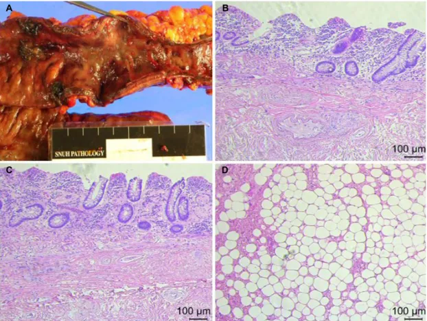
(Fig. 3). 상기 검사 소견을 모두 종합하였을 때, 환자는 정맥 경화성 대장염(phlebosclerotic colitis)으로 진단되었고, 철 결핍성 빈혈과 만성 신질환에 동반된 빈혈에 대하여 경구 철 분제와 에리스로포이에틴(erythropoietin) 자극제 처방 후 퇴 원하였다. 이후 외래에서 추적 관찰하였으나 빈혈에 호전이 없었으며 혈색소 감소를 동반한 대량 혈변이 반복되어 5개월 후 대장아전절제술(subtotal colectomy)을 시행하였다. 수술 검체에서 육안적으로 대장 벽의 비후와 함께 궤양 및 대장 팽대의 소실이 관찰되었으며, 현미경적으로 점막의 탈락, 점 막고유층의 유리질화 및 점막하층의 섬유화, 혈관 내 석회화, 림프절의 대상성 비대가 관찰되었고 장간막 내 지방층염과 만 성 염증이 동반된 섬유 지방 조직이 확인되었다(Fig. 4). 환자 는 수술 후 합병증 없이 퇴원하였으며, 이후 증상 재발 없이 외래 추적 관찰 중에 있다.
진단: 정맥경화성 대장염
허혈성 대장염(ischemic colitis)은 주로 고령 및 기저 질환 이 있는 환자에서 호발하는 질환으로, 증상은 갑작스럽게 발 생하는 복통, 설사, 혈변 등이 있다.1정맥경화성 대장염은 허 혈성 대장염의 한 형태로 장간막 정맥의 석회화가 동반된 대 장 정맥의 울혈로 발생하는 드문 질환이다. 아직 정확한 병인
Soh H and Chun J. Phlebosclerotic Colitis
115
Vol. 73 No. 2, February 2019 A B
Fig. 1. Contrast-enhanced abdominal computed tomography (CT). (A, B) CT images showed bowel wall thickening with intramural calcification in the ascending and transverse colon with calcifications in the colic vein.
A B
C D
Fig. 2. Colonoscopic findings. (A-C) Loss of haustral marking and friable mucosa with multiple exudative ulcers from the ascending to proximal transverse colon and (D) dark colored mucosa with whitish scars in the mid-sigmoid colon was noted.
116
소호심, 천재영. 정맥경화성 대장염The Korean Journal of Gastroenterology
Fig. 4. Pathologic findings of surgical specimen. (A) Gross findings of the resected colon showed thickened colon wall with loss of haustra and several ulcers with exudates. (B, C) Microscopic findings of the colon revealed mucosal sloughing, hyalinized lamina propria and submucosal fibrosis with dystrophic calcification in blood vessels (H&E, ×100). (D) Microscopic findings of the omentum showed fibroadipose tissue with chronic inflammation and panniculitis in omental fat (H&E, ×100).
Fig. 3. Pathologic findings of biopsy specimen from the colonic mucosa showed mucosal sloughing and hyalinized lamina propria, consistent with ischemic colitis (H&E, ×50).
과 발병 기전이 명확하게 규명되지는 않았으나, 장간막 정맥 의 석회화로 인한 대장 정맥의 배출 장애가 허혈성 장염을
유발하는 것으로 생각되며,2,3 장간막 정맥 석회화는 당뇨, 심 장부전, 만성 신기능 저하 및 간경화 등의 만성 질환에 의하여 장기간 복강내 정맥압이 상승하여 발생하는 이차적인 현상으 로 생각된다.3-5이 외에도 화학적 자극이나 약제, 대장 내강의 압력 증가 등이 제안되고 있으나 아직 확실하게 밝혀진 병인 은 없는 상태이다.6-8
정맥경화성 대장염의 주된 증상은 만성 복통과 설사이며 중증인 경우 만성 빈혈을 동반한 혈변, 장폐색 및 장천공으로 나타나기도 한다.9,10 주로 우측 대장을 침범하나 전 대장을 침범하였다는 증례보고도 있었으며 침범된 대장의 부위와 길 이에 따라 다양한 임상 양상을 보인다.11 방사선 검사에서는 복부 단순촬영에서 석회화가 관찰되기도 하며 복부 전산화단 층촬영에서 장간막 정맥의 다발성 석회화 및 장벽의 비후가 발견된다. 혈관조영술 소견에서는 드러몬드 연동맥(marginal artery of Drummond)의 협착과 직혈관(vasa recta)의 사행 성 주행 및 직혈관을 따라 동반되는 정맥의 확장이 관찰된다.2 대장 내시경에서는 주로 우측 대장을 침범하는 점막의 청자색 또는 암적색 침착과 내강의 협착, 다발성의 미란 또는 궤양
A B
C D
Soh H and Chun J. Phlebosclerotic Colitis
117
Vol. 73 No. 2, February 2019
및 결장반월판 주름의 소실이 발견된다.12 병리 소견은 다양하 나 대체적으로 말초 정맥벽의 비후와 섬유화, 유리화 및 석회 화가 발견되며 장 점막의 비후와 점막하 섬유화가 동반된다.3 정맥경화성 대장염의 치료는 그 중등도와 질병의 경과, 혈 류 공급 정도와 장 점막의 손상에 따라 보존적 또는 수술적 치료를 결정하게 된다. 과거에는 대부분 수술적 치료를 요하 였으나 최근 점막 손상이 심하지 않을 경우 보존적 치료로도 호전된다고 보고된 바 있다.13 수술적 치료로는 침범 정도에 따라 부분대장절제술, 대장아전절제술 또는 대장전절제술을 시행하며 수술 후 비교적 좋은 예후를 보이는 것으로 알려져 있다.14
REFERENCES
1. Brandt LJ, Boley SJ. AGA technical review on intestinal ischemia.
American gastrointestinal association. Gastroenterology 2000;
118:954-968.
2. Yao T, Iwashita A, Hoashi T, et al. Phlebosclerotic colitis: value of radiography in diagnosis--report of three cases. Radiology 2000;214:188-192.
3. Choi JM, Lee KN, Kim HS, et al. Idiopathic phlebosclerotic colitis:
a rare entity of chronic ischemic colitis. Korean J Gastroenterol 2014;63:183-186.
4. Kang HY, Noh R, Kim SM, Shin HD, Yun SY, Song IH. Phlebosclerotic colitis in a cirrhotic patient with portal hypertension: the first case in Korea. J Korean Med Sci 2009;24:1195-1199.
5. Song JH, Kim JI, Jung JH, et al. A case of phlebosclerotic colitis in a hemodialysis patient. Korean J Gastroenterol 2012;59:
40-43.
6. Chang KM. New histologic findings in idiopathic mesenteric phle- bosclerosis: clues to its pathogenesis and etiology--probably in- gested toxic agent-related. J Chin Med Assoc 2007;70:227-235.
7. Yeh HJ, Lin PY, Kao WY, Kun CH, Chang CC. Idiopathic mesenteric phlebosclerosis associated with long-term use of Chinese herbal medicine. Turk J Gastroenterol 2018;29:140-142.
8. Hiramatsu K, Sakata H, Horita Y, et al. Mesenteric phlebo- sclerosis associated with long-term oral intake of geniposide, an ingredient of herbal medicine. Aliment Pharmacol Ther 2012;
36:575-586.
9. Kato T, Miyazaki K, Nakamura T, Tan KY, Chiba T, Konishi F.
Perforated phlebosclerotic colitis--description of a case and re- view of this condition. Colorectal Dis 2010;12:149-151.
10. Iwashita A, Yao T, Schlemper RJ, et al. Mesenteric phlebo- sclerosis: a new disease entity causing ischemic colitis. Dis Colon Rectum 2003;46:209-220.
11. Chen MT, Yu SL, Yang TH. A case of phlebosclerotic colitis with in- volvement of the entire colon. Chang Gung Med J 2010;33:
581-585.
12. Hu P, Deng L. Phlebosclerotic colitis: three cases and literature review. Abdom Imaging 2013;38:1220-1224.
13. Yu CJ, Wang HH, Chou JW, et al. Phlebosclerotic colitis with non- surgical treatment. Int J Colorectal Dis 2009;24:1241-1242.
14. Fang YL, Hsu HC, Chou YH, Wu CC, Chou YY. Phlebosclerotic col- itis: a case report and review of the literature. Exp Ther Med 2014;7:583-586.