PGHN
Original Article
Association between Gastric pH and Helicobacter pylori Infection in Children
Ji-Hyun Seo, Heung Keun Park, Ji Sook Park, Jung Sook Yeom, Jae-Young Lim, Chan-Hoo Park, Hyang-Ok Woo, Hee-Shang Youn, Jin-Su Jun, Gyung-Hyuck Ko*, Seung-Chul Baik
†,
Woo-Kon Lee
†, Myung-Je Cho
†, and Kwang-Ho Rhee
†Department of Pediatrics, Gyeongsang National University Hospital and Institute of Health Sciences, Gyeongsang National University School of Medicine, Departments of *Pathology and †Microbiology, Institute of Health Sciences, Gyeongsang National University School of Medicine, Jinju, Korea
Purpose: To assess gastric pH and its relationship with urease-test positivity and histological findings in children with Helicobacter pylori infection.
Methods: Fasting gastric juices and endoscopic antral biopsy specimens were collected from 562 children and sub- jected to the urease test and histopathological examination. The subjects were divided into 3 age groups: 0-4, 5-9, and 10-15 years. The histopathological grade was assessed using the Updated Sydney System, while the gastric juice pH was determined using a pH meter.
Results: The median gastric juice pH did not differ significantly among the age groups (p=0.655). The proportion of individuals with gastric pH >4.0 was 1.3% in the 0-4 years group, 6.1% in the 5-9 years group, and 8.2% in 10-15 years (p=0.101). The proportions of moderate and severe chronic gastritis, active gastritis, and H. pylori infiltration increased with age (p<0.005). Urease-test positivity was higher in children with hypochlorhydria (77.8%) than in those with normal gastric pH (31.7%) (p<0.001). Chronic and active gastritis were more severe in the former than the latter (p<0.001), but the degree of H. pylori infiltration did not differ (20.9% vs. 38.9%; p=0.186).
Conclusion: Gastric pH while fasting is normal in most children regardless of age. Urease-test positivity may be related to hypochlorhydria in children, and hypochlorhydria is in turn related to H. pylori infection.
Key Words: Urease test, Helicobacter pylori, Child, Gastric juice, Hypochlorhydria
Received:July 25, 2015, Revised:September 1, 2015, Accepted:September 15, 2015
Corresponding author: Hee-Shang Youn, Department of Pediatrics, Gyeongsang National University Hospital, 79 Gangnam-ro, Jinju 52727, Korea. Tel: +82-55-750-8158, Fax: +82-55-752-9339, E-mail: [email protected]
Copyright ⓒ 2015 by The Korean Society of Pediatric Gastroenterology, Hepatology and Nutrition
This is an openaccess article distributed under the terms of the Creative Commons Attribution NonCommercial License (http://creativecommons.org/licenses/by-nc/4.0/) which permits unrestricted noncommercial use, distribution, and reproduction in any medium, provided the original work is properly cited.
INTRODUCTION
Helicobacter pylori causes a chronic, persistent in-
fection that may lead to chronic gastritis, atrophic gastritis, intestinal metaplasia, and even gastric ad- enocarcinoma [1,2]. Primary H. pylori infection oc-
curs during early childhood, with most adults in de- veloping countries eventually becoming carriers [3,4]. Although the stomach is considered a sterile organ because of the acidic conditions (pH<4) [5], H. pylori can survive in the human stomach by acti- vating a cytoplasmic urease, which converts urea in- to carbon dioxide and ammonia and thereby in- creases the pH of the environment [6]. The urease test used to diagnose H. pylori infection is based on this activity of the microbe: the urease test uses phe- nol red, which changes from yellow to pink or red as the pH increases [7]. In children, the diagnosis of H.
pylori infection is usually based on the findings of histological examination and the urease test [8].
A previous study identified the sampling site of gastric biopsy examination, histopathologic find- ings, and the H. pylori load as factors that influence the results of the urease test [9]. Further, several conditions can lead to false-negative results in the urease test, the two most common of which are the recent use of proton-pump inhibitors (PPIs) and the presence of intestinal metaplasia [7]. Both these conditions are associated with low gastric acidity.
Thus, the pH of gastric juice may be related to the re- sults of the urease test. In the present study, we as- sessed the pH of gastric juice in children aged 0-15 years and investigated the correlation between gas- tric pH and the results of the urease test and histo- pathologic examination.
MATERIALS AND METHODS
Study population
The institutional review board approved the re- search protocols in the present retrospective study (GNUHIRB-2015-07-016), and 562 children with up- per abdominal pain who underwent endoscopy of the upper digestive tract and gastric juice aspira- tion via endoscopy at the Department of Pediatrics, Gyeongsang National University Hospital (Jinju, Korea), were enrolled. Endoscopy was performed af- ter overnight fasting, and the indication of endos- copy was upper abdominal pain. No participant was taking PPIs before endoscopy, and no active bleeding
was observed during endoscopy. The study pop- ulation was stratified into 3 age groups: 0-4 years (n=75), 5-9 years (n=244), and 10-14 years (n=243).
Measurement of gastric pH
The pH of gastric juice was measured using a Metter Toledo Delta 320 pH meter (Metter-Toledo International Inc., Columbus, OH, USA) immedi- ately after it was thawed. To ensure accuracy, the pH was adjusted using 3 buffer systems (pH 4.00, 7.00, and 10.01). Hypochlorhydria was defined as a gastric juice pH of ≥4.0 [5].
Urease test
A clinical diagnosis of H. pylori infection was ob- tained by conducting the urease test and histopatho- logical analysis on gastric antral biopsy samples.
Urease tests were performed in the endoscopy room according to a previous method [9]. Briefly, three bi- opsy specimens from the antrum were incubated in a 2% urea broth (20 g/L urea, 0.04 g/L phenol red, 0.2 g/L KH2PO4, 0.5 g/L NaCl; pH 6.8), and if color change occurred in the following 24 hours, the biopsy was deemed to be urease test positive.
Histopathologic findings
Histopathological examination was performed us- ing biopsy specimens that were fixed in 10% buffered formalin overnight, processed for paraffin embed- ding, cut into 4- to 5-μm thick sections, and stained with hematoxylin-eosin. The histological results were interpreted using the Updated Sydney System [10]. For this, the degrees of chronic gastritis, active gastritis, and H. pylori infiltration were classified as normal, mild, moderate, or marked.
Statistical analysis
Data were analyzed using IBM SPSS Statistics ver.
21.0 for Windows (IBM Co., Armonk, NY, USA).
Variation in urease-test positivity, histopathologic findings, and gastric pH depending on age group was evaluated. Statistically significant differences in ure- ase-test positivity and gastric pH among the 3 age groups and in urease-test positivity and the degrees
Fig. 1. Comparison of gastric juice pH among the 3 age groups.
The median pH was 1.77 in the 0-4 years group, 1.59 in the 5-9 years group, and 1.55 in the 10-15 years group (p=0.655).
Table 1. Comparison of Urease-test Positivity and Histopathologic Findings between Subjects with Normal Gastric pH and Hypochlorhydria
pH of gastric juice <4.0 pH of gastric juice ≥4.0 p-value Age group (yr)
0–4 5–9 10–15
74 (98.7) 229 (93.9) 223 (91.8)
1 (1.3) 15 (6.1) 20 (8.2)
0.101
Urease test Positive Negative
167 (31.7) 359 (68.3)
28 (77.8) 8 (22.2)
<0.001
Histopathologic findings Chronic gastritis Normal Mild Moderate Severe
6 (1.1) 403 (76.6) 93 (17.7) 24 (4.6)
0 (0) 12 (33.3) 16 (44.4) 8 (22.2)
<0.001
Active gastritis Normal Mild Moderate Severe
427 (81.2) 79 (15.0) 16 (3.0) 4 (0.8)
14 (38.9) 15 (41.7) 7 (19.4)
0 (0)
<0.001
Helicobacter pylori infiltration Normal
Mild Moderate Severe
416 (79.1) 85 (16.2) 22 (4.2) 3 (0.6)
22 (61.1) 14 (38.9)
0 (0) 0 (0)
0.186
Values are presented as number (%).
of chronic and active gastritis and H. pylori infiltra- tion between those with normal gastric pH and hy- pochlorhydria were determined by the χ2 test and generalized linear models. Nonparametric tests were used to analyze the mean gastric juice pH in different age groups. p-values of <0.05 were considered stat- istically significant.
RESULTS
Gastric juice pH
The median pH of the gastric juice was 1.77 in the 0-4 years group, 1.59 in the 5-9 years group, and 1.55 in the 10-15 years group (Fig. 1). No differences were found in the range of gastric pH among the 3 age groups (p=0.655). The proportion of individuals with hypochlorhydria was 1.3% (1/75) in the 0-4 years group, 6.1% (15/244) in the 5-9 years group, and 8.2% (20/243) in the 10-15 years group (p=0.101;
Table 1).
Comparison of urease-test positivity and histopathologic findings between subjects with hypochlorhydria and normal gastric pH
Table 1 shows a comparison of urease-test pos-
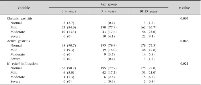
Table 2.Distribution of the Degrees of Chronic and Active Gastritis and Helicobacter pylori Infiltration Depending on Age
Variable Age group
p-value
0–4 years 5–9 years 10–15 years
Chronic gastritis Normal Mild Moderate Severe
2 (2.7) 63 (84.0) 10 (13.3) 0 (0)
1 (0.4) 190 (77.9) 43 (17.6) 10 (4.1)
3 (1.2) 162 (66.7) 56 (23.0) 22 (9.1)
0.003
Active gastritis Normal Mild Moderate Severe
68 (90.7) 7 (9.3) 0 (0) 0 (0)
195 (79.9) 39 (16.0) 9 (3.7) 1 (0.4)
178 (73.3) 48 (19.8) 14 (5.8) 3 (1.2)
0.046
H. pylori infiltration Normal
Mild Moderate Severe
68 (90.7) 6 (8.0) 1 (1.3) 0 (0)
195 (79.9) 42 (17.2) 6 (2.5) 1 (0.4)
175 (72.0) 51 (21.0) 15 (6.2) 2 (0.8)
0.021
Values are presented as number (%).
itivity and histopathologic findings between subjects with normal gastric pH and hypochlorhydria.
The rate of urease-test positivity was 31.7% among subjects with normal gastric pH and 77.8% among those with hypochlorhydria (p<0.001). Urease-test positivity showed a positive correlation with gastric acidity or pH (R=0.237, p<0.001).
Subjects with hypochlorhydria had moderate to severe degrees of chronic and active gastritis while those with normal gastric pH did not (p<0.001 for both, Table 1). H. pylori was detected in 38.9% of the subjects with hypochlorhydria, and this proportion was higher than the proportion of subjects with nor- mal gastric pH in whom H. pylori was detected (20.9%).
Urease-test positivity and histopathologic findings
The rate of urease-test positivity was 36.0% in the 0-4 years group, 32.0% in the 5-9 years group, and 37.0% in the 10-15 years group. This parameter did not differ significantly among the 3 age groups (p=0.487). The proportions of individuals with mod- erate and severe chronic gastritis (p=0.003), active gastritis (p=0.046), and H. pylori infiltration (p=0.021) increased with age (Table 2).
DISCUSSION
In the present study, the range of gastric juice pH was similar in the 3 age groups, and the median gas- tric juice pH was <2.0 in all age groups. A previous study reported that gastric acidity increased with age and reached adult levels by age 14 years [11]. It found that the ranges of gastric pH while fasting were 3-4 in neonates, 1.5-3 in infants, 1-3 in pre- schoolers, 1-2 in school-going children, and 0.5-2 in adolescents and adults [11].
In this study, 93.6% of the subjects had normal gastric pH, while 6.4% had hypochlorhydria (pH > 4.0). This proportion of subjects with hypochlorhy- dria was lower than that in a study on Bangladeshi children aged 2-5 years, in which 70.0% of 30 chil- dren with H. pylori infection and 43.3% of 30 children without H. pylori infection had hypochlorhydria [12].
These differences in the proportions of hypochlorhy- dria in children might be related to differences in H.
pylori infection rates in the general population and socioeconomic conditions between the countries un- der study. In the present study, the range of ure- ase-test positivity was 32.0-37.0% in children aged 0-15 years, but in the Bangladeshi study, the preva- lence of H. pylori infection was about 60% in children
aged 0-5 years [13]. The proportion of subjects with hypochlorhydria in the present study increased with age, from 1.3% in the 0-4 years group to 8.2% in the 10-15 years group, although without statistical significance.
In a previous study, adult individuals with acute H. pylori infection were found to have transient hypo- chlorhydria [14]. Further, among male Japanese pa- tients, H. pylori showed a stronger inhibitory effect on acid secretion [15]. Nonetheless, the relation be- tween hypochlorhydria and H. pylori infection in children remains controversial. Park et al. [16] re- ported that H. pylori infection was significantly more frequent in children with hypochlorhydria (pH > 4) than in those with normal gastric pH (≤4). Sarker et al. reported that hypochlorhydria was observed in 70.0% of children with H. pylori infection [12] and that gastric acid output improved after treatment of the infection [17]. However, Nagita et al. [11] re- ported no significant correlation between the histo- logic density of H. pylori and gastric juice pH.
In normal healthy adults, age has an independent positive effect on acid secretion, while H. pylori in- fection has an independent negative effect [18]. The proportion of hypochlorhydria was found to increase with age in the present study. This finding may be re- lated to the higher degree of chronic gastritis and ac- tive gastritis and higher urease-test positivity ob- served in the older subjects than the younger ones.
Hypochlorhydria in adult patients was found to re- sult from H. pylori-associated corpus atrophy [19]. H.
pylori infection occurs in early childhood, and chron- ic H. pylori infection induces atrophy of the gastric mucosa [20]. However, the incidence of atrophic gastritis and intestinal metaplasia is lower in chil- dren than in adults [21,22]. In fact, no subject in the present study had atrophic gastritis or intestinal metaplasia. A previous study showed that although there was no difference in the proportion of moder- ate and severe degrees of chronic and acute gastritis between the antrum and body in children, the pos- itivity rate in the urease test was higher for gastric body samples (49.4%) than antrum samples (85.1%) [9]. Another pediatric study found that the pH of gas-
tric juice was higher in the urease-test positive group (2.52±1.45) than the negative group (1.80±0.52) [11]. Further, the pH was significantly correlated with the degree of chronic gastritis and was sig- nificantly higher in urease-test positive subjects [11]. In our study as well, we compared urease-test positivity and histopathologic findings between chil- dren with normal gastric acidity and those with hypochlorhydria. The rate of urease-test positivity was higher in the latter than the former. Additionally, moderate to severe degrees of active gastritis and chronic gastritis were observed in children with hy- pochlorhydria than in those with normal gastric acidity, and the presence of H. pylori was more fre- quently observed in children with hypochlorhydria than in those without hypochlorhydria, although without statistical significance. These results collec- tively suggest that hypochlorhydria in children is re- lated to H. pylori infection.
Subjects with H. pylori-related body gastritis were previously found to have hypochlorhydria and a low density of H. pylori colonization [20]. In the present study, moderate and severe degrees of H. pylori in- filtration were observed in subjects with normal gas- tric pH even though the presence of H. pylori was higher in subjects with hypochlorhydria (38.9%) than those with normal gastric pH (20.9%). We did not evaluate the histopathologic findings of the gas- tric body and did not know the exact duration of H.
pylori infection. Further investigation is needed to better understand the association between the de- gree of H. pylori infiltration and hypochlorhydria.
Chronic H. pylori-related gastritis with hypochlo- rhydria was previously found to be related to false negative results in the urea breath test [20], and as mentioned in the introduction, PPI use is known to produce false negative results in the urease test [7].
Although hypochlorhydria is observed in acute H. py- lori infection and chronic atrophic gastritis of the gastric body and with the use of PPIs, the results of the urease test may be different: positive in acute H.
pylori infection and negative in chronic atrophic gas- tritis and use of PPIs. In the present study, we were not able to determine the cause of hypochlorhydria
in children with negative urease test results and no H. pylori infection.
The present study has some limitations. First, it was a retrospective study. Further, we could not in- vestigate the exact reason for hypochlorhydria in the children who did not take PPIs and did not have H.
pylori infection. Second, we did not measure the acid output. Finally, we did not evaluate the pH of gastric juice in healthy children.
In summary, the pH of gastric juice while fasting seems to be normal in children regardless of age.
Urease-test positivity is related to hypochlorhydria in children who do not take PPIs. Our results suggest that hypochlorhydria is related to H. pylori infection in children, although further studies are needed for prove the pathogenesis of hypochlorhydria in chil- dren and the clinical course of H. pylori-infected chil- dren with hypochlorhydria.
ACKNOWLEDGEMENTS
The biospecimens used in this study were provided by the Gyeongsang National University Hospital, a member of the National Biobank of Korea supported by the Ministry of Health and Welfare Affairs. All samples were obtained following approval from the Gyeongsang National University Hospital Institutional Review Board.
This study was funded by a grant from the National R&D Program for Cancer Control of the Ministry of Health & Welfare of the Republic of Korea (0820050).
REFERENCES
1. Queiroz DM, Carneiro JG, Braga-Neto MB, Fialho AB, Fialho AM, Goncalves MH, et al. Natural history of Helicobacter pylori infection in childhood: eight-year follow-up cohort study in an urban community in north- east of Brazil. Helicobacter 2012;17:23-9.
2. Shiotani A, Cen P, Graham DY. Eradication of gastric cancer is now both possible and practical. Semin Cancer Biol 2013;23:492-501.
3. Malaty HM, El-Kasabany A, Graham DY, Miller CC, Reddy SG, Srinivasan SR, et al. Age at acquisition of
Helicobacter pylori infection: a follow-up study from in- fancy to adulthood. Lancet 2002;359:931-5.
4. Rhee KH, Youn HS, Baik SC, Lee WK, Cho MJ, Choi HJ, et al. Prevalence of Helicobacter pylori infection in Korea. J Korean Soc Microbiol 1990;25:475-90.
5. Hill M. Normal and pathological microbial flora of the upper gastrointestinal tract. Scand J Gastroenterol Suppl 1985;111:1-6.
6. Berger A. Scientists discover how helicobacter survives gastric acid. BMJ 2000;320:268.
7. Uotani T, Graham DY. Diagnosis of Helicobacter pylori using the rapid urease test. Ann Transl Med 2015;3:9.
8. Gold BD, Colletti RB, Abbott M, Czinn SJ, Elitsur Y, Hassall E, et al; North American Society for Pediatric Gastroenterology and Nutrition. Helicobacter pylori in- fection in children: recommendations for diagnosis and treatment. J Pediatr Gastroenterol Nutr 2000;31:490-7.
9. Seo JH, Youn HS, Park JJ, Yeom JS, Park JS, Jun JS, et al. Influencing factors to results of the urease test:
age, sampling site, histopathologic findings, and den- sity of Helicobacter pylori. Pediatr Gastroenterol Hepa- tol Nutr 2013;16:34-40.
10. Dixon MF, Genta RM, Yardley JH, Correa P. Classifica- tion and grading of gastritis. The updated Sydney System. International Workshop on the Histopathol- ogy of Gastritis, Houston 1994. Am J Surg Pathol 1996;20:1161-81.
11. Nagita A, Amemoto K, Yoden A, Aoki S, Sakaguchi M, Ashida K, et al. Diurnal variation in intragastric pH in children with and without peptic ulcers. Pediatr Res 1996;40:528-32.
12. Sarker SA, Sultana S, Sattar S, Ahmed T, Beglinger C, Gyr N, et al. Influence of Helicobacter pylori infection on gastric acid secretion in pre-school Bangladeshi children. Helicobacter 2012;17:333-9.
13. Sarker SA, Mahalanabis D, Hildebrand P, Rahaman MM, Bardhan PK, Fuchs G, et al. Helicobacter pylori:
prevalence, transmission, and serum pepsinogen II concentrations in children of a poor periurban commun- ity in Bangladesh. Clin Infect Dis 1997;25:990-5.
14. Harris PR, Serrano CA, Villagrán A, Walker MM, Thomson M, Duarte I, et al. Helicobacter pylori-as- sociated hypochlorhydria in children, and development of iron deficiency. J Clin Pathol 2013;66:343-7.
15. Iijima K, Ohara S, Koike T, Sekine H, Shimosegawa T.
Gastric acid secretion of normal Japanese subjects in re- lation to Helicobacter pylori infection, aging, and gender. Scand J Gastroenterol 2004;39:709-16.
16. Park JH, Kim SY, Kim DW, Lee WG, Rhee KH, Youn HS. Correlation between Helicobacter pylori infection and vitamin C levels in whole blood, plasma, and gastric
juice, and the pH of gastric juice in Korean children. J Pediatr Gastroenterol Nutr 2003;37:53-62.
17. Sarker SA, Davidsson L, Mahmud H, Walczyk T, Hurrell RF, Gyr N, et al. Helicobacter pylori infection, iron absorption, and gastric acid secretion in Banglade- shi children. Am J Clin Nutr 2004;80:149-53.
18. Annibale B, Capurso G, Delle Fave G. The stomach and iron deficiency anaemia: a forgotten link. Dig Liver Dis 2003;35:288-95.
19. Harford WV, Barnett C, Lee E, Perez-Perez G, Blaser MJ, Peterson WL. Acute gastritis with hypochlorhy- dria: report of 35 cases with long term follow up. Gut
2000;47:467-72.
20. McColl KE, el-Omar E, Gillen D. Interactions between H. pylori infection, gastric acid secretion and anti-se- cretory therapy. Br Med Bull 1998;54:121-38.
21. Ricuarte O, Gutierrez O, Cardona H, Kim JG, Graham DY, El-Zimaity HM. Atrophic gastritis in young chil- dren and adolescents. J Clin Pathol 2005;58:1189-93.
22. Langner M, Machado RS, Patrício FR, Kawakami E.
Evaluation of gastric histology in children and adoles- cents with Helicobacter pylori gastritis using the Update Sydney System. Arq Gastroenterol 2009;46:
328-32.