ABSTRACT
In treating the ventral pathology of spine, ligating the segmental vessels is sometimes necessary. This may cause spinal cord ischemia, and concerns of neurologic injury have been presented. However, spinal cord ischemic injury after sacrificing segmental vessels during spine surgery is very rare. Reports of this have been scarce in the literature and most of these complications occur after multi-level segmental vessel ligation. Here we report a case of a patient with postoperative anterior spinal artery syndrome, which occurred after ligating one level segmental vessels during spinal surgery for a T8 vertebral pathologic fracture. Despite its rarity, the risk of spinal cord ischemic injury after segmental vessel ligation is certainly present. Surgeons must keep in mind such risk, and surgery should be planned under a careful risk-benefit consideration.
Keywords: Spinal cord ischemia; Anterior spinal artery syndrome; Spine; Surgery;
Postoperative complication
INTRODUCTION
The spinal cord receives blood from 3 intrinsic arteries: 1 anterior spinal artery (ASA) and 2 posterior spinal arteries. These arteries are supplied by multiple radicular arteries, and numerous anastomoses between these arteries protect the spinal cord from ischemia.
However, it is well known that the vascular supply of the spinal cord is disproportionately distributed. Compared to the cervical and lumbar region, the thoracic spinal cord is poorly collateralized that compromise of blood flow potentially creates a great risk of ischemia.7 ASA syndrome is caused by ischemia of the ASA. ASA syndrome typically presents with an abrupt onset of bilateral weakness, flaccid paraplegia, dissociative anesthesia (loss of pain and temperature sensation while sparing of proprioception and vibration sense below the level of the lesion), and autonomic dysfunction involving the bladder and bowel.15 This is one of the most commonly reported complications after aortic aneurysm surgery.
In surgeries to treat pathology of the anterior thoracic spine, segmental arteries are sometimes ligated to earn a wide exposure and/or to reduce intraoperative bleeding.
Case Report
Received: Jul 17, 2020 Revised: Sep 4, 2020 Accepted: Sep 15, 2020 Address for correspondence:
Sungjoon Lee
Department of Neurosurgery, Samsung Medical Center, Sungkyunkwan University School of Medicine, 81 Irwon-ro, Gangnam-gu, Seoul 06351, Korea.
E-mail: potata98@gmail.com
Copyright © 2020 Korean Neurotraumatology Society
This is an Open Access article distributed under the terms of the Creative Commons Attribution Non-Commercial License (https://
creativecommons.org/licenses/by-nc/4.0/) which permits unrestricted non-commercial use, distribution, and reproduction in any medium, provided the original work is properly cited.
ORCID iDs John Kwon
https://orcid.org/0000-0003-0014-7689 Hae Yu Kim
https://orcid.org/0000-0002-6588-050X Sungjoon Lee
https://orcid.org/0000-0002-1675-0506 Conflict of Interest
The authors have no financial conflicts of interest.
John Kwon 1, Byeong sam Choi2, Hae Yu Kim 2, and Sungjoon Lee 1
1 Department of Neurosurgery, Samsung Medical Center, Sungkyunkwan University School of Medicine, Seoul, Korea
2Department of Neurosurgery, Inje University Haeundae Paik Hospital, Busan, Korea
Anterior Spinal Artery Syndrome
Occurring after One Level Segmental
Artery Ligation during Spinal Surgery
Considering the fact that the distance between the radiculomedullary arteries which are sources of blood supply of the thoracic spinal cord is considerable,7 the risk of unexpected cut-off of the blood supply and cord ischemia seems prominent. However, limited reports are present in the literature documenting ASA syndrome in association with spinal surgery.
In studies of large clinical series of spinal deformity correction, reported incidence of spinal cord ischemia secondary to multi-level segmental artery ligation ranged between 0% to 0.75%.1,10,14 Herein, we describe a rare case of ASA syndrome caused by one level segmental vessel ligation during spinal surgery.
CASE REPORT
A 71-year old man was referred to our clinic for the sudden onset back pain with weakness in both legs and sensory change. The pain was located at mid thoracic area, and was positively related to motion. It started 5 days prior to the visit and the neurologic changes occurred the following day. Neurologic examination revealed grade IV motor power on both legs and hypoesthesia was present below the level of the xiphoid process. The patient was able to walk with the aid of a cane. He had been taking anti-arrhythmic medications for a year, and had no other past medical history.
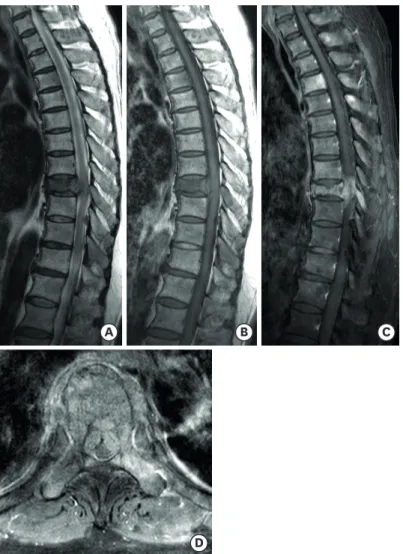
Plain radiographs and magnetic resonance imaging (MRI) taken from the other hospital revealed a T8 acute compression fracture with cord compression (FIGURE 1). Under an impression of the T8 pathologic fracture, we ran a series of tests to find the primary origin of the spinal metastasis. All laboratory tests including tumor markers were within normal limits. Chest and abdominal computed tomography scans did not show any mass lesions in solid organs. We discussed the plan for diagnosis with oncologists. Since the patient presented with neurologic symptoms in both lower extremities, we came to an agreement that planning an early spine surgery of T8 decompression and stabilization and making the pathologic diagnosis through tumor tissue removed during the surgery would be reasonable.
Further systemic work ups such as whole body positron emission tomography was planned after confirmation of the pathologic diagnosis. No steroid therapy was performed during the preoperative period.
A spinal embolization was performed prior to the operation. As a small size of the ASA was visualized on the left T8 segmental artery angiography (FIGURE 2), only the right T8 segmental artery was embolized. On the following day, we carried out a total laminectomy of T8 and pedicle screw fixation from T6 to T10. Tumors located at the ventral side of the dura sac were partially removed to achieve circumferential decompression of the spinal cord.
For this procedure, we resected both T8 roots. The operation was uneventful. No significant changes were observed on neurophysiologic intraoperative monitoring (NIOM) throughout the operation. Immediate postoperative neurologic examination showed no change in neurologic status compared to the preoperative state.
Around 8 hours after the operation, the patient suddenly became paraplegic. Neurologic examination revealed motor power grade 0 on both lower extremities. Pain sensation was lost below the level of the xiphoid process, but touch sense and proprioception were intact. Under a suspicion of operation site hematoma, follow-up MRI was taken immediately. However, we could not identify any lesions causing the patient's paraplegia. No notable events such as hypotension during intraoperative and postoperative period was identified. Therefore,
A B C
D
FIGURE 1. Preoperative T-spine magnetic resonance imaging. (A) T2 sagittal, (B) T1 sagittal, (C, D) T1 enhance sagittal and axial images. A pathologic compression fracture of T8 is observed. Enhancing epidural mass at the index level compresses the spinal cord.
FIGURE 2. Preoperative angiography of the right T8 segmental artery. Anterior spinal artery is seen (black arrow heads).
medical treatments, including hydration and high dose steroid injection, were administered and the patient was put on overnight observation.
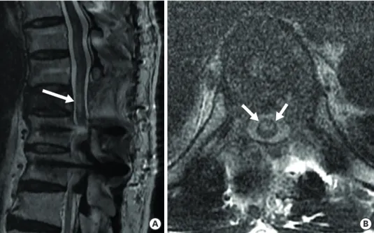
The patient did not show any neurologic improvements the next morning. We discussed this situation with the neurology and radiology departments and concluded that the neurologic features were similar to ASA syndrome. Follow-up T2-weighted sagittal and axial T-spine MRI revealed a high signal change at the anterior half of the spinal cord around the T8 level which confirmed ischemia or infarction of the anterior spinal cord (FIGURE 3A & B). Anticoagulation with low molecular heparin and antiplatelet medication were added. Mean arterial blood pressure was monitored and kept above 70 mmHg to maintain spinal cord perfusion pressure.
However, the patient's neurologic status did not recover. One week later, the remaining touch sense and proprioception of both lower extremities had also disappeared. The spinal pathology confirmed the diagnosis of multiple myeloma. Throughout 6 months follow-up, the patient remained paraplegic without any neurologic improvements.
DISCUSSION
Concerns of spinal cord ischemia are present in surgeries which affect the blood flow of segmental arteries. In thoracic/thoracoabdominal aortic aneurysm surgeries, the reported risk of spinal cord injury secondary to decreased cord perfusion ranges up to 20%.3 In spinal surgeries, the risk of ischemic spinal cord injury is much lower. Winter et al.14 retrospectively reviewed their 1,197 cases of corrective spinal deformity surgeries, and reported no instance of paraplegia related to segmental vessel ligation. The authors concluded that there is virtually no risk in segmental vessel ligation if the vessel ligation is unilateral, performed on the convexity of a scoliosis and at mid-vertebral body level, and hypotensive anesthesia is avoided. Later, Bridwell et al.1 reported 4 cases of major neurologic complication which were related with ligating unilateral convex segmental vessels among their 1,090 surgical cases and
A B
FIGURE 3. Postoperative magnetic resonance imaging taken at one day post-surgery. (A) T2 sagittal and (B) T2 axial images. High signal change at T8 level, the anterior half of the spinal cord, is seen (white arrows). This is a compatible finding with spinal cord ischemia at the anterior spinal artery territory.
showed the actual risk of ischemic cord injury present in the spine surgery, despite its rarity.
Few reports supporting the presence of this risk have followed.10,11
Spinal cord ischemia related to segmental artery ligation has been deeply studied by a group of Japanese surgeons in regard to total en bloc spondylectomy. To perform this operation, triple-level bilateral segmental artery embolization prior is usually recommended.6 As Doita et al.2 described in their report, sacrificing multi-level segmental arteries can compromise spinal cord perfusion and cause ischemic injury to the spinal cord. Therefore, it is necessary to verify safety issues of the preoperative segmental artery embolization. Through several animal studies using dogs, researchers have shown that neurologic deficits caused by spinal cord ischemia was common when ligating 5 or more levels of bilateral segmental arteries4 and less than 3 level ligation of the segmental arteries reduced spinal cord blood flow but did not compromised cord function.4,9,13 A further animal study using dogs confirmed that this 3 level safety zone is valid when the level of the Adamkiewicz artery is included in the ligating level.5 Clinical results of total en bloc spondylectomy backed up this result. Several authors have confirmed that preoperative 3 level ligations of segmental arteries (the index level, one level above, and one below) did not cause neurologic deficits by spinal cord ischemia.6,8,12 To our best knowledge, there have been no reports of spinal cord ischemia after sacrificing less than 3 levels of segmental arteries in the literature. In this case, we considered that the left T8 segmental artery was less likely to be the Adamkiewicz artery due to its small size. Even if incorrect, we assumed that the risk of spinal cord ischemia by its ligation would be low based on evidence described previously. However, despite its rarity, this case shows that the risk is substantial and spinal cord ischemic injury can happen even in one level segmental artery ligation. Therefore, the necessity of segmental artery ligation must be thoroughly evaluated, and it should be performed only in inevitable situations.
For preventive measures, NIOM comprised of motor and somatosensory evoked potentials could be considered; however, this cannot be fully relied upon. Apart from limitations regarding sensitivity and specificity, the pathophysiology of spinal cord ischemia makes it hard to detect during surgery. Patients of spinal cord ischemia have various symptom onset to peak times which range from a few minutes to 48 to 72 hours.15 In accordance with this case, some case reports have shown that patients diagnosed with ASA syndrome after spinal surgeries were free of neurological symptoms in the immediate postoperative period and became symptomatic several hours later.11 In these cases, there were no changes in NIOM during the operation. No neurologic deficit was observed in the intraoperative wake up test.11 This suggests that temporary measures to predict postoperative neurologic deficits, such as temporary clipping of the radicular artery, could be ineffective. Preoperative spinal angiography may be helpful; however, considering a good number of surgeries for spinal metastasis are performed in emergency settings, it would be impractical to enforce angiography to be performed in every case.
Modalities for managing patients with ASA syndrome after ligating segmental arteries are limited. Since anastomosis of the ligated segmental arteries to restore the blood flow is not a viable option, management goals should focus on managing blood pressure to maintain spinal perfusion. Antiplatelet or anticoagulation medication could be used to prevent vascular occlusions or embolisms; however, as seen in our case, the prognosis is expected to be poor unless substantial blood flow restoration is achieved. Although we did not perform this in our case, continuous cerebrospinal fluid (CSF) drainage may be worth considering as a treatment option. A body of literature has presented its effectiveness in the risk reduction
of spinal cord ischemic injury during and after thoracic/thoracoabdominal aortic aneurysm surgery.3 Despite the specific conditions between aortic aneurysm surgery and our case being different, we think the role of CSF drainage should be sought thoroughly in patients with similar clinical settings, as shown in this report.
CONCLUSION
Although very rare, spinal cord infarction can happen after one level segmental artery ligation during spinal surgery. Surgeons must keep in mind such risk, and surgery should be planned under a careful risk-benefit consideration.
REFERENCES
1. Bridwell KH, Lenke LG, Baldus C, Blanke K. Major intraoperative neurologic deficits in pediatric and adult spinal deformity patients. Incidence and etiology at one institution. Spine (Phila Pa 1976) 23:324- 331, 1998
PUBMED | CROSSREF
2. Doita M, Marui T, Nishida K, Kurosaka M, Yoshiya S, Sha N. Anterior spinal artery syndrome after total spondylectomy of T10, T11, and T12. Clin Orthop Relat Res 405:175-181, 2002
PUBMED | CROSSREF
3. Epstein NE. Cerebrospinal fluid drains reduce risk of spinal cord injury for thoracic/thoracoabdominal aneurysm surgery: a review. Surg Neurol Int 9:48, 2018
PUBMED | CROSSREF
4. Fujimaki Y, Kawahara N, Tomita K, Murakami H, Ueda Y. How many ligations of bilateral segmental arteries cause ischemic spinal cord dysfunction? An experimental study using a dog model. Spine (Phila Pa 1976) 31:E781-E789, 2006
PUBMED | CROSSREF
5. Kato S, Kawahara N, Tomita K, Murakami H, Demura S, Fujimaki Y. Effects on spinal cord blood flow and neurologic function secondary to interruption of bilateral segmental arteries which supply the artery of Adamkiewicz: an experimental study using a dog model. Spine (Phila Pa 1976) 33:1533-1541, 2008 PUBMED | CROSSREF
6. Kawahara N, Tomita K, Murakami H, Demura S. Total en bloc spondylectomy for spinal tumors: Surgical techniques and related basic background. Orthop Clin North Am 40:47-63, 2009
PUBMED | CROSSREF
7. Martirosyan NL, Feuerstein JS, Theodore N, Cavalcanti DD, Spetzler RF, Preul MC. Blood supply and vascular reactivity of the spinal cord under normal and pathological conditions. J Neurosurg Spine 15:238-251, 2011
PUBMED | CROSSREF
8. Murakami H, Kawahara N, Tomita K, Demura S, Kato S, Yoshioka K. Does interruption of the artery of Adamkiewicz during total en bloc spondylectomy affect neurologic function? Spine (Phila Pa 1976) 35:E1187-E1192, 2010
PUBMED | CROSSREF
9. Nambu K, Kawahara N, Kobayashi T, Murakami H, Ueda Y, Tomita K. Interruption of the bilateral segmental arteries at several levels: influence on vertebral blood flow. Spine (Phila Pa 1976) 29:1530-1534, 2004 PUBMED | CROSSREF
10. Orchowski J, Bridwell KH, Lenke LG. Neurological deficit from a purely vascular etiology after unilateral vessel ligation during anterior thoracolumbar fusion of the spine. Spine (Phila Pa 1976) 30:406-410, 2005 PUBMED | CROSSREF
11. Stöckl B, Wimmer C, Innerhofer P, Kofler M, Behensky H. Delayed anterior spinal artery syndrome following posterior scoliosis correction. Eur Spine J 14:906-909, 2005
PUBMED | CROSSREF
12. Tomita K, Kawahara N, Murakami H, Demura S. Total en bloc spondylectomy for spinal tumors:
improvement of the technique and its associated basic background. J Orthop Sci 11:3-12, 2006 PUBMED | CROSSREF
13. Ueda Y, Kawahara N, Tomita K, Kobayashi T, Murakami H, Nambu K. Influence on spinal cord blood flow and function by interruption of bilateral segmental arteries at up to three levels: experimental study in dogs. Spine (Phila Pa 1976) 30:2239-2243, 2005
PUBMED | CROSSREF
14. Winter RB, Lonstein JE, Denis F, Leonard AS, Garamella JJ. Paraplegia resulting from vessel ligation.
Spine (Phila Pa 1976) 21:1232-1233, 1996 PUBMED | CROSSREF
15. Yadav N, Pendharkar H, Kulkarni GB. Spinal cord infarction: clinical and radiological features. J Stroke Cerebrovasc Dis 27:2810-2821, 2018
PUBMED | CROSSREF