- 174 -
대한폐경학회지 제17권 제3호 2011
Vol. 17, No. 3, December. 2011 http://dx.doi.org/10.6118/jksm.2011.17.3.174
접수일: 2011년 9월 4일, 심사일: 2011년 9월 4일, 게재확정일: 2011년 10월 28일
주관책임자: Tae-Hee Kim, Department of Obstetrics and Gynecology, Soonchunhyang University Bucheon Hospital, 1174, Jung-dong, Wonmi-gu, Bucheon 420-020, Korea
Tel: (032) 621-5055, Fax: (032) 621-5015, e-mail: [email protected]
Cervical Endometriosis in a Post-menopausal Woman: A Case Report
Departments of Obstetrics and Gynecology1, Pathology2, College of Medicine, Soonchunhyang University Bucheon Hospital, Bucheon, Korea
Junsik Park, M.D.
1, Tae-Hee Kim, M.D., Ph.D.
1, Hae-Hyeog Lee, M.D., Ph.D.
1, Woo Seok Lee, M.D., Ph.D.
1, Soo-Ho Chung, M.D.
1, Sang-Mo Park, M.D.
2=Abstract=
Cervical endometriosis is defined as the presence of endometrial glands and stroma at the cervix. This is rare and sometimes asymptomatic. Most of these are diagnosed by incidental findings within histopathology. As the presence of cytological features do not guarantee the presence of cervical endometriosis, it is difficult to diagnose this disorder prior to surgery. We recently encountered a case of cervical endometriosis in a post-menopausal woman who was not receiving hormone therapy. As a reminder to clinicians about this neglected issue, we report a case of cervical endometriosis with a literature review. (J Korean Soc Menopause 2011;17:174-177)
Key Words: Cervix uteri, Endometriosis, Postmenopause
Introduction
Endometriosis is defined as the presence of endometrial glands and stroma at extra-uterine sites. These ectopic endo- metrial implants are usually located on the ovaries. But it can be observed on the uterus and its ligaments, in the abdominal cavity, on the cervix, pleura, and very rarely in the lungs, brain, and eyes. Namely, it can occur nearly anywhere in the body.
Many patients with cervix endometriosis are asymptomatic.
By reviewing literature, we can find a diverse range of persistent symptoms which is abnormal pap smear, post-coital bleeding, pelvic pain, intermenstrual bleeding and massive hemorrhage.1∼5 Cervix endometriosis may be a source of glandular cells in cervical smears and hence a cause of abnormal smear result. So it can lead to misjudge as cervix cancer. A considerable number of patients reported in the literature only had abnormal smear results and were diagnosed during colposcopy or by histopa- thological examinations of their biopsy or hysterectomy speci- mens. But, adenocarcinoma arising from cervix endometriosis has been reported in literature.6,7 This provides the reason to
interested in the disease in detail.
In 1928 cervical endometriosis was first mentioned by Fels.
After that, many case reports had been accumulated in li- terature. However there is paucity of knowledge in literature regarding cervix endometriosis. Limited awareness of the cli- nical appearance of the disease may account for its apparent rarity.
We recently encountered a case of cervical endometriosis in post-menopause woman. To remind clinician of the neglected issue of endometriosis of the cervix, we report a case of cer- vical endometriosis with literature review.
Case Report
54-year-old woman was referred to Soonchunhyang Uni- versity Hospital with the suspicion of huge myoma uteri from local clinics. Having had two full term vaginal deliveries, she has been menopause state three years ago without hormone therapy. Routine pap smear showed reactive cellular changes with inflammation and human papillomavirus (HPV) deoxy- ribonucleic acid (DNA) Chip test was negative. Ultrasound
Junsik Park, et al
- 175 -
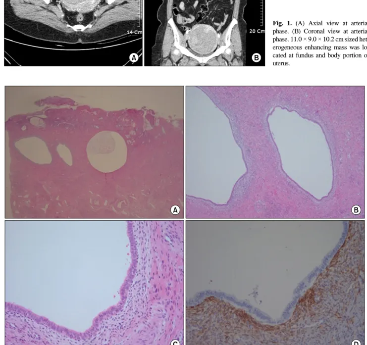
Fig. 1. (A) Axial view at arterial phase. (B) Coronal view at arterial phase. 11.0 × 9.0 × 10.2 cm sized het- erogeneous enhancing mass was lo- cated at fundus and body portion of uterus.
Fig. 2. (A∼C) × 10, × 40, × 200 H&E. Low magnification show several small and large endometrial glands with surrounding stroma admixed with endocercvical glands at the cervix. (D) Endometrial stromal cells exhibits CD10 expression.
Cervical Endometriosis in a Post-menopausal Woman: A Case Report
- 176 - examination and computed tomography (CT) revealed 11.0 × 9.0 × 10.2 cm sized mass in uterine fundus and body portion (Fig. 1). On physical examination, the patient was generally well, weighed 74.9 kg and not pale. General examination was unremarkable. Hematological examination was within normal range. The patient underwent laparoscopy assisted vaginal hysterectomy on June 14, 2011. At operation, there was no evidence of endometriosis. Grossly the uterus was diffusely enlarged, weighed 735 gm. Microscopically, both endometrial glands and stroma present at the cervical stroma (Fig. 2). Path- ologic findings indicate that there was endometriosis at cervix, atrophy at endometrium, and leiomyoma at myometrium.
Discussion
English-language published literatures from 1970 to 2011 were searched on the PubMed database using the keywords
‘cervix endometriosis’. A total of 45 papers related to cervix endometriosis were retrieved from the database. From these papers, the incidence of cervix endometriosis has been reported to be between 1.6% to 2.4%.8 Endometriosis was estrogen dependent disease. So cervix endometriosis in post-menopause women has been much rarely occurred. Even though the post-menopause endometriosis is rarely associated with cancer, gynecologists should pay attention to management for this disease. Kim et al. have presented two case of endometriosis detected in post-menopause women and a case of serous adenocarcinoma arising from ovarian endometriosis after menopause.9,10
There is no symptoms in all women with cervix endo- metriosis. Abnormal pap smear result is a common presentation.
Baker et al. described a series of 20 cases of superficial endometriosis of the cervix in 1999.11 The majority of the patients were referred because of abnormal uterine cervical lesions like endocervical glandular dysplasia, adenocarcinoma in situ (ACIS) or, rarely, an invasive carcinoma of the cervix.
And cervix endometriosis was incidentally discovered. Cervix endometriosis might be the cause of abnormal smears, because the cytomorphological features of endometriosis cells change with the cyclical hormonal changes, and sometimes revealed crowded, overlapping glandular cells with loss of cell polarity and rosette formations. These cytological features would be sufficient to overlap with precancerous and cancerous glandular
lesions. Therefore if one is unaware of the presence of cervix endometriosis, the condition can be diagnosed as ‘atypical glandular cells’.12
Several theories have been proposed to explain pathogenesis of cervix endometriosis. Many investigators commonly account for the pathogenesis of cervix endometriosis as implantation of cast-off endometrial fragments on the previously traumatized lesion of cervix. Cervix endometriosis is generally considered as rare lesion. But with widespread use of invasive cervical procedure an increased incidence of cervical endometriosis can be expected. However, as in our case, cervix endometriosis can be encountered in patients who never underwent procedures traumatizing the cervix. This might be explained by Hoang et al hypothesised that cervix endometriosis could develop in mullerian rests which persist in the stroma of the cervix.13 In summary, not uncommonly, cervix endometriosis is diagnosed after operation. The clinical feature is very diverse.
In our case, the woman was post-menopause state without hormone therapy. There was only reactive cellular change in her pap smear result. She never underwent traumatic procedure of cervix. Like this case, to most post-menopause women, cervix endometriosis would be the only incidental finding. But cervix endometriosis in post-menopause women was rare and interesting finding. Many post-menopause women would un- dergo operation because of other pathology and after operation cervix endometriosis could be incidentally diagnosed by his- topathology. With increased use of hormone therapy, cervix endometriosis in post-menopause could be shown different clinical features. We thought that studying cervix endometriosis in post-menopause women with or without hormone therapy would be interesting.
References
1. Kim TH, Lee HH. Hemoperitoneum during pregnancy with en- dometriosis; report of four cases. Iran J Reprod Med 2010; 8:
90-3.
2. Iwase A, Goto M, Kurotsuchi S, Harata T, Kaseki S, Kikkawa F.
Successful management of a massive hemorrhage due to rupture of cystic cervical endometriosis by a loop electrosurgical ex- cision procedure. Fertil Steril 2008; 89: 991, e13-5.
3. Phadnis SV, Doshi JS, Ogunnaike O, Coady A, Padwick M, Sanusi FA. Cervical endometriosis: a diagnostic and manage-
Junsik Park, et al
- 177 -
= 국문초록 =
자궁경부에서 발생하는 자궁내막증은 자궁내막샘과 간질이 자궁경부에 존재하는 드문 질환이다. 이 질환은 특별한 증상 이 없는 경우가 많아서 대부분의 경우 우연히 조직병리소견으로 진단된다. 그래서 수술 전에는 자궁경부에서 발생하는 자궁내막증을 진단하기 힘들다. 우리는 최근 호르몬 치료를 시행하고 있지 않은 폐경 여성에게서 진단된 자궁경부의 자궁내막증을 경험하였다. 임상의사들이 간과하고 넘어갈 수 있는 주제인 자궁경부의 자궁내막증을 한번 더 살펴보고자 문헌 고찰과 함께 자궁경부에서 발견된 자궁내막증 1예를 보고하는 바이다.
중심단어: 자궁경부, 자궁내막증, 폐경
ment dilemma. Arch Gynecol Obstet 2005; 272: 289-93.
4. Wong FW, Lim CE, Karia S, Santos L. Cervical endometriosis:
case series and review of literature. J Obstet Gynaecol Res 2010;
36: 916-9.
5. Yokota N, Yoshida H, Sakakibara H, Inayama Y, Hirahara F. A severe vaginal hemorrhage caused by cervical endometriosis.
Am J Obstet Gynecol 2008; 199: e12-3.
6. Chang SH, Maddox WA. Adenocarcinoma arising within cer- vical endometriosis and invading the adjacent vagina. Am J Obstet Gynecol 1971; 110: 1015-7.
7. Noda K, Kimura K, Ikeda M, Teshima K. Studies on the histo- genesis of cervical adenocarcinoma. Int J Gynecol Pathol 1983;
1: 336-46.
8. Veiga-Ferreira MM, Leiman G, Dunbar F, Margolius KA.
Cervical endometriosis: facilitated diagnosis by fine needle as- piration cytologic testing. Am J Obstet Gynecol 1987; 157:
849-56.
9. Kim TH, Lee HH, Chung SH, Kwak JJ, Park HS. Endometriosis detected in postmenopausal women not receiving menopausal hormone therapy: two case reports. J Korean Soc Menopause 2010; 16: 176-80.
10. Kim TH, Lee HH, Chung SH, Kwak JJ, Lee BI, Hong YP.
Serous adenocarcinoma arising from ovarian endometriosis af- ter menopause. Korean J Obstet Gynecol 2010; 53: 365-70.
11. Baker PM, Clement PB, Bell DA, Young RH. Superficial endo- metriosis of the uterine cervix: a report of 20 cases of a process that may be confused with endocervical glandular dysplasia or adenocarcinoma in situ. Int J Gynecol Pathol 1999; 18: 198-205.
12. Szyfelbein WM, Baker PM, Bell DA. Superficial endometriosis of the cervix: A source of abnormal glandular cells on cervico- vaginal smears. Diagn Cytopathol 2004; 30: 88-91.
13. Hoang NM, Smadja A, Orcel L. Endometriosis of the uterine cervix. A hypothesis on its histogenesis. J Gynecol Obstet Biol Reprod (Paris) 1987; 16: 587-93.