Effectiveness of the albumin-bilirubin score as a prognostic factor for early recurrence after
curative hepatic resection for hepatocellular carcinoma
Yun Ho Lee, Yang Seok Koh, Young Hoe Hur, Chol Kyoon Cho, Hee Joon Kim, and Eun Kyu Park
Department of Surgery, Chonnam National University Hwasun Hospital and Medical School, Gwangju, Korea
Backgrounds/Aims: The albumin–bilirubin (ALBI) score has been validated as a predictor of disease-free survival and overall survival in hepatocellular carcinoma (HCC). The purpose of this study was to assess the ALBI score as a risk factor for early recurrence (ER) after curative liver resection in HCC. Methods: Patients who underwent liver re- section with curative intent for HCC without previous treatment between January 2004 and December 2014 were in- cluded in this retrospective study. The utility of the ALBI score in predicting ER and late recurrence (LR) was evaluated.
Results: A total of 465 HCC patients were enrolled; multivariate analysis identified ALBI grade ≥2 (p=0.003) as a risk factor for ER, in addition to hepatitis B virus surface antigen (HBsAg)-positive status (p<0.001), tumor size ≥3.5cm (p≤0.001), lymph-vascular invasion (p=0.001), and the presence of satellite lesions (p=0.009). In subgroup analysis for ALBI grade 1, Model for End-stage Liver Disease score >9 (p=0.046), HBsAg positive status (p=0.004), tumor size ≥3.5 cm (p<0.001), lymph-vascular invasion (p=0.001), presence of satellite lesions (p=0.002), and poor tumor differentiation (p=0.007) were independent risk factors for ER; however, in subgroup analysis for ALBI grade 2, no significant associations with ER were found. Kaplan-Meier curve analysis showed that long-term survival in HCC with ER was significantly shorter than in patients with LR. Conclusions: The ALBI score was a preoperative risk factor for ER and may be useful in determining appropriate management according to liver function when recurrence develops. (Ann Hepatobiliary Pancreat Surg 2018;22:335-343)
Key Words: Albumin bilirubin; Hepatocellular carcinoma; Risk factor; Liver resection
Received: August 22, 2018; Revised: October 21, 2018; Accepted: October 22, 2018 Corresponding author: Yang Seok Koh
Department of Surgery, Chonnam National University Hwasun Hospital and Medical School, 322 Seoyang-ro, Chonnam 58128, Korea Tel: +82-61-379-7646, Fax: +82-61-379-7661, E-mail: [email protected]
Copyright Ⓒ 2018 by The Korean Association of Hepato-Biliary-Pancreatic Surgery
This is an Open Access article distributed under the terms of the Creative Commons Attribution Non-Commercial License (http://creativecommons.org/
licenses/by-nc/4.0) which permits unrestricted non-commercial use, distribution, and reproduction in any medium, provided the original work is properly cited.
Annals of Hepato-Biliary-Pancreatic Surgery ∙ pISSN: 2508-5778ㆍeISSN: 2508-5859
INTRODUCTION
Hepatocellular carcinoma (HCC) is common, and is one of the leading causes of death worldwide.1 Liver resection is standard treatment for HCC, followed by liver trans- plantation, but is a major cause of death in long term fol- low-up due to recurrence, even after liver resection.2-4 The overall recurrence rate is reportedly 54% to 63% after HCC treatment.5,6 Other studies have shown an early re- currence rate of 38%7 less than a year after radical hep- atectomy, and it is known that tumor stage, presence of microsatellites, microvascular invasion, liver cirrhosis, multinodularity, hepatitis activity, and alpha-fetoprotein (AFP) level are associated with early recurrence.8-11 It is very important to determine the risk factors for early re- currence and to diagnose early postoperative recurrence
with strict follow-up because transarterial chemoemboli- zation (TACE), radiofrequency ablation (RFA), or reoper- ation can increase survival duration.12
The albumin–bilirubin (ALBI) score has been high- lighted as a predictor of postoperative hepatic failure and long-term survival after hepatic resection, and is a new tool for the assessment of liver failure and function.12 According to another study, ALBI grade was associated with significant differences in disease-free survival after hepatectomy for HCC.13
We hypothesized a relationship between ALBI score and early recurrence (ER). To date, no study has de- termined whether ALBI grade is associated with re- currence within a year (we defined this as ER). Therefore, this study aimed to investigate the usefulness of ALBI score for relapse within a year.
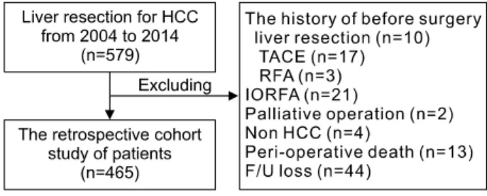
Fig. 1. Flow chart of patient selection procedures in this study. HCC, hepatocellular carcinoma; TACE, transarterial chemoembolization; RFA, radiofrequency ablation; IORFA, intraoperative radiofrequency ablation.
MATERIALS AND METHODS
Patients
Between January 2004 and December 2014, all consec- utive patients who underwent liver resection with curative intent for HCC at the Hwasun Chonnam National University Hospital were considered for this retrospective study. The inclusion criteria were: initial liver resection with curative intent performed at the authors’ center; no treatment for HCC before liver resection; and no other si- multaneous malignancies.
Diagnosis and definitions
We defined ER as recurrence within a year after cura- tive liver resection for HCC, based on a study by Tung- Ping Poon et al.12 Diagnosis of HCC was based on histo- logical evidence after surgery. Major liver resection was defined as resection of at least three Couinaud liver segments.14 In pathologic reports, vascular invasion in- cluded gross as well as microscopic invasion of vessels.
Microscopic vascular invasion is defined by tumor within a vascular space lined by endothelium, identified only on microscopy in the capsule or noncapsular fibrous septa, or liver tissue surrounding the tumor. The ALBI score is a validated formula used for rigorous statistical analysis of HCC patients, based on bilirubin and albumin levels.15 In this study, the ALBI score was calculated using the fol- lowing formula with preoperative laboratory analysis:
0.66× log10 (total bilirubin [mol/l]) –0.085 (albumin[g/l])
The ALBI score was stratified as grade 1 (–2.60 or less), grade 2 (–2.59 to –1.39), or grade 3 (greater than –1.39).
Follow-up
All patients were followed up at 1 month after liver re- section, followed by every 3 months in the first year and every 3-6 months in subsequent years. Routine tests such as serum AFP levels, serum biochemistry, chest X-ray, abdominal ultrasound, and abdominal computed tomog- raphy or magnetic resonance imaging were performed at every follow–up. Patients with relapses were treated with liver resection, percutaneous ethanol injection, RFA, TACE, or sorafenib, depending on liver functional status, extent of disease, and overall health and economic status.
Statistical analysis
Statistical analysis was performed using SPSS® version 22.0 (IBM, Armonk, NY, USA). Student's t-test, the Mann-Whitney U test, and the 2 test were used to com- pare continuous and categorical variables, as appropriate.
Multivariate analysis was performed using a logistic re- gression model to identify independent predictors of early recurrence. The Kaplan-Meier method was used to esti- mate overall survival. Data are expressed as number of patients, ratio (%), or odds ratio (OR) and 95% con- fidence interval (CI), as indicated. A p-value <0.05 was considered statistically significant.
RESULTS
Of 579 patients who underwent hepatic resection during the study period, 114 were excluded for the following rea- sons: 30 received treatment for HCC before liver re- section, 21 underwent intraoperative RFA, 2 had surgery with non-curative intent, 4 had other malignant tumors, 13 died during the perioperative period, and 44 were lost to follow-up. The remaining 465 patients were enrolled in the study (Fig. 1).
Incidence and characteristics of cancer recurrence
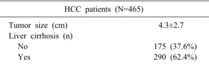
Patient demographics and clinicopathological features are listed in Table 1. Among the 465 patients, 398 were male (85.6%) and 67 were female (14.4%). The mean pa- tient age was 59.2 years. Overall, 290 patients (62.4%) had recurrence after liver resection with curative intent.
ER was observed in 140 patients (30.1%) in the first year after liver resection and 150 patients (32.3%) had re-
Table 1. Baseline clinicopathological characteristics HCC patients (N=465)
Sex (n)
F 67 (14.4%)
M 398 (85.6%)
Age (years) 59.2±10.0 Hepatic resection (n)
Major 147 (31.6%)
Minor 318 (68.4%)
Overall recur (n)
No 175 (37.6%)
Yes 290 (62.4%)
≤1 yr recur (n)
No 325 (69.9%)
Yes 140 (30.1%)
>1 yr recur (n)
No 315 (67.7%)
Yes 150 (32.3%)
Pre-operative blood laboratory Platelet count (103/μL) 169.2±65.1
AST (IU/L) 47.5±44.0
ALT (IU/L) 45.2±48.4
Albumin (g/dL) 4.3±0.5 Total bilirubin (mg/dL) 0.8±0.4 Prothrombin time(INR) 1.1±0.4 Pre-operative AFP (ng/ml) 1636.5±6861.4 Pre-operative ICGR 15 (%) 10.7±7.3
Child-Pugh Score
A 456 (98.1%)
B 9 (1.9%)
C 0
ALBI grade(preoperative)
1 365 (78.5%)
2 98 (21.1%)
3 2 (0.4%)
ALBI grade(POD7)
1 75 (16.2%)
2 381 (81.9%)
3 19 (1.9%)
MELD score
≤9 441 (94.8%)
10-19 23 (4.9%)
30-39 1 (0.2%)
≥40 0
HBsAg
Negative 165 (35.5%)
Positive 300 (64.5%)
Anti-HCV
Negative 416 (89.5%)
Positive 49 (10.5%)
alcoholic liver cirrhosis (n)
No 434 (93.3%)
Yes 31 (6.7%)
Tumor number (n)
≤3 459 (98.7%)
>3 6 (1.3%)
Table 1. Continued
HCC patients (N=465)
Tumor size (cm) 4.3±2.7 Liver cirrhosis (n)
No 175 (37.6%)
Yes 290 (62.4%)
AST, aspartate aminotransferase; ALT, alanine amino- transferase; AFP, alpha-fetoprotein; ICGR15, indocyanine green retention test at 15 minutes; ALBI, albumin–bilirubin;
MELD, model for end-stage liver disease; HBsAg, hepatitis B surface antigen; Anti-HCV, Anti-hepatitis C virus; POD7, Postoperative day 7
currence after the first year (late recurrence, LR).
Recurrence was not observed in 175 patients (37.6%). The majority (98.1%) had Child-Pugh (CP) grade A disease and the remaining 1.9% had CP grade B disease. The Model for End-stage Liver Disease (MELD) score was ≤ 9 in 94.8% of patients, between 10 and 19 in 4.9%, and between 30 and 39 in 0.2%. The preoperative ALBI score classified 78.5% of the patients as grade 1, 21.1% as grade 2, and 0.4% as grade 3.
Univariate and multivariate analysis of risk factors for ER and LR
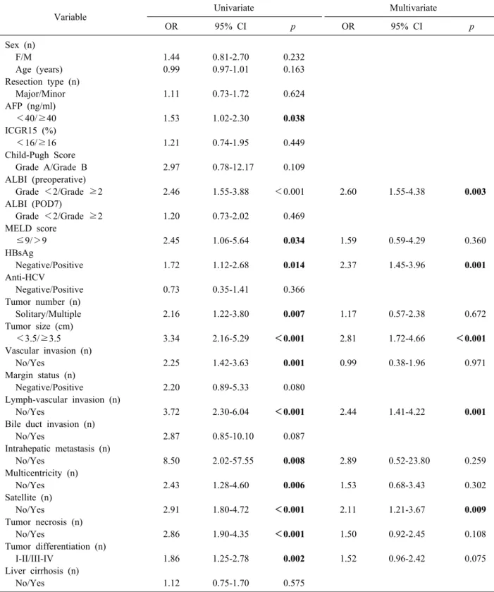
Univariate analysis to identify prognostic factors affect- ing ER showed that AFP ≥40 (p=0.038, OR: 1.53, 95%
CI: 1.02-2.30), preoperative ALBI grade ≥2 (p<0.001, OR: 2.46, 95% CI: 1.55-3.88), MELD score >9 (p=
0.034, OR: 2.45,95% CI: 1.06-5.64), hepatitis B virus sur- face antigen (HBsAg)-positive status (p=0.014, OR: 1.72, 95% CI: 1.12–2.68), presence of multiple tumors (p=
0.007, OR: 2.16, 95% CI: 1.22-3.80), tumor size ≥3.5 cm (p<0.001, OR: 3.34, 95% CI: 2.16-5.29), vascular in- vasion (p=0.001, OR: 2.25, 95% CI: 1.42-3.63), lymph- vascular invasion (p<0.001, OR: 3.72, 95% CI: 2.30- 6.04), intrahepatic metastasis (p<0.008, OR: 8.50, 95%
CI: 2.02-57.55), multicentricity (p<0.006, OR: 2.43, 95%
CI: 1.28-4.60), presence of satellite lesions (p<0.001, OR: 2.91, 95% CI: 1.80-4.72), tumor necrosis (p<0.001, OR: 2.86, 95% CI: 1.90-4.35), and tumor differentiation (p=0.002, OR: 1.86, 95% CI: 1.25-2.78) were significant risk factors (Table 2). After these variables were included in multivariate analysis, ALBI grade ≥2 (p<0.003, OR:
2.60, 95% CI: 1.55-4.38), HBsAg-positive status (p=0.001, OR: 2.37, 95% CI: 1.45-3.96), tumor size ≥3.5
Table 2. Univariate and multivariate analysis of risk factors for early recurrence (≤1 year) after curative liver resection in HCC patients
Variable Univariate Multivariate
OR 95% CI p OR 95% CI p
Sex (n)
F/M 1.44 0.81-2.70 0.232
Age (years) 0.99 0.97-1.01 0.163
Resection type (n)
Major/Minor 1.11 0.73-1.72 0.624
AFP (ng/ml)
<40/≥40 1.53 1.02-2.30 0.038
ICGR15 (%)
<16/≥16 1.21 0.74-1.95 0.449
Child-Pugh Score
Grade A/Grade B 2.97 0.78-12.17 0.109
ALBI (preoperative)
Grade <2/Grade ≥2 2.46 1.55-3.88 <0.001 2.60 1.55-4.38 0.003
ALBI (POD7)
Grade <2/Grade ≥2 1.20 0.73-2.02 0.469
MELD score
≤9/>9 2.45 1.06-5.64 0.034 1.59 0.59-4.29 0.360
HBsAg
Negative/Positive 1.72 1.12-2.68 0.014 2.37 1.45-3.96 0.001
Anti-HCV
Negative/Positive 0.73 0.35-1.41 0.366
Tumor number (n)
Solitary/Multiple 2.16 1.22-3.80 0.007 1.17 0.57-2.38 0.672
Tumor size (cm)
<3.5/≥3.5 3.34 2.16-5.29 <0.001 2.81 1.72-4.66 <0.001
Vascular invasion (n)
No/Yes 2.25 1.42-3.63 0.001 0.99 0.38-1.96 0.971
Margin status (n)
Negative/Positive 2.20 0.89-5.33 0.080
Lymph-vascular invasion (n)
No/Yes 3.72 2.30-6.04 <0.001 2.44 1.41-4.22 0.001
Bile duct invasion (n)
No/Yes 2.87 0.85-10.10 0.087
Intrahepatic metastasis (n)
No/Yes 8.50 2.02-57.55 0.008 2.89 0.52-23.80 0.259
Multicentricity (n)
No/Yes 2.43 1.28-4.60 0.006 1.53 0.68-3.43 0.302
Satellite (n)
No/Yes 2.91 1.80-4.72 <0.001 2.11 1.21-3.67 0.009
Tumor necrosis (n)
No/Yes 2.86 1.90-4.35 <0.001 1.50 0.92-2.45 0.108
Tumor differentiation (n)
I-II/III-IV 1.86 1.25-2.78 0.002 1.52 0.96-2.42 0.075
Liver cirrhosis (n)
No/Yes 1.12 0.75-1.70 0.575
AFP, alpha-fetoprotein; ICGR15, indocyanine green retention test at 15 minutes; ALBI, albumin–bilirubin; MELD, model for end-stage liver disease; HBsAg, hepatitis B surface antigen; Anti-HCV, Anti-hepatitis C virus; POD7, Postoperative day 7
cm (p≤0.001, OR: 2.81, 95% CI: 1.72-4.66), lymph- vas- cular invasion (p=0.001, OR: 2.44, 95% CI: 1.41- 4.22), and presence of satellite lesions (p=0.009, OR: 2.11, 95%
CI: 1.21-3.67) were found to be independently predictive of ER.
Univariate analysis to identify prognostic factors affect-
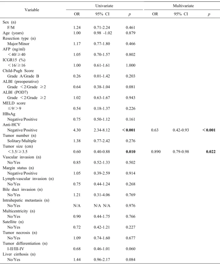
Table 3. Univariate and multivariate analysis of risk factors for late recurrence (>1 year) after curative liver resection in HCC patients
Variable Univariate Multivariate
OR 95% CI p OR 95% CI p
Sex (n)
F/M 1.24 0.71-2.24 0.461
Age (years) 1.00 0.98 -1.02 0.879
Resection type (n)
Major/Minor 1.17 0.77-1.80 0.466
AFP (ng/ml)
<40/≥40 1.05 0.70-1.57 0.802
ICGR15 (%)
<16/≥16 1.00 0.61-1.61 1.000
Child-Pugh Score
Grade A/Grade B 0.26 0.01-1.42 0.203
ALBI (preoperative)
Grade <2/Grade ≥2 0.64 0.38-1.04 0.081
ALBI (POD7)
Grade <2/Grade ≥2 1.02 0.63-1.67 0.943
MELD score
≤9/>9 0.54 0.18-1.37 0.226
HBsAg
Negative/Positive 0.75 0.50-1.12 0.161
Anti-HCV
Negative/Positive 4.30 2.34-8.12 <0.001 0.63 0.42-0.93 <0.001
Tumor number (n)
Solitary/Multiple 1.38 0.77-2.42 0.276
Tumor size (cm)
<3.5/≥3.5 0.60 0.40-0.88 0.010 0.890 0.79-0.98 0.022
Vascular invasion (n)
No/Yes 0.85 0.52-1.33 0.502
Margin status (n)
Negative/Positive 1.05 0.39-2.59 0.914
Lymph-vascular invasion (n)
No/Yes 0.75 0.44-1.24 0.268
Bile duct invasion (n)
No/Yes 1.21 0.31-4.06 0.769
Intrahepatic metastasis (n)
No/Yes N/A N/A N/A 0.976
Multicentricity (n)
No/Yes 0.90 0.44-1.75 0.766
Satellite (n)
No/Yes 0.72 0.42-1.21 0.227
Tumor necrosis (n)
No/Yes 1.09 0.74-1.60 0.677
Tumor differentiation (n)
I-II/III-IV 0.68 0.46-1.01 0.060
Liver cirrhosis (n)
No/Yes 1.44 0.96-2.17 0.084
AFP, alpha-fetoprotein; ICGR15, indocyanine green retention test at 15 minutes; ALBI, albumin–bilirubin; MELD, model for end-stage liver disease; HBsAg, hepatitis B surface antigen; Anti-HCV, Anti-hepatitis C virus; POD7, Postoperative day 7
ing LR showed that anti-hepatitis C virus (HCV)-positive status (p<0.001, OR: 4.30, 95% CI: 2.34-8.12) and tumor size ≥3.5 cm (p=0.010, OR: 0.60, 95% CI: 0.40-0.88)
were significant risk factors. On multivariate analysis, an- ti-HCV-positive status (p<0.001 OR: 0.63, 95% CI:
0.42-0.93) and tumor size ≥3.5 cm (p=0.022, OR: 0.89,
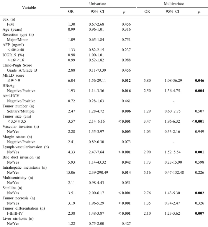
Table 4. Univariate and multivariate analysis of risk factors for early recurrence (≤1 year) in ALBI grade 1 after curative liver resection in HCC patients
Variable Univariate Multivariate
OR 95% CI p OR 95% CI p
Sex (n)
F/M 1.30 0.67-2.68 0.456
Age (years) 0.99 0.96-1.01 0.316
Resection type (n)
Major/Minor 1.09 0.65-1.84 0.751
AFP (ng/ml)
<40/≥40 1.33 0.82-2.15 0.237
ICGR15 (%) 0.98 1.00-1.01
<16/≥16 0.99 0.52-1.82 0.988
Child-Pugh Score
Grade A/Grade B 2.88 0.11-73.39 0.456
MELD score
≤9/>9 6.04 1.56-29.11 0.012 5.80 1.08-36.29 0.046
HBsAg
Negative/Positive 1.93 1.14-3.36 0.016 2.50 1.36-4.75 0.004
Anti-HCV
Negative/Positive 0.72 0.28-1.63 0.461 -
Tumor number (n)
Solitary/Multiple 2.47 1.28-4.72 0.006 1.29 0.60 2.75 0.507
Tumor size (cm)
<3.5/≥3.5 3.57 2.14 6.16 <0.001 3.47 1.96-6.32 <0.001
Vascular invasion (n)
No/Yes 2.28 1.35-3.97 0.003 1.03 0.33-2.16 0.949
Margin status (n)
Negative/Positive 2.41 0.89-6.30 0.073 -
Lymph-vascularinvasion (n)
No/Yes 4.33 2.47-7.64 <0.001 2.90 1.52 5.54 0.001
Bile duct invasion (n)
No/Yes 5.93 1.14-43.32 0.042 1.73 0.23-15.90 0.598
Intrahepatic metastasis (n)
No/Yes 15.06 2.39-290.49 0.014 5.16 0.47-132.48 0.226
Multicentricity (n)
No/Yes 2.11 0.98-4.43 0.051
Satellite (n)
No/Yes 3.51 2.00-6.17 <0.001 2.76 1.43-5.30 0.002
Tumor necrosis (n)
No/Yes 3.19 1.96-5.29 <0.001 1.35 0.74-2.47 0.326
Tumor differentiation (n)
I-II/III-IV 2.38 1.48-3.87 <0.001 2.10 1.23-3.62 0.007
Liver cirrhosis (n)
No/Yes 1.22 0.75-2.00 0.427
AFP, alpha-fetoprotein; ICGR15, indocyanine green retention test at 15 minutes; MELD, model for end-stage liver disease;
HBsAg, hepatitis B surface antigen; Anti-HCV, Anti-hepatitis C virus
95% CI: 0.79-0.98) were significant risk factors (Table 3).
Subgroup analysis by ALBI grade
We performed subgroup analysis by preoperative ALBI grade 1 and 2 after excluding 2 patients of ALBI grade
3 (Table 4). In the ALBI grade 1 subgroup, MELD score
>9 (p=0.012, OR: 6.04, 95% CI: 1.56-29.11), HBsAg- positive status (p=0.016, OR: 1.93, 95% CI: 1.14-3.36), presence of multiple tumors (p=0.006, OR: 2.47, 95% CI:
1.28-4.72), tumor size ≥3.5 cm (p<0.001, OR: 3.57,
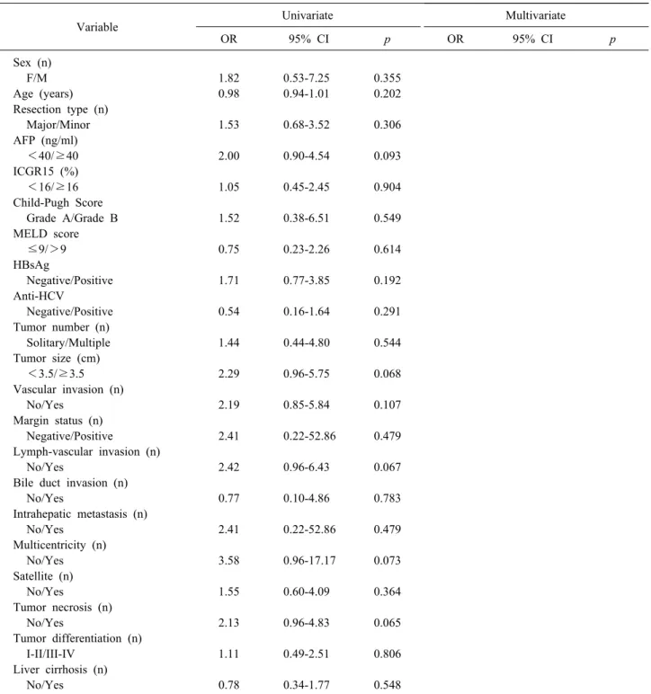
Table 5. Univariate and multivariate analysis of risk factors for early recurrence (≤1 year) in ALBI grade 2 after curative liver resection in HCC patients
Variable Univariate Multivariate
OR 95% CI p OR 95% CI p
Sex (n)
F/M 1.82 0.53-7.25 0.355
Age (years) 0.98 0.94-1.01 0.202
Resection type (n)
Major/Minor 1.53 0.68-3.52 0.306
AFP (ng/ml)
<40/≥40 2.00 0.90-4.54 0.093
ICGR15 (%)
<16/≥16 1.05 0.45-2.45 0.904
Child-Pugh Score
Grade A/Grade B 1.52 0.38-6.51 0.549
MELD score
≤9/>9 0.75 0.23-2.26 0.614
HBsAg
Negative/Positive 1.71 0.77-3.85 0.192
Anti-HCV
Negative/Positive 0.54 0.16-1.64 0.291
Tumor number (n)
Solitary/Multiple 1.44 0.44-4.80 0.544
Tumor size (cm)
<3.5/≥3.5 2.29 0.96-5.75 0.068
Vascular invasion (n)
No/Yes 2.19 0.85-5.84 0.107
Margin status (n)
Negative/Positive 2.41 0.22-52.86 0.479
Lymph-vascular invasion (n)
No/Yes 2.42 0.96-6.43 0.067
Bile duct invasion (n)
No/Yes 0.77 0.10-4.86 0.783
Intrahepatic metastasis (n)
No/Yes 2.41 0.22-52.86 0.479
Multicentricity (n)
No/Yes 3.58 0.96-17.17 0.073
Satellite (n)
No/Yes 1.55 0.60-4.09 0.364
Tumor necrosis (n)
No/Yes 2.13 0.96-4.83 0.065
Tumor differentiation (n)
I-II/III-IV 1.11 0.49-2.51 0.806
Liver cirrhosis (n)
No/Yes 0.78 0.34-1.77 0.548
AFP, alpha-fetoprotein; ICGR15, indocyanine green retention test at 15 minutes; MELD, model for end-stage liver disease;
HBsAg, hepatitis B surface antigen; Anti-HCV, Anti-hepatitis C virus
95% CI: 2.14-6.16), vascular invasion (p=0.003, OR:
2.28, 95% CI: 1.35-3.97), lymph-vascular invasion (p
<0.001, OR: 4.33, 95% CI: 2.47-7.64), bile duct invasion (p=0.042, OR: 5.93, 95% CI: 1.14-43.32), intrahepatic metastasis (p=0.014, OR: 15.06, 95% CI: 2.39-290.49),
presence of satellite lesions (p<0.001, OR: 3.51, 95% CI:
2.00-6.17), tumor necrosis (p<0.001, OR: 3.19, 95% CI:
1.96-5.29), and tumor differentiation (p<0.001, OR: 2.38, 95% CI: 1.48-3.87) were associated with a high risk of ER on univariate analysis; MELD score >9 (p=0.046,
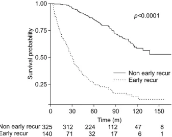
Fig. 2. Overall survival of early recurrence (≤1 year) after curative hepatectomy in hepatocellular carcinoma patients.
OR: 5.80, 95% CI: 1.08-36.29), HBsAg-positive status (p=0.004, OR: 2.50, 95% CI: 1.36-4.75), tumor size ≥3.5 cm (p<0.001, OR: 3.47, 95% CI: 1.96-6.32), lymph-vas- cular invasion (p=0.001, OR: 2.90, 95% CI: 1.52-5.54), presence of satellite lesions (p=0.002, OR: 2.76, 95% CI:
1.43-5.30), and tumor differentiation (p=0.007, OR: 2.10, 95% CI: 1.23-3.62) were found to be independent risk factors in multivariate analysis. In the ALBI grade 2 sub- group, no risk factors were associated with ER (Table 5).
The Kaplan-Meier curve showed that significantly low- er overall survival in ER patients than non-ER patients (Fig. 2).
DISCUSSION
In this study, the risk factors for ER after curative liver resection in HCC patients were AFP ≥40, preoperative ALBI grade ≥2, HBsAg-positive status, presence of mul- tiple tumors, large tumor size, vascular invasion, lymph-vascular invasion, intrahepatic metastasis, multi- centricity, presence of satellite lesions, tumor necrosis, and poor tumor differentiation, consistent with the find- ings in previous studies. The risk factors reported in pre- vious studies suggest that intrahepatic metastases are the main cause of early intrahepatic recurrence.4,16 In this study, ALBI score was found to be associated with ER, which was not reported in previous studies.
ALBI score has been identified as one of the best in- dicators of liver function. Most studies have shown that the ALBI score may be a better predictor of liver function
than the CP score, MELD score, and ICGR15 and is therefore more useful as a predictor of postoperative hep- atic failure.17 Moreover, several studies have found that ALBI is associated with prognostic factors for disease-free survival and overall survival.13 Kanda et al.18 reported that ALBI grade 2 patients were more likely to have shorter disease-specific and disease-free survival after radical gas- trectomy, compared with that for ALBI grade 1 patients.
Additionally multivariable analysis identified ALBI grade 2 as an independent prognostic factor for disease-free survival.
We performed subgroup analysis based on the assump- tion that liver function is associated with ER. In ALBI grade 1 patients, the risk factors for ER were tumor size
≥3.5 cm, venous/lymphatic invasion, presence of satellite lesions, and poor tumor differentiation; however, grade 2 patients showed no associations with ER. It can be as- sumed that ER is mainly affected by histological factors in ALBI grade 1 patients, which means liver function is favorable. However in ALBI grade 2 patients, no risk fac- tors were associated with ER. According to these results, liver function impairment is mainly associated with ER when liver function is not favorable. Lise et al.3 reported that CP class was an independent prognostic factor for disease-free survival and overall survival in multivariate analysis, while liver function impairment may be asso- ciated with recurrence of HCC. Hirokawa et al.19 reported that ER after curative hepatectomy in HCC patients was associated with ICGR15 >16%, and that recurrence pat- terns and risk factors vary by liver function status. These results suggest that liver function may be associated with ER.
Our study has some limitations. First, this study was conducted at a single institution, and the retrospective study design might have resulted in bias. Second, the most common cause of HCC in this study was hepatitis B virus, but HCC in Western countries is associated with other causes such as hepatitis C virus infection. Therefore, more studies are required according to etiological populations.
Despite these limitations, this study showed that ALBI grade was associated with ER after curative liver resection for HCC and could be useful as a preoperative predictor of ER. The ALBI grade can also be used to determine the likelihood of resection or transplantation because liver function can be assessed.
In conclusion, preoperative ALBI grade ≥2 is a pre- operative risk factor for ER, along with other well-known factors, and requires more thorough follow-up. ALBI grade may be useful in determining appropriate manage- ment according to liver function when recurrence develops.
REFERENCES
1. Park JH, Koh KC, Choi MS, Lee JH, Yoo BC, Paik SW, et al.
Analysis of risk factors associated with early multinodular recur- rences after hepatic resection for hepatocellular carcinoma. Am J Surg 2006;192:29-33.
2. Chen MF, Hwang TL, Jeng LB, Wang CS, Jan YY, Chen SC.
Postoperative recurrence of hepatocellular carcinoma. Two hun- dred five consecutive patients who underwent hepatic resection in 15 years. Arch Surg 1994;129:738-742.
3. Lise M, Bacchetti S, Da Pian P, Nitti D, Pilati PL, Pigato P.
Prognostic factors affecting long term outcome after liver re- section for hepatocellular carcinoma: results in a series of 100 Italian patients. Cancer 1998;82:1028-1036.
4. Poon RT, Fan ST, Lo CM, Liu CL, Wong J. Long-term survival and pattern of recurrence after resection of small hepatocellular carcinoma in patients with preserved liver function: implications for a strategy of salvage transplantation. Ann Surg 2002;235:
373-382.
5. Lee SY, Konstantinidis IT, Eaton AA, Gönen M, Kingham TP, D'Angelica MI, et al. Predicting recurrence patterns after re- section of hepatocellular cancer. HPB (Oxford) 2014;16:943-953.
6. Tabrizian P, Jibara G, Shrager B, Schwartz M, Roayaie S.
Recurrence of hepatocellular cancer after resection: patterns, treatments, and prognosis. Ann Surg 2015;261:947-955.
7. Shah SA, Greig PD, Gallinger S, Cattral MS, Dixon E, Kim RD, et al. Factors associated with early recurrence after resection for hepatocellular carcinoma and outcomes. J Am Coll Surg 2006;202:275-283.
8. Cheng Z, Yang P, Qu S, Zhou J, Yang J, Yang X, et al. Risk factors and management for early and late intrahepatic re- currence of solitary hepatocellular carcinoma after curative
resection. HPB (Oxford) 2015;17:422-427.
9. Imamura H, Matsuyama Y, Tanaka E, Ohkubo T, Hasegawa K, Miyagawa S, et al. Risk factors contributing to early and late phase intrahepatic recurrence of hepatocellular carcinoma after hepatectomy. J Hepatol 2003;38:200-207.
10. Li T, Fan J, Qin LX, Zhou J, Sun HC, Qiu SJ, et al. Risk factors, prognosis, and management of early and late intrahepatic re- currence after resection of primary clear cell carcinoma of the liver. Ann Surg Oncol 2011;18:1955-1963.
11. Wu JC, Huang YH, Chau GY, Su CW, Lai CR, Lee PC, et al.
Risk factors for early and late recurrence in hepatitis B-related hepatocellular carcinoma. J Hepatol 2009;51:890-897.
12. Tung-Ping Poon R, Fan ST, Wong J. Risk factors, prevention, and management of postoperative recurrence after resection of hepatocellular carcinoma. Ann Surg 2000;232:10-24.
13. Harimoto N, Yoshizumi T, Sakata K, Nagatsu A, Motomura T, Itoh S, et al. Prognostic significance of combined albumin-bilir- ubin and tumor-node-metastasis staging system in patients who underwent hepatic resection for hepatocellular carcinoma.
Hepatol Res 2017;47:1289-1298.
14. Pol B, Campan P, Hardwigsen J, Botti G, Pons J, Le Treut YP.
Morbidity of major hepatic resections: a 100-case prospective study. Eur J Surg 1999;165:446-453.
15. Johnson PJ, Berhane S, Kagebayashi C, Satomura S, Teng M, Reeves HL, et al. Assessment of liver function in patients with hepatocellular carcinoma: a new evidence-based approach-the ALBI grade. J Clin Oncol 2015;33:550-558.
16. Yamamoto J, Kosuge T, Takayama T, Shimada K, Yamasaki S, Ozaki H, et al. Recurrence of hepatocellular carcinoma after surgery. Br J Surg 1996;83:1219-1222.
17. Wang YY, Zhong JH, Su ZY, Huang JF, Lu SD, Xiang BD, et al. Albumin-bilirubin versus Child-Pugh score as a predictor of outcome after liver resection for hepatocellular carcinoma. Br J Surg 2016;103:725-734.
18. Kanda M, Tanaka C, Kobayashi D, Uda H, Inaoka K, Tanaka Y, et al. Preoperative albumin-bilirubin grade predicts recur- rences after radical gastrectomy in patients with pT2-4 gastric cancer. World J Surg 2018;42:773-781.
19. Hirokawa F, Hayashi M, Asakuma M, Shimizu T, Inoue Y, Uchiyama K. Risk factors and patterns of early recurrence after curative hepatectomy for hepatocellular carcinoma. Surg Oncol 2016;25:24-29.