Introduction
The hamstring muscles refer to four muscles lo- cated in the posterior region of the thigh: the semi- membranosus, the semitendinosus, the biceps femoris long head and the biceps femoris short head (Woodley and Mercer, 2005). An inadequate hamstring muscle length has historically been thought of as a possible cause of hamstring injury (Rolls and George, 2004).
Hamstring flexibility is recommended during routine physical examinations and in deciding athlete read- iness to return-to-play following injury (Croisier et al, 2002; Drezner, 2003; Mendiguchia and Brughelli, 2011).
Clinicians routinely assess hamstring muscle length in people with injuries to their musculoskeletal system
(Magee, 2006). Methods to assess hamstring flexibility include the straight-leg-raising (SLR) test, the sit and reach (SR) test, the standing toe-touch test and the passive knee extension (PKE) test and the active knee extension (AKE) test (Baltaci et al, 2003; Booher and Thibodeau, 1985; Gajdosik and Lusin, 1983).
The specificity of the SLR test has been ques- tioned, as it is also widely used as a neurological test (Malanga and Nadler, 2006). In addition, Liebenson et al (2009) suggested that neural mobility limits the SLR test. Further, the SLR test causes elongation of the sciatic nerve and associated structures. Finally, pelvic rotation may influence the validity of SLR an- gle measurements (Bohannon, 1982). The validity of SR test is also considered to be poor (Clark, 2008).
Corresponding author: Oh-yun Kwon [email protected]
Reliability of the Active Knee Extension Test With a Pressure Biofeedback Unit
Chang-ho Kim1, MSc, PT, Gyeong-tae Gwak1, BPT, PT, Oh-yun Kwon2,3, PhD, PT
1Dept. of Physical Therapy, The Graduate School, Yonsei University
2Dept. of Physical Therapy, College of Health Science, Yonsei University
3Dept. of Ergonomic Therapy, The Graduate School of Health and Environment, Yonsei University
Abstract
1)Background: The active knee extension (AKE) test commonly used to assess the flexibility of the hamstring muscles. Many researchers have tested the reliability of the AKE test; however, no published studies have examined the intrarater and interrater reliability of the AKE test using a PBU.
Objects: The purpose of this study was to determine the intrarater and interrater reliability of the AKE test performed with a pressure biofeedback unit (PBU) on healthy subjects.
Methods: Sixteen healthy male participants volunteered and gave informed consent to participate in this study. Two raters conducted AKE tests independently with a PBU. Each knee was measured twice, and the AKE testing was repeated one week after the first round of testing.
Results: The interrater reliability’s intraclass correlation coefficients (ICC2,1) were .887∼.986 for the right knees and .915∼.988 for the left knees. In addition, the intrarater (test-retest) reliability (ICC3,1) values ranged between .820∼.915 and .820∼.884 for Raters 1 and 2, respectively. The values for the standard error of mesurement were low for all tests (.81∼2.97˚); the calculated minimum detectable change was 2.24∼8.21˚.
Conclusion: These findings suggest that the AKE test performed with a PBU had excellent interrater and intrarater reliability for assessing hamstring flexibility in healthy young males.
Key Words: Active knee extension test; Hamstring flexibility; Pressure biofeedback unit; Reliability.
Some factors can contribute to changes in-the result, such as-the differences in the proportions of the up- per and lower limbs, the flexibility of the spine and scapular abduction (Perin et al, 2015). The standing toe-touch test assesses gross motion, such as hip and vertebral flexion. Because of the concurrent mo- tion of each of the segments involved (i.e., the lum- bar vertebrae, the sacroiliac joint, and the hip joint), it is impossible to measure the amount of motion that occurs at each segment. Therefore, this test cannot accurately assess hamstring length (Kippers and Parker, 1987). The PKE test was performed in a study in which the subjects laid supine while a re- searcher passively positioned the hip to be tested at 90 degrees of flexion and the other hip remained in a neutral position. Then, another researcher passively extended the knee of the tested leg until the subject complained of pain or the researcher felt tightness in the hamstring. The intratester reliability for this method was .98 (intra correlation coefficient; ICC1,1).
In light of the evidence presented above, we chose to assess the interrater reliability of measuring ham- string length using the AKE test described by Gadjosik and Lusin (1983). This test has been shown to demonstrate good intrarater reliability (.99) in meas- uring hamstring length (Gadjosik and Lusin, 1983).
The advantage of this test is that significant amounts of movement at the hip joint, sacroiliac joint, and lum- bar spine are controlled through stabilization (Gadjosik and Lusin, 1983). However, there is no evidence that pelvic rotation is well controlled using this method.
Learning and teaching accurate control pelvic movement is difficult because it is limited in its ability to detect fine activities of the deeply located lumbopelvic area. For this reason, feedback tools are often used. A pressure biofeedback unit (PBU) is a non-invasive method that is economical, and it can be easily used anywhere since it is portable (Park and Lee, 2013). Furthermore, a PBU has been used to monitor the motion of the lumbopelvic region (Cynn et al, 2006; von Garnier et al, 2009). Therefore, the PBU can be used for preventing lumbopelvic mo-
tion during AKE test.
However, no published studies have examined the interrater reliability of the AKE test using a PBU.
Therefore, the purpose of this study was to establish the intra and interrater reliability of the AKE test using a PBU.
Methods
Subjects
The convenience sampling of 16 healthy male par- ticipants with-“normal”-muscle strength and a nor- mal range of motion in the hips and knees was conducted. All participants have not known impair- ment of the musculoskeletal system affecting the lumbar or lower extremities for at least 1 year and received instructions regarding the required re- striction of excessive physical activity. All procedures were explained to the subjects, and. informed consent was obtained from all the subjects. The subjects were instructed to avoid any sporting activities on the day of the assessments. All data were gathered in the department of physical therapy at Yonsei University Wonju campus in South Korea between 9:00 and 11:00 a.m. at room temperature (22˚∼24˚).
This study was approved by the institutional review board of Yonsei University.
Active knee extension testing using a pressure biofeedback unit
The PBU is a pressure transducer consisting of a there-chamber air-filled pressure bag connected to a sphygmomanometer gauge by a catheter. Moving body segments causes volume changes in the pressure bag, which is registered on the device (Storheim K, 2002).
Before beginning the AKE test, four anatomical references were marked with a black pen while the subject remained in a natural standing position. The anatomical landmarks were the anterior superior iliac spines, and the posterior superior iliac spines were marked bilaterally. The anatomical references were
used to determine the settings of the PBU. All AKE tests using the PUB performed in this study were conducted according to the AKE test described by Gajdosik and Lusin (1983). A PBU (Pressure Biofeedback unit, HEALIENCE, Korea) was used to monitor lumbopelvic movement during the AKE test.
The same PBU was used throughout the study to avoid between-device differences (von Garnier et al, 2009). To assure the exactness of the PBU measure- ments, the unit was pretested by loading the bio- feedback unit bag for 24 hours with 4 ㎏. A unit is considered appropriate if the device loses no more than .5 ㎜Hg during the 24-hour period (von Garnier et al, 2009). The accuracy of the apparatus was found to be ±3 ㎜Hg (Storheim et al, 2002).
Two physiotherapists conducted all the AKE tests with a PBU independently. Each subject was as- sessed in the supine position on an examination table with both lower extremities extended. Both anterior superior iliac spines were aligned with the vertical target bars, and a strap was placed over the anterior superior spines of the ilia to stabilize the pelvis passively. The lower extremity not being measured was strapped to the table across the lower third of the thigh using an adjustable belt (Gajdosik and Lusin, 1983) (Figure 1).
The PBU was positioned under the lumbar spine so that the lower border of the pressure bag was aligned with the posterior superior iliac spine marks.
To minimize the displacement of the pressure bag during the AKE test, the distal end of the pressure
bag was taped to the subject.
The pressure bag was filled with air to create a pressure of 40 ㎜Hg, and active lumbo-pelvic stabili- zation was accomplished by internal fixation.
Abdominal contraction was performed at a slightly increased pressure of about 0∼2 ㎜Hg during the AKE test (Park and Lee, 2013). With the lumbopel- vic stabilized using a PBU and the ankle relaxed in plantar flexion, the subject actively extended the knee while maintaining contact with the target bar.
A bubble inclinometer was used to measure popliteal angle during the AKE test. The popliteal angle is defined as the degree of knee flexion from terminal knee extension. The participants were instructed to rest for 10 minutes to minimize the effect of the previous measurement of the previous measurement.
The popliteal angle was recorded twice, and the mean angle of the AKE test was used for analysis.
All participants attended two testing sessions one week apart to determine the test-retest reliability of the method. The AKE testing was carried out by Raters 1 and 2 independently in two separate ses- sioins (Tests 1 and 2) (Figure 2). Both assessor were physical therapists with over one year of expe- rience in physical therapy and underwent specific training to conduct AKE test using the proposed method. The order in which the rater assessed the participants was randomly assigned in the first ses- sion and maintained thereafter.
Statistical analysis
SPSS version 18.0 (SPSS Inc., Chicago, IL, USA) was used for the statistical analyses, which was treated as a descriptive analysis (i.e., using means and standard deviation; SD). The Shapiro-Wilk test was used to check the data normality. Paired t-tests were conducted to compare differences between test and retest measurements within and between raters.
The intra-rater and inter-rater reproducibility of the AKE test using a PBU was determined by com- puting intraclass correlation coefficients (ICCs). The ICC value of the standard error of measurement Figure 1. Active knee extension with PBU.
(SEM) was calculated to show the magnitude of dif- ference between the measurements (Shrout and Fleiss, 1979). The SEM was calculated using a pre- vious study’s formula: SEM=SDavg (√1−ICC) (Gabbe, 2004). A smaller SEM value suggests great- er agreement between measurements (Atkinson and Nevill, 1998). The minimum detectable change (MDC) scores were calculated using the following formula:
MDC=1.96×SEM×√2 (Macedo and Magee, 2008).
Results
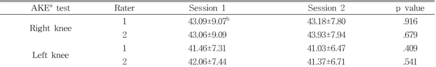
The mean age, weight, and height (mean±SD) of the participant were 23.5±2.8 years, 74.0±1.4 ㎏, and 171.9±2.1 ㎝, respectively. The mean, SD and P val- ue are shown for the popliteal angle according to measurement methods shown in Tables 1 and 3. No significant difference between raters was noted for either of the lower limb scores (Table 1). There was no significant difference in the AKE measurements using a PBU between sessions (Table 3). The AKE test interrater reproducibility was excellent, with ICC values ranging from .887 to .988 for both raters (Table 2). The AKE measurements of Raters 1 and
2 were compared between the first and second test- ing sessions. The test-retest reproducibility in this study was excellent, with ICC values ranging from .820 to .915 for both raters (Table 4).
Discussion
The original AKE test was designed to minimize pelvic motion by securing the pelvis with a strap (Gajdosik and Lusin, 1983). The AKE test is now considered to be the gold standard for assessing hamstring flexibility (Davis et al, 2008). This study showed that the AKE test performed using a PBU is reproducible as a method for measuring hamstring muscle flexibility in healthy young adults. In fact, no difference was found between the test-retest measurements.
It is important to correct AKE test values biased by active lumbopelvic movement to attain a precise as- sessment of hamstring flexibility. With the aid of a lumbopelvic stabilizing device, we showed that an as- sessor can conduct the AKE test more precisely with- out the need to move the lumbopelvic area. Interrater reproducibility ICC2,1 values of .986 and .988 were Figure 2. Process of the reproducibility of the AKE test with PBU.
found for the right and left knees, respectively, in the first session. To improve the precision of the meas- urement, using a PBU produced a SEM ranging from .81∼2.97 for the right and left knees and the MDC score for this method, ranging form 2.24∼8.21 degrees, indicated that Lumbopelvic stabilization is one of the considerations for precise measurement and may help to minimize measurement error when evaluating ham- string muscle flexibility using the AKE test with PBU.
Intrarater reproducibility in this study was ex-
cellent, with ICC3,1 values ranging from .820∼.915 for both raters. From a clinical point of view, the high intrarater and interrater reliability values obtained in this study lead us believe that this test can be used with minimal bias. Our results is in agreement with earlier studies. Neto et al (2015) found an intrarater ICC of .91 for AKE in the dominant leg and .91 for the nondominant leg. Another study reported a sim- ilar value for the interrater ICC2,1 values of .87 for the dominant knee and .81 for the nondominant knee
AKEa measurements Session Rater 1 Rater 2 p value
Right knee 1 43.09±9.07b 43.06±9.09 .992
2 43.18±7.80 43.93±7.94 .789
Left knee 1 41.46±7.31 42.06±7.44 .821
2 41.03±6.47 41.37±6.71 .884
aactive knee extension, bmean±standard deviation.
Table 1. Mean and standard deviation of the popliteal angle according to different measurement methods (N=16)
AKEa measurements Session ICC2,1b ICC2,1(95% CIc) SEMd MDCe
Right knee 1 .986 .960∼.995 .99 2.74
2 .887 .707∼.959 2.68 7.41
Left knee 1 .988 .966∼.996 .81 2.24
2 .915 .775∼.970 1.91 5.28
aactive knee extension, bintraclass correlation coefficient, cconfidence interval, dstandard error of measurement, eminimum detectable change.
Table 2. Interrater reproducibility of the active knee extension (AKE) test
AKEa test Rater Session 1 Session 2 p value
Right knee 1 43.09±9.07b 43.18±7.80 .916
2 43.06±9.09 43.93±7.94 .679
Left knee 1 41.46±7.31 41.03±6.47 .409
2 42.06±7.44 41.37±6.71 .541
aactive knee extension, bmean±standard deviation.
Table 3. Mean and standard deviation of the popliteal angle according to different measurement methods (N=16)
AKEa test Rater ICC3,1b ICC3,1(95% CIc) SEMd MDCe
Right knee 1 .915 .775∼.970 2.45 6.77
2 .884 .699∼.958 2.90 8.01
Left knee 1 .820 .559∼.933 2.89 7.93
2 .820 .559∼.933 2.97 8.21
aactive knee extension, bintraclass correlation coefficient, cconfidence interval, dstandard error of measurement, eminimum detectable change.
Table 4. Test-retest reproducibility of Raters 1 and 2
(Hamid et al, 2013).
Active lumbopelvic stabilization is another consid- eration to achieve even more precise assessments and may assist in minimizing measurement errors when evaluating hamstring muscle flexibility using the AKE test.
However, the AKE test does not account for the potential effect of active lumbopelvic movement. It is not enough to stabilize the spine and pelvis with a strap when the end point of knee extension is accom- plished as the hamstring muscle is stretched, as the stretching force is delivered to the ischial tuberosity, causing the pelvis to rotate (Bohannon, 1982). Further, while Gajdosik and Lusin (1983) reported that the AKE test had a Pearson product-moment correlation coefficient of .99 for both lower extremities, the high coefficient value can be explained by the use of dif- ferent statistical analyses. In addition, the time be- tween the first AKE test and the retest was not long enough in their study; both AKE tests were con- ducted on the same day just 30 minutes apart, which may have caused systematic bias or influenced the reliability (Gajdosik and Lusin, 1983).
This study also has some limitations. In spite of the excellent interrater and intrarater reproducibility, a wide range of confidence interval was reported.
Future research with larger samples may be neces- sary to determine reliability estimates with greater precision. In addition, as the subject is supposed to be able to contract the abdomen and knee extensor muscles during the test, this test is unsuitable for patients with neuromuscular disorders. Finally, ab- normal movements were visually observed during the AKE test with a PBU, we consider the lack of kine- matic data for objective control of movement is a limitation of the present study.
Conclusion
The purpose of this study was to establish the in- tra and interrater reliability of the AKE test using a
PBU. This study demonstrated that the AKE test can be performed more precisely using a PBU, as this method has excellent interrater and intrarater reproducibility. Despite several limitations, the results of this study suggest that using a PBU was effec- tive for testing hamstring muscle flexibility and for minimization of measurement errors in the AKE test.
Overall, this study provides useful information that can assist physical trainers, researchers, and clini- cians select the proper tools to assess hamstring muscle flexibility.
References
Atkinson G, Nevill AM. Statistical methods for as- sessing measurement error (reliability) in varia- bles relevant to Sports Medicine. Sports Med.
1998;26(4):217-238.
Baltaci G, Un N, Tunay V, et al. Comparison of three different sit and reach tests for measurement of hamstring flexibility in female university students.
Br J Sports Med. 2003;37(1):59-61.
Bohannon RW. Cinematographic analysis of the pas- sive straight-leg raising test for hamstring muscle length. Phys Ther. 1982;62(9):1269-1274.
Booher JM, Thibodeau GA. Athletic Injury Assessment.
4th ed. New York, McGraw-Hill, 1985:457-506.
Clark RA. Hamstring injuries: Risk assessment and injury prevention. Ann Acad Med Singapore. 2008;
37(4):341-346.
Croisier JL, Forthomme B, Namurois MH, et al.
Hamstring muscle strain recurrence and strength performance disorders. Am J Sports Med. 2002;
30(2):199-203.
Cynn HS, Oh JS, Kwon OY, et al. Effects of lumbar stabilization using a pressure biofeedback unit on muscle activity and lateral pelvic tilt during hip abduction in sidelying. Arch Phys Med Rehabil. 2006;87(11):1454-1458.
Davis DS, Quinn RO, Whiteman CT, et al. Concurrent validity of four clinical tests used to measure hamstring
flexibility. J Strength Cond Res. 2008;22(2):583-588.
https://doi.org/10.1519/jsc.0b013e31816359f2 Drezner JA. Practical management: Hamstring muscle
injuries. Clin J Sport Med, 2003;13(1):48-52.
Gabbe BJ. Reliability of common lower extremity musculoskeletal screening tests. Phys Ther Sport.
2004;5(2):90-97.
Gajdosik R, Lusin G. Hamstring muscle tightness.
Reliability of an active-knee-extension test.
Phys Ther. 1983;63:1085-1090.
Hamid MS, Ali MR, Yusof A. Interrater and intrarater reliability of the active knee extension (AKE) Test among Healthy Adults. J Phys Ther Sci. 2013;
25(8):957-961. https://doi.org/10.1589/jpts.25.957 Kippers V, Parker AW. Toe-touch test. A measure
of its validity. Phys Ther. 1987;67(11):1680-1684.
Liebenson C, Karpowicz AM, Brown SH, et al. The active straight leg raise test and lumbar spine stability. PM.R. 2009;1(6):530-535. https://doi.org/
10.1016/j.pmrj.2009.03.007
Macedo LG, Magee DJ. Differences in range of mo- tion between dominant and nondominant sides of the upper and lower extremities. J Manipulative Physiol Ther. 2008;31(8):577-582. https://doi.org/
10.1016/j.jmpt.2008.09.003
Magee DJ. Orthopedic Physical Assessment. 4th ed.
Philadelphia, Saunders elsevier, 2006:696-697.
Malanga GA, Nadler S. Musculoskeletal Physical Examination: An evidence-based approach.
Philadelphia, Hanley & Belfus, 2006:251-279.
Mendiguchia J, Brughelli M. A return-to-sport algo- rithm for acute hamstring injuries. Phys Ther Sport. 2011;12(1):2-14. https://doi.org/10.1016/
j.ptsp.2010.07.003
Neto T, Jacobsohn L, Carita AI, et al. Reliability of the active-knee-extension and straight-leg-raise
tests in subjects with flexibility deficits. J Sport Rehabil. 2015; e pub only. https://doi.org/10.1123/
jsr.2014-0220
Park DJ, Lee SK. What is a suitable pressure for the abdominal drawing-in maneuver in the su- pine position using a pressure biofeedback unit?
J Phys Ther Sci. 2013;25(5):527-530. https://do- i.org/10.1589/jpts.25.527
Perin A, Ulbricht L, Neves EB. Contribution of dif- ferent body segments in sit and reach test. mo- tricidade 2015;11(2):153-162.
Rolls A, George K. The relationship between ham- string muscle injuries and hamstring muscle length in young elite footballers. Phys Ther Sport. 2004;5(4):179-187.
Shrout PE, Fleiss JL. Intraclass correlations: Uses in assessing rater reliability. Psychol Bull. 1979;
86(2):420-428.
Storheim K, Bo K, Pederstad O, et al. Intra-tester reproducibility of pressure biofeedback in meas- urement of transversus abdominis function.
Physiother Res Int. 2002;7(4):239-249.
von Garnier K, Koveker K, Rackwitz B, et al.
Reliability of a test measuring transversus ab- dominis muscle recruitment with a pressure bio- feedback unit. Physiotherapy. 2009;95(1):8-14.
https://doi.org/10.1016/j.physio.2008.10.003
Woodley SJ, Mercer SR. Hamstring Muscles:
Architecture and innervation. Cells Tissues Organs. 2005;179(3):125-141.
This article was received August 10, 2017, was reviewed August 10, 2017, and was accepted September 11, 2017.