J. Exp. Biomed. Sci. 2013, 19(2): 153~157 pISSN : 1738-3226
Performance of Quantitative Real-Time PCR for Detection of Tuberculosis in Granulomatous Lymphadenitis Using
Formalin-Fixed Paraffin-Embedded Tissue
Jijgee Munkhdelger 1 , Khalilullah Mia-Jan 1 , Dongsup Lee 2 , Sangjung Park 3 , Sunghyun Kim 3 , Yeonim Choi 3 , Hye-Young Wang 4 , Bo-Young Jeon 3 , Hyeyoung Lee 3,† and Kwang Hwa Park 1,†
1
Department of Pathology, Wonju College of Medicine, Yonsei University, Wonju 220-701, Korea
2
Department of Clinical Laboratory Science, Hyejeon College, Hongseong, Chungnam 350-702, Korea
3
Department of Biomedical Laboratory Science, College of Health Sciences, Yonsei University, Wonju 220-710, Korea
4
M&D, Inc., Wonju Eco Environmental Technology Center, Wonju, Gangwon-do 220-710, Korea
Although culture is the gold standard method to identify mycobacteria, its use in tuberculous lymphadenitis (TBL) is limited due to formalin fixation of the submitted specimens. We evaluated the performance of quantitative real-time PCR (q-PCR) for Mycobacterium Tuberculosis (MTB) in granulomatous lymphadenitis using formalin-fixed paraffin- embedded (FFPE) tissues. From 2000 to 2010, a total number of 117 cases of lymph node samples with granulomatous inflammation which were surgically removed and fixed in formalin were studied. Hematoxylin & Eosin (H&E) and Ziehl-Neelsen-stained (ZN) slides were reviewed. qPCR using Real TB-Taq
®was performed for all cases to identify Mycobacterium tuberculosis. Thirteen non-tuberculous lymphadenopathy cases were used as negative control. Cervical lymph nodes were more frequently affected (60%, 70/117) than other sites. ZN stain for acid fast bacilli was positive in 19 (16.24%) cases. qPCR for tuberculosis was positive in 92 (78.63%) cases. Caseous necrosis was found in 103 (88.03%) cases. While the ZN stain and qPCR were both negative in all control cases, the qPCR showed a significantly higher positive rate (78.63% vs. 16.24%) compared to ZN stain in histologically diagnosed TBL. Quantitative real-time PCR proves to be more sensitive than ZN stain for diagnosis of tuberculous lymphadenitis.
Key Words: Granulomatous lymphadenopathy, Mycobacterium tuberculosis, Real-time polymerase chain reaction
Tuberculosis (TB) remains a major threat to human health.
Although the incidence rate of TB has been declining for several years, 8.7 million new cases and 1.4 million mortalities were estimated in 2011 (Huang et al., 2009).
Peripheral tuberculous lymphadenitis (TBL), previously
termed "scrofula" is the most common form of extra- pulmonary TB. In countries with high incidence of TB, the TBL is the most common cause of lymphadenopathy (Ahmed et al., 2011).
Diagnosis of TBL still has its own difficulties. Granulo- matous inflammation, caseous necrosis and Langhans giant cells, which are the main histopathologic findings are not specific to only TB, but can be found in other diseases, such as sarcoidosis, fungal infections, cat scratch disease, toxo- plasmosis, collagen vascular diseases and some malignancies.
A confirmative diagnosis of disease therefore relies on detection of the pathogen. Ziehl-Neelsen (ZN) stain and culture, which are conventional methods for detecting Mycobacterium tuberculosis complex, have their own disadvantages. Because of the paucibacillary nature of the
Brief Communication
*
Received: March 13, 2013 / Revised: April 13, 2013 Accepted: April 25, 2013
†
Corresponding author: Hyeyoung Lee. Department of Biomedical Laboratory Science, College of Health Sciences, Yonsei University 1 Yonseidae-gil, Wonju 220-710, Korea.
Tel: +82-33-760-2740, Fax: +82-33-760-2561 e-mail: [email protected]
†
Corresponding author: Kwang Hwa Park. Department of Pathology, Wonju College of Medicine, Yonsei University 162 Ilsandong, Wonju 220-701, Korea.
Tel: +82-33-731-6590, Fax: +82-33-741-1556 e-mail: [email protected]
○
CThe Korean Society for Biomedical Laboratory Sciences. All rights reserved.
specimens, the sensitivity is low in both methods. The sensitivity ranges between 15~47% and 35~65% in ZN stain and culture, respectively (Singh et al., 2000). Moreover, the culture takes several weeks to reveal the results, causing delay in the initial treatment.
Recently, the nucleic acid amplification tests (NAAT), especially polymerase chain reaction (PCR) has become a frequently used molecular test in TB diagnosis and its accuracy in rapid diagnosis of pulmonary tuberculosis is well accepted (Greco et al., 2006; Yang et al., 2011;
Linasmita et al., 2012). Nevertheless, the use of PCR in diagnosis of TBL is ambiguous and not well studied (Linasmita et al., 2012). Although previous studies on PCR for diagnosis of TBL have generally shown modestly higher diagnostic accuracy compared to performance of conventional methods, their results are highly variable and inconsistent (Daley et al., 2007). Nonetheless, studies done on formalin fixed paraffin embedded (FFPE) lymph node tissues are scarce (Kwon et al., 2000; Park et al., 2003;
Johansen et al., 2004; Selva et al., 2004; Schulz et al., 2005).
Commercial PCR kits give greater diagnostic accuracy than in-house assays. Among them, the Gen-Probe AMTD (Gen-Probe Inc, San Diego, CA, USA) and Roche Amplicor (Roche Diagnostics, Branchburg, NJ, USA) are widely used commercial detection kits with relatively higher accuracy (Daley et al., 2007).
In this study, we aimed to evaluate the performance of quantitative real-time PCR (qPCR) kit, Real TB-Taq
®(M&D Inc, Wonju, Korea) in diagnosis of TBL, using formalin-fixed paraffin embedded (FFPE) tissues.
One hundred seventeen cases of granulomatous lymph- adenitis were retrieved from the Department of Pathology, Wonju Christian Hospital, between January 2000 and December 2010. Approval was obtained from the In- stitutional Ethics Committee of Yonsei University Wonju College of Medicine (YWMR-12-4-076). All cases were diagnosed as tuberculosis based on histologic findings. Other causes of granulomatous lymphadenitis such as sarcoidosis, rheumatoid arthritis, fungal infections, and cat scratch disease were ruled out by pathologic findings, histochemical staining and clinical information. All cases were stained with hematoxylin and eosin (H&E) and ZN stains and
reviewed by pathologist to identify caseous necrosis and acid-fast bacilli. For the negative control, we used thirteen cases of non-tuberculous lymphadenopathy (toxoplasmosis, fungal infection, Hodgkin's lymphoma, and sarcoidosis).
Five 5 μm tissue sections were cut from the paraffin block and put into Eppendorf tube. 1.2 ml of xylene was added to each tube and the samples were gently mixed for 20 minutes at 65℃. Samples were centrifuged at 13,000 rpm for 5 minutes and the supernatants were discarded.
Residual xylene was removed by washing the samples twice with 1.2 ml of 100% ethanol for 5 minutes and centrifuging at 13,000 rpm for 5 minutes. The pellets were dried at 37℃ for 10 minutes. HELEX-100 (5% in d.w.;
Bio-Rad Laboratories, Hercules, CA, USA) was added to each sample and mixed for 1 minute and incubated at 56℃
for 15 minutes. The sample were then incubated at 100℃
for 10 minutes in boiling water bath and centrifuged at 13,000 rpm for 3 minutes.
We performed qPCR using Real TB-Taq
®kit (M&D Inc, Wonju, Korea) according to the manufacturer's instruction.
In brief, qPCR was performed in a 25 μl reaction using the ABI Prism 7500 (Applied Biosystems, Foster City, CA, USA). Each 25 μl reaction mixture contained 2.5 μl of primer-probe mix, 12.5 μl of 2 × Real-time PCR Master mix (TOYOBO, Osaka, Japan) and 5 μl of DNA templates.
The thermal cycling conditions were 10 min at 95℃, followed by 40 cycles of 30s at 95℃, 2 min at 60℃.
Thermocycling, fluorescence data collection, and data analysis were performed with a 7500 fast real-time PCR system (v. 2.0.2) (ABI) according to the manufacturer's instructions, with the passive reference dye, ROX, turned off.
The results of qPCR were compared with those of ZN stain and histological features of the clinical specimens of the individual patients. Statistical analysis was performed using Microsoft
®Office Excel (2010) program.
From the total 117 chronic granulomatous lymphadenitis
patients, 40 were men and 77 were women with mean age of
41.5 years (range 0~81 years). The most frequent involved
sites were cervical (n=70, 59.83%), axillary (n=17, 14.53%),
supraclavicular (n=12, 10.26%), intra-abdominal (n=6,
5.13%), pulmonary (n=4, 3.42%), inguinal (n=4, 3.42%)
and submandibular, thymic, pancreatic and pericholedochal (n=1, 0.85%, respectively). Four patients were diagnosed with malignancy (stomach, colon, lung and submandibular gland) in the same year.
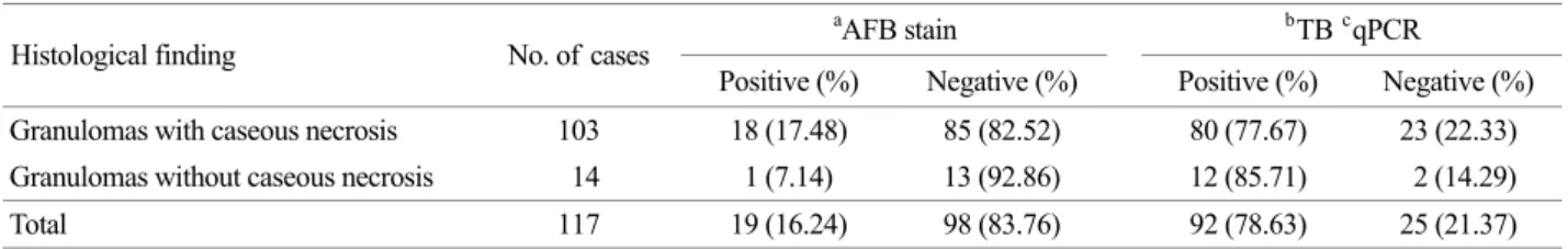
Histologically, all 117 cases showed chronic granulo- matous inflammation. Most of granulomatous lymphadenitis cases showed caseous necrosis (n=103, 88.03%). From the total 117 cases, 19 (16.24%) cases were AFB positive on ZN stain (Table 1). Most of the AFB positive cases (n=18, 94.74%) showed caseous necrosis.
The negative control cases included toxoplasmosis (n=4), fungal infection (n=4), Hodgkin's lymphoma (n=3), and sarcoidosis (n=2). All control cases were AFB negative.
From the 117 patients studied, 92 (78.63%) cases were positive for Real TB-Taq qPCR. From 103 cases with caseous necrosis, 18 (17.48%) cases were AFB positive and 80 (76.19%) cases were qPCR positive. From 14 cases without caseous necrosis, 1 (7.14%) case was AFB positive and 12 (85.71%) cases were qPCR positive. All 13 cases of non-tuberculous lymphadenopathy serving as negative control were negative in qPCR. Our PCR results did not show a single false positive result.
A comparison of the results of q-PCR and AFB staining is shown in Table 2. From the 92 qPCR positive cases, 13 (14.13%) cases were AFB positive and from 19 AFB positive cases, 13 (68.42%) were qPCR positive.
The lymph node tuberculosis is the most common form of extra pulmonary tuberculosis. The number of previous studies testing NAAT performance in nonrespiratory specimens, especially in lymph node tissue is relatively few and their results are highly heterogeneous due to differences in methods, and therefore limiting the clinical applicability
of their results and precluding summary estimate of their sensitivity and specificity (Daley et al., 2007; Linasmita et al., 2012). Most of the studies used fine needle aspiration (FNA) samples, while the sensitivity of biopsy PCR is significantly higher than that of the FNA PCR (Linasmita et al., 2012). Furthermore, performance of qPCR for detection of mycobacteria in lymph node specimen is not well studied (Bruijnesteijn Van Coppenraet et al., 2004; Causse et al., 2011; Linasmita et al., 2012). Additionally qPCR has the advantage of delivering quick results and lack of possibility of contamination that occurs during gel electrophoresis in conventional PCR. To test the performance of qPCR for detection of mycobacteria, we used Real TB-Taq
®kit in surgically excised FFPE lymph node tissues.
The previous studies evaluated the performance of PCR for diagnosis of TBL mostly on a limited number of samples (Daley et al., 2007). In our study, we performed qPCR analysis for Mycobacterium tuberculosis in 117 routinely submitted FFPE tissue blocks from histologically confirmed chronic granulomatous inflammation cases together with 13 negative-control cases of non-tuberculous lymphadenopathy.
Because lymph node specimens routinely go through formalin fixation after the submission to the pathology Table 1. Comparison of cases by existence of caseous necrosis, AFB stain results and TB-qPCR results
a
AFB stain
bTB
cqPCR
Histological finding No. of cases
Positive (%) Negative (%) Positive (%) Negative (%) Granulomas with caseous necrosis 103 18 (17.48) 85 (82.52) 80 (77.67) 23 (22.33) Granulomas without caseous necrosis 14 1 (7.14) 13 (92.86) 12 (85.71) 2 (14.29)
Total 117 19 (16.24) 98 (83.76) 92 (78.63) 25 (21.37)
a
AFB : Acid fast bacilli
b
TB : Tuberculosis
c
qPCR : Quantitative real-time polymerase chain reaction
Table 2. Comparison between the TB qPCR results and AFB stain results
c
AFB stain, number of cases
a
TB
bqPCR,
number of cases Positive Negative
Positive 13 79
Negative 6 19
a
TB : Tuberculosis
b
qPCR : Quantitative real-time polymerase chain reaction
c