INTRODUCTION
Primary pancreatic lymphoma (PPL) is defined as an extra- nodal lymphoma arising in the pancreas with the bulk of the tumor localized to the pancreas (1). PPL is a very rare disease, accounting for less than 0.5% of pancreatic tumors (1). Since PPLs are reported to have a better prognosis compared to car- cinomas, and chemotherapy is the mainstay of the treatment, it is crucial to differentiate PPL from pancreatic carcinoma (2).
Most reports on PPL are single case reports. The objectives of this study were to investigate the characteristics of PPL in Korean patients, and to report our experience with this rare disease.
MATERIALS AND METHODS
We retrospectively evaluated four patients who were patho- logically diagnosed with PPL from January 1, 1997 to De- cember 31, 2007 at Seoul National University Hospital. The diagnosis of PPL was based on the criteria defined by Daw- son et al. (3) (Table 1). The endpoints of this study were death of the patient or June 30, 2008. Gender, age at diagnosis, clas- sification of the lymphoma, cell lineage, presenting symp-
toms and signs, laboratory results, location of the tumor in the pancreas, characteristics of computed tomography (CT) imaging, involvement of lymph nodes, extrapancreatic organ involvement, stage, treatment, and outcome of the patients were evaluated. The follow-up and survival information were obtained by contacting the Resident Service Division of the Ministry of Public Administration and Security, Seoul, Korea, and by reviewing medical records. All analyses were done using SPSS for Windows Ver. 11.0 (SPSS Inc., Chicago, IL, USA). This study was approved by the institutional review board at our hospital (IRB number 1001-011-305).
RESULTS
Diagnosis, clinical presentation, and laboratory findings
A total of 4 patients were collected (Table 2). Three patients were male. The diagnoses were: diffuse large B cell lymphoma (n=2), Ki-1 (+) anaplastic large cell lymphoma (n=1), and Burkitt lymphoma (n=1). Three cases were of B cell lineage (diffuse large B cell lymphoma and Burkitt lymphoma); one case was of T cell lineage (Ki-1 [+]anaplastic large cell lym- phoma) (Fig. 1). The median age at diagnosis was 46.5 yr
536
Won Jae Yoon, Yong Bum Yoon, Youn Joo Kim, Ji Kon Ryu, and Yong-Tae Kim
Department of Internal Medicine, Seoul National University College of Medicine, Seoul, Korea
Address for Correspondence Yong Bum Yoon, M.D.
Division of Gastroenterology, Department of Internal Medicine, Seoul National University College of Medicine, 101 Daehang-no, Jongno-gu, Seoul 110-744, Korea Tel : +82.2-2072-3346, Fax : +82.2-765-8265 E-mail : [email protected]
Primary Pancreatic Lymphoma in Korea-A Single Center Experience
The aim of this study was to report a single center experience of primary pancreat- ic lymphoma (PPL) in Korea. We analyzed the clinicopathological data from four PPL patients (three male, median age 36 yr) diagnosed from 1997 to 2007 at Seoul National University Hospital. The diagnoses were: diffuse large B cell lymphoma (n=2), Ki-1 (+) anaplastic large cell lymphoma (n=1), and Burkitt lymphoma (n=1).
Presenting symptoms and signs were: abdominal pain (n=4), pancreatitis (n=2), weight loss (n=2) and abdominal mass (n=1). No patient underwent surgery. The Ann Arbor stages of the patients were: IEA (n=1), IIEA (n=1), and IVEB (n=2). Two patients underwent treatment. The stage IEA patient underwent chemotherapy and radiation therapy that resulted in a complete remission. The stage IVEB patient who underwent chemotherapy relapsed. This patient underwent subsequent peripheral blood stem cell transplantation and is alive at 30 months. Two patients (stages IVEB and IIEA) without treatment died at 0.8 and 7.0 months, respectively. For PPL pati- ents, chemotherapy-based treatment, and addition of radiation therapy, if possible, may offer good prognosis.
Key Words : Pancreas; Lymphoma
Received : 3 March 2009 Accepted : 16 June 2009
ⓒ 2010 The Korean Academy of Medical Sciences.
This is an Open Access article distributed under the terms of the Creative Commons Attribution Non-Commercial License (http://creativecommons.org/licenses/by-nc/3.0) which permits unrestricted non-commercial use, distribution, and reproduction in any medium, provided the original work is properly cited.
(range 11-80 yr). All patients presented with abdominal pain;
two patients had evidence of pancreatitis. Other symptoms and signs were weight loss (n=2) and a palpable abdominal mass (n=1). No patient presented with fever or night sweats.
No patient had evidence of HIV infection; no patient had a history of organ transplantation.
The serum lactate dehydrogenase (LDH) was measured in three patients. One patient had an elevated serum LDH (>225 IU/L). The serum CEA and CA 19-9 were measured in two patients. No patient had an elevated serum CEA or CA 19- 9. No patient had an elevated serum bilirubin. All patients underwent ultrasound-guided percutaneous biopsy of the pan- creas for tissue acquisition. There were no complications after the biopsy. No patient underwent surgical procedures, either for diagnostic or therapeutic purposes. Two of four patients underwent bone marrow examination; no patient had bone marrow involvement.
1. Neither superficial lymphadenopathy nor enlargement of mediasti- nal lymph nodes on chest radiography
2. A normal leukocyte count in peripheral blood
3. Main mass in the pancreas with lymph-nodal involvement confined to the peripancreatic region
4. No hepatic or splenic involvement
Table 1. Diagnostic criteria for PPL defined by Dawson et al. (3)
PPL, primary pancreatic lymphoma; M, male; F, female; DLBL, diffuse large B cell lymphoma; ALCL, Ki-1 (+) anaplastic large cell lymphoma; BM, bone marrow.
Case Presenting symptoms
and signs
Serum LDH (100-225 IU/L)
Sex Age
(yr) Diagnosis Serum CEA
(0-5 ng/mL)
Serum CA 19-9 (0-37 U/mL)
BM involvement
1 M 30 DLBL Pancreatitis 173 1.0 20 None
2 F 80 DLBL Abdominal pain, palpable Not done Not done Not done Not done
mass, weight loss
3 M 63 ALCL Abdominal pain 179 1.7 8 Not done
4 M 11 Burkitt lymphoma Pancreatitis, weight loss 500 Not done Not done None
Table 2. Diagnosis, clinical presentation, and laboratory findings of PPL patients
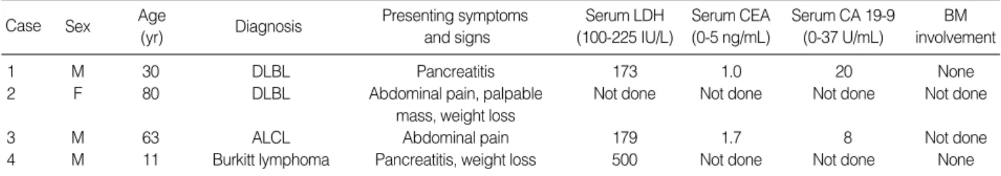
Fig. 1. Pathology findings of case 4. (A) Photomicrograph shows starry sky appearance and small non-cleaved cells (H&E, ×400).
(B) The specimen shows immunoreactivity for Ki-67 and (C) bcl-6.
A B
C
Radiological findings and stages
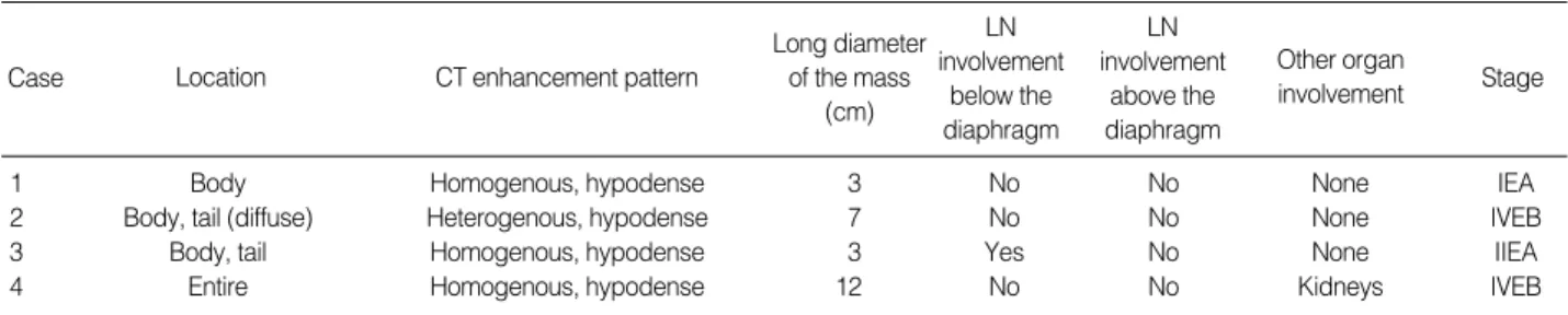
The CT findings are summarized in Table 3. The locations of the lesions were body and tail (n=2), body (n=1), and dif- fuse involvement of the entire pancreas (n=1). Three patients demonstrated homogenous hypodense pancreatic lesions, and one patient demonstrated a heterogenous hypodense pancre- atic lesion. The median long diameter of the mass on CT was 5 cm (range 3-12 cm). Two patients who presented with pan- creatitis showed minimal pancreatic duct dilatation on CT, whereas one of the two patients who did not present with pan- creatitis showed minimal main pancreatic duct dilatation and the other showed no main pancreatic duct dilatation. One patient showed lymph node enlargement on the same side of the diaphragm. No patient demonstrated lymph node en- largement above the diaphragm. One patient demonstrated extrapancreatic organ involvement in both kidneys (Fig. 2).
The Ann Arbor stages of the patients were: IEA (n=1), IIEA (n=1), and IVEB (n=2).
Treatment and prognosis
The patient with stage IEA disease underwent chemother- apy and radiation therapy; this patient had a complete remis-
sion after treatment. The patient with stage IVEB disease had chemotherapy; this patient showed relapse in the bone mar- row, central nervous system, liver, and the kidneys 6.8 months after the diagnosis. The patient underwent peripheral blood stem cell transplantation and is alive at 30 months. Two pati- ents (stages IVEB and IIEA) refused to be treated and did not have any treatment, and both died 0.8 and 7.0 months
Fig. 2. CT findings of case 4. Homogenous, hypodense tumorous lesion is replacing almost the entire pancreas. (A) Dilatation of the main pancreatic duct is noted. Multiple nodules are also found in both kidneys. (B) CT taken 1 yr after peripheral blood stem cell transplanta- tion shows disappearance of lesions in the pancreas and kidneys.
A B
Case
LN involvement
below the diaphragm Long diameter
of the mass (cm) CT enhancement pattern
Location
LN involvement
above the diaphragm
Other organ
involvement Stage
1 Body Homogenous, hypodense 3 No No None IEA
2 Body, tail (diffuse) Heterogenous, hypodense 7 No No None IVEB
3 Body, tail Homogenous, hypodense 3 Yes No None IIEA
4 Entire Homogenous, hypodense 12 No No Kidneys IVEB
Table 3. CT findings and stages of PPL patients
CT, computed tomography; PPL, primary pancreatic lymphoma; LN, lymph node.
Case Treatment Course Follow-up Status
(months)
1 CHOP+RT Complete remission 45 No evidence of disease
2 None Dead 0.8 Dead
3 None Dead 7.0 Dead
4 COPADM Relapsed in BM, 30 No evidence
CNS, liver and kidneys of disease at 6.8 months.
Underwent auto-PBSCT Table 4. Treatment and prognosis of PPL patients
PPL, primary pancreatic lymphoma; CHOP, cyclophosphamide, dox- orubicin, vincristine, and prednisone; RT, radiation therapy; COPADM, cyclophosphamide, vincristine, prednisone, adriamycin, high-dose metho- trexate; BM, bone marrow; CNS, central nervous system; PBSCT, periph- eral blood stem cell transplantation.
after diagnosis, respectively (Table 4).
DISCUSSION
PPL is a rare disease. In a review by Saif in 2006, fewer than 150 cases of PPL have been reported in English literature at that time (4). Most are single case reports. Single center expe- riences usually have included less than 15 cases (2, 5-11). A nationwide study from Japan reported on 19 cases of PPL (12).
In our study, four patients were diagnosed over an 11-yr peri- od. To the best of our knowledge, five cases of primary pan- creatic lymphoma have been reported in Korea (13-17).
The clinical presentation of PPL is usually nonspecific. B symptoms (weight loss, fever, and night sweats) are uncom- mon. Therefore, the clinical presentation will usually help little in distinguishing PPL patients from those with other types of pancreatic tumors (18). Patient with a large, palpa- ble pancreatic mass, presenting with abdominal pain with- out jaundice, mass homogenous on imaging, with elevated serum LDH may be suspected to have PPL (5, 19). The most common presenting symptom of PPL is abdominal pain, fol- lowed by abdominal mass, weight loss, jaundice, acute pan- creatitis, small bowel obstruction, and diarrhea (4). In our experience, all patients presented with abdominal pain, and two patients had pancreatitis. No patient developed jaundice in our study. B symptoms other than weight loss (i.e., fever and night sweats) have been reported to be rare in PPL pati- ents. Although weight loss was present in two patients, fever and night sweats were not present in any of our patients.
The head of the pancreas is reported to be the most com- mon location of PPL (2, 7, 12). However, the body/tail region is reported to be more frequently affected in some reports (5).
In our study, one of four patients had pancreatic head involve- ment; this patient showed diffuse involvement of the entire pancreas.
There are reports of PPL arising in the setting of immun- odeficiency, such as AIDS or organ transplant (20, 21). In our study, however, no patient had evidence of AIDS or history of organ transplantation.
The majority of PPLs are of the B-cell type (2, 5-7). T- cell PPLs are rare, as described in Japanese series (12). In the Japanese report, although not statistically significant, the 1- yr survival for B-cell PPL (51.9%) was higher than that for T-cell PPL (0%) (12). Among the four patients in our study, only one patient had a T-cell lineage. This patient (initial stage IIEA) did not undergo any treatment, and died seven months after the diagnosis.
There have been controversies over the treatment of PPLs.
However, chemotherapy, not surgery, is accepted as the main- stay of the treatment (4, 18). Using complex treatment app- roaches, cure rates of over 30% of PPL patients by chemo- therapy was reported as early as the 1980s (22). With advances in chemotherapy such as monoclonal antibodies, complete
remission can be expected in up to 85% of patients with dif- fuse large B cell lymphoma (23). It is advocated that surgery should be reserved for cases where percutaneous or endoscop- ic biopsies are not diagnostic, or treatment with chemother- apy and/or radiation therapy is not successful (4, 18). In our study, no patient underwent surgical procedures. The stage IEA patient, who underwent chemotherapy and radiation therapy, achieved a long-term complete remission. The other treated patient, who had stage IVEB disease, relapsed after initial treatment. After subsequent treatment, this patient is alive at 30 months. The treated patients are alive without evidence of disease for more than two years.
In conclusion, PPL is a rare disease. Since chemotherapy is the mainstay of the treatment, differentiation from other pancreatic malignancies is crucial. Nonsurgical biopsy modal- ities appear adequate for the diagnosis of PPL. Chemothera- py-based treatment, and addition of radiation therapy, if pos- sible, may provide a good prognosis.
REFERENCES
1. Hamilton SR, Lauri LA. Pathology and genetics of tumours of the digestive system. In: World Health Organization Classification of Tumours. Lyon: IARC Press; 2000.
2. Nayer H, Weir EG, Sheth S, Ali SZ. Primary pancreatic lymphomas:
a cytopathologic analysis of a rare malignancy. Cancer 2004; 102:
315-21.
3. Dawson IM, Cornes JS, Morson BC. Primary malignant lymphoid tumours of the intestinal tract. Report of 37 cases with a study of fac- tors influencing prognosis. Br J Surg 1961; 49: 80-9.
4. Saif MW. Primary pancreatic lymphomas. JOP 2006; 7: 262-73.
5. Bouvet M, Staerkel GA, Spitz FR, Curley SA, Charnsangavej C, Hage- meister FB, Janjan NA, Pisters PW, Evans DB. Primary pancreatic lymphoma. Surgery 1998; 123: 382-90.
6. Lin H, Li SD, Hu XG, Li ZS. Primary pancreatic lymphoma: report of six cases. World J Gastroenterol 2006; 12: 5064-7.
7. Ezzat A, Jamshed A, Khafaga Y, Rahal M, Linjawi T, Martin J, Taha I. Primary pancreatic non-Hodgkin’s lymphomas. J Clin Gastroen- terol 1996; 23: 109-12.
8. Tuchek JM, De Jong SA, Pickleman J. Diagnosis, surgical interven- tion, and prognosis of primary pancreatic lymphoma. Am Surg 1993;
59: 513-8.
9. Webb TH, Lillemoe KD, Pitt HA, Jones RJ, Cameron JL. Pancreat- ic lymphoma. Is surgery mandatory for diagnosis or treatment? Ann Surg 1989; 209: 25-30.
10. Behrns KE, Sarr MG, Strickler JG. Pancreatic lymphoma: is it a sur- gical disease? Pancreas 1994; 9: 662-7.
11. Mansour GM, Cucchiaro G, Niotis MT, Fetter BF, Moore J, Rice RR, Branum GD, Meyers WC. Surgical management of pancreatic lymphoma. Arch Surg 1989; 124: 1287-9.
12. Nishimura R, Takakuwa T, Hoshida Y, Tsujimoto M, Aozasa K.
Primary pancreatic lymphoma: clinicopathological analysis of 19 cases from Japan and review of the literature. Oncology 2001; 60:
322-9.
13. Nam SY, Chung JP, Kim E, Lee JH, Kim KW, Jeong HJ, Chung JB, Hahn JS, Kang JK, Park IS. A case of primary T-cell lymphoma of the pancreas. Korean J Gastroenterol 1994; 26: 892-8.
14. Song JH, Chung WJ, Lee JW, Kuon KS, Shin YW, Park JW, Kim JM. Non-Hodgkin’s lymphoma of the pancreas presenting with acute pancreatitis as the first manifestation. Korean J Gastroenterol 1994;
26: 758-63.
15. Lee KS, Park KS, Lee SY, Bae IH, Kim SJ, Han GS, Cha SH, Lee OJ. A case of primary pancreatic T-cell lymphoma. J Korean Radi- ol Soc 2006; 55: 271-4.
16. Lee MK, Jeon SW, Lee YD, Seo HE, Cho CM, Kim SG, Yoon YK.
A case of primary pancreatic non-Hodgkin’s lymphoma. Korean J Intern Med 2006; 21: 123-6.
17. Hwang JE, Park CH, Cho YC, Kim SK, Kim HS, Choi SK, Rew JS, Lee WS. A case of primary pancreatic lymphoma that manifested with acute pancreatitis and obstructive jaundice. Korean J Gastroin-
test Endosc 2009; 38: 176-9.
18. Mortenson MM, Katz MH, Tamm EP, Bhutani MS, Wang H, Evans DB, Fleming JB. Current diagnosis and management of unusual pan- creatic tumors. Am J Surg 2008; 196: 100-13.
19. Merkle EM, Bender GN, Brambs HJ. Imaging findings in pancreat- ic lymphoma: differential aspects. AJR Am J Roentgenol 2000; 174:
671-5.
20. Jones WF, Sheikh MY, McClave SA. AIDS-related non-Hodgkin’s lymphoma of the pancreas. Am J Gastroenterol 1997; 92: 335-8.
21. Cario E, Runzi M, Metz K, Layer P, Goebell H. Diagnostic dilemma in pancreatic lymphoma. Case report and review. Int J Pancreatol 1997; 22: 67-71.
22. Brown PC, Hart MJ, White TT. Pancreatic lymphoma, diagnosis and management. Int J Pancreatol 1987; 2: 93-9.
23. Coiffier B. Monoclonal antibody as therapy for malignant lymphomas.
C R Biol 2006; 329: 241-54.