INTRODUCTION
Treatment with pharmacologic doses of glucocorticoids frequently results in significant osteoporosis (1). Patients on high-dose glucocorticoid therapy are susceptible to bone loss and vertebral fractures within weeks to months of initiation of therapy (2). It is generally accepted that glucocorticoids decrease bone formation and increase bone resorption in vitro as well as in vivo (3-5). In contrast to the consistent finding of reduced osteoblastic activity, the exact mechanism respon- sible for the effects of glucocorticoids on bone resorption is less clear. It may involve both direct effects on osteoclasts (4) as well as indirect effects mediated through osteoblasts (6).
Despite these side effects, glucocorticoids are necessary for the treatment of a variety of medical diseases. Deflazacort is a synthetic glucocorticoid with antiinflammatory and anti- immune properties similar to those reported for prednisone and has been reported to be relatively bone sparing (7). Al- though long-term clinical trials comparing deflazacort with prednisone are not available, an initial study suggests that trabecular bone loss is less with deflazacort compared to pred- nisone (8). Deflazacort and other glucocorticoids have simi-
lar inhibitory actions on aspects of bone formation in cultures of intact calvariae and osteoblast-enriched cells. Information on precise effects of deflazacort on bone cell function, espe- cially osteoclasts, is quite limited. Therefore, the present study was undertaken to compare the effect of deflazacort and dexamethasone on aspects of osteoclast formation in mouse marrow culture. Recently osteoprotegerin (OPG) and its ligand (RANKL) were cloned and demonstrated to be critical regulators of osteoclastogenesis (9, 10). OPG is a novel receptor that blocks osteoclast formation. Either in a cell membrane-bound or in a soluble form, RANKL stimu- lates osteoclastogenesis and osteoclastic bone resorption.
OPG and RANKL are expressed by marrow stromal cells, osteoblast-like cell lines and primary cultures of osteoblasts derived from mouse calvaria. Recent studies demonstrated that OPG and RANKL gene expressions in osteoblastic cells are regulated by various calciotropic hormones and cytokines (11). Furthermore, Hofbauer et al. has identified that glucocor- ticoids promote osteoclastogenesis by inhibiting OPG and concurrently stimulating RANKL production by osteoblas- tic lineage cells, thereby enhancing bone resorption (12).
In order to find out differences of deflazacort in terms of Hoyeon Chung, Young Soon Kang*, Chang Sun Hwang*, In Kul Moon*, Chang Hoon Yim, Kyu Hong Choi�, Ki Ok Han, Hak Chul Jang, Hyun Koo Yoon, In Kwon Han
Department of Medicine/Endocrinology, Laboratory of Endocrinology*, Department of Obstetrics and Gynecology�, Sungkyunkwan University School of Medicine, Samsung Cheil Hospital, Seoul, Korea
Received : 26 December 2000 Accepted : 8 August 2001
Address for correspondence Hoyeon Chung, M.D.
Department of Medicine/Endocrinology, Samsung Cheil Hospital, 1-19 Mukjeong-dong, Jung-ku, Seoul 100-380, Korea
Tel : +82.2-2000-7366, Fax : +82.2-2273-8502 E-mail : [email protected]
769
Deflazacort Increases Osteoclast Formation in Mouse Bone Marrow Culture and the Ratio of RANKL/OPG mRNA Expression in Marrow Stromal Cells
Information on precise effects of deflazacort on bone cell function, especially osteoclasts, is quite limited. Therefore, the present study was undertaken to test effects of deflazacort on osteoclast-like cell formation in mouse bone marrow cultures and on the regulation of osteoprotegerin (OPG) and its ligand (RANKL) mRNA expressions by RT-PCR in the ST2 marrow stromal cells. TRAP-positive mononuclear cells increased after the treatment of deflazacort at 10-9to 10-7 M alone for 6 days in a dose-dependent manner. Number of TRAP-positive multi- nucleated cells (MNCs) increased significantly with combined treatment of deflaza- cort at 10-7 M and 1,25-(OH)2D3at 10-9 M compared to that of cultures treated with 1,25-(OH)2D3alone (p<0.05). Exposure to deflazacort at 10-7 M in the pres- ence of 1,25-(OH)2D3at 10-9 M in the last 3-day culture had greater stimulatory effect on osteoclast-like cell formation than that of the first 3-day culture did. De- flazacort at 10-10-10-6 M downregulated OPG and upregulated RANKL in mRNA levels in a dose-dependent manner. These observations suggest that deflaza- cort stimulate osteoclast precursor in the absence of 1,25-(OH)2D3and enhance differentiation of osteoclasts in the presence of 1,25-(OH)2D3. These effects are, in part, thought to be mediated by the regulation of the expression of OPG and RANKL mRNA in marrow stromal cells.
Key Words : Glucocorticoids; Osteoclasts; OPG; RANKL
bone effects, we have investigated the effects of deflazacort on osteoclast-like cell formation in mouse bone marrow cul- tures, and on OPG and RANKL mRNA expression in ST2 cells.
MATERIALS AND METHODS Bone marrow culture system
Six- to 9-week-old male ICR mice were sacrificed and bone marrow was obtained as described by Takahashi et al. (13).
Briefly, tibiae and femora were dissected free of adherent soft tissues, both epiphyses were cut off with scissors, and the marrow cavities were vigorously flushed out with -mini- mum essential medium ( -MEM, Gibco BRL, Grand Island, NY, U.S.A.) using a syringe. Freshly isolated whole bone marrow cells were cultured in -MEM containing 10% fetal calf serum (FCS, Gibco BRL) at 1.5×10⁶cells/mL in 48-well plates (Nunc, Denmark). Cultures were fed every 3 days by replacing half of the media. 10-9 M of 1,25-(OH)2D3(Cal- biochem, U.S.A.) and various concentrations of deflazacort (Gruppo Lepetit, Italy) were added at the beginning of the culture and at each media change. All cultures were main- tained at 37℃ in a humidified atmosphere of 5% CO2and 95% air. After various periods of culture, cells were fixed with citrate-acetone-formaldehyde fixative, and stained for TRAP using an acid phosphatase kit (Sigma). TRAP-positive cells containing three or more nuclei were scored as osteoclasts microscopically. The results were expressed as the mean± SEM for four cultures.
Semiquantitative RT-PCR
Osteoclast formation-supporting stromal cell line, ST2 cells (RIKEN cell bank, Tsukuba, Japan) were cultured in 6-well plates containing -MEM supplemented with 10%
heat-inactivated fetal calf serum (FCS) at 37℃ in a humidi- fied atmosphere of 5% CO2and 95% air. When the effect of deflazacort was to be tested, ST2 cells were serum-starved in -MEM containing 0.5% FCS for a period of 24 hr, fol- lowed by the addition of deflazacort at final concentrations ranging from 10-10to 10-6M. Total RNA was extracted from the ST2 cells with Tri-reagent (Molecular Research Center, Cincinnati, OH, U.S.A.) and then used for reverse transcrip- tase PCR (RT-PCR) assay. PCR reactions were done using gene-specific PCR primers and Taq polymerase (SR producs, U.K.) in a thermal cycler (Perkin-Elmer Corp., Norwalk, CT, U.S.A.). Specific primer sets were designed from pub- lished cDNA sequences: murine OPG (sense: 5、-AACCCC AGAGCGAAACAC-3、; antisense: 5、-AAGAAGGCCTCT TCACAC-3、), murine RANKL (sense: 5、-GGTCGGGCA ATTCTGAATT-3、; antisense: 5、-GGGGAATTACAAAG TGCACCAG-3、), -actin (sense: 5、-GTGGGCCGCCCT
AGGCACCAG-3、; antisense: 5、-CACTTTGATGTCACG CACGATTTC-3、). The amplified samples were run on a 1.5- 2.0% agarose gel, stained with ethidium bromide, and photo- graphed under UV illumination. Optical density was deter- mined using a digital image processing and analysis program (Vilber Lourmate, France). Ratios of OPG or RANKL/ -actin PCR product density were determined for semiquantitation of mRNA expression. The conditions for the PCR reaction for mouse OPG cDNA of 219 bp was 95℃ for 50 sec, 54℃ for 20 sec, and 72℃ for 20 sec for 35 cycles. The conditions for PCR analysis of RANKL were the same as for OPG except an annealing temperature of 62℃. PCR was performed for 27 cycles for -actin.
Statistical analysis
Statistical analysis was performed using Student’s t-test to evaluate differences between the sample of interest and its respective control. For analysis of dose response, multiple measurement ANOVA was used. A p value of <0.05 was considered significant.
RESULTS
Effects of glucocorticoids on the formation of osteo- clast-like cells
TRAP-positive multinucleated cells (MNCs) did not in- crease in cultures treated with deflazacort alone, whereas TRAP-positive mononuclear cells increased (data not shown).
Number of TRAP-positive multinucleated cells/culture 100
80
60
40
20
0
dexamethasone - 10-9 10-8 10-7 - - - deflazacort - - - - 10-9 10-8 10-7M
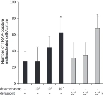
Fig. 1. Effects of increased concentration of glucocorticoids on the formation of TRAP-positive multinucleated cells. TRAP-posi- tive cells were counted after mouse marrow culture in the pres- ence of 1,25-(OH)2D3, 10-9 M for 6 days. Data are expressed as the mean±SEM for four cultures (*: p<0.05).
* *
Fig. 1 shows the effects of increased concentration of gluco- corticoids on the formation of TRAP-positive MNCs in the presence of 1,25-(OH)2D3(10-9 M) for 6 days. The addition of dexamethasone or deflazacort to the cultures resulted in
enhancement of TRAP-positive MNC formation induced by 1,25-(OH)2D3. A significant increase was observed at the concentration of 10-7M dexamethasone or deflazacort (p<
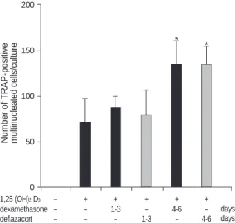
0.05). To characterize the time course for the effects of gluco- corticoids on osteoclast formation, mouse bone marrow cul- tures were treated with dexamethasone or deflazacort for vary- ing time. The number of TRAP-positive MNCs by 10-9 M 1,25-(OH)2D3and 10-7 M glucocorticoids reached a nearly maximal level with treatment of dexamethasone or deflaza- cort in the last 3 days of culture (Fig. 2).
Regulation of OPG and RANKL mRNA expression by deflazacort in ST2 cell
OPG, RANKL, and -actin mRNA expression levels were assessed by RT-PCR in ST2 cell cultures that were treated with or without deflazacort for 8 hr. The amounts of OPG and RANKL mRNA were compared with the amount of -actin mRNA. As shown in Fig. 3, the level of OPG mRNA expres- sion decreased significantly after treatment with deflazacort at and above the concentration of 10-7 M. On the contrary, stimulation with deflazacort for 8 hr increased the RANKL mRNA levels in a dose-dependent manner (Fig. 4).
DISCUSSION
Deflazacort has a similar effect on osteoclast formation com-
Number of TRAP-positive multinucleated cells/culture 200
150
100
50
0
1,25 (OH)2D3 - + + + + +
dexamethasone - - 1-3 - 4-6 -
deflazacort - - - 1-3 - 4-6
days days
Fig. 2. Effects of varying treatment time with glucocorticoids on the TRAP-positive multinucleated cells in mouse bone marrow cultures in the presence of 1,25-(OH)2D3, 10-9 M for 6 days. During the indicated times, cells were cultured with 10-7M of glucocor- ticoids (*: p<0.05).
Optical density ratio
1.6
1.2
0.8
0.4
0
0 10-10 10-9 10-8 10-7 10-6
Fig. 3. mRNA expression of OPG and -actin assessed by RT- PCR in ST2 cells. ST2 cells were precultured and then stimulat- ed, with or without deflazacort, for 8 hr. (A) Photographs of the PCR analysis from a representative experiment. (B) Mean optical density ratio values for OPG, normalized to -actin of triplicate independent experiments, to document the consistency of the results (*: p<0.01).
Deflazacort (M) -actin
OPG
A
B
Optical density ratio
1.0
0.8
0.6
0.4
0.2
0
0 10-10 10-9 10-8 10-7 10-6
Fig. 4. mRNA expression of RANKL and -actin assessed by RT- PCR in ST2 cells. ST2 cells were precultured and then stimulat- ed, with or without deflazacort, for 8 hr. (A) Photographs of the PCR analysis from a representative experiment. (B) Mean optical density ratio values for RANKL, normalized to -actin of triplicate independent experiments, to document the consistency of the results (*: p<0.01).
Deflazacort (M) -actin
RANKL
A
B
* *
* *
*
pared with those of dexamethasone (14). No differences in osteoclast formation of deflazacort and dexamethasone were detected in this study (Fig. 1). The number of TRAP-posi- tive mononuclear cells was also increased by treatment with deflazacort alone, suggesting that deflazacort may induce proliferation or differentiation of osteoclast precursors in mouse bone marrow cultures. Time-course effect for TRAP- positive MNC formation showed that deflazacort had its greatest stimulatory effects on osteoclast formation during the later stages of the cultures, the period of differentiation, rather than during the proliferation phase of mouse bone marrow cultures (Fig. 2). Although it is possible that deflaza- cort stimulated osteoclast-like cell formation by directly act- ing on hematopoietic cells, based on our results, it is more likely that deflazacort mediates its effects through stromal cells in enhancing osteoclast formation in mouse bone mar- row cultures. Most of the TRAP-positive mononuclear cells and MNCs were found adjacent to colonies of osteoblast-like cells stained for alkaline phosphatase. Alkaline phosphatase positive cells appeared almost on day 3 (13).
The balance between osteoblast and osteoclast functions is regulated systemically by a variety of hormones and locally by the production of paracrine factors by osteoblasts or bone marrow stromal cells that regulate osteoclast function (15, 16). The development of active osteoclasts in vitro requires intimate contact between osteoblastic stromal cells and osteo- clast precursors (17). RANKL, receptor activator of NF-kappa B (RANK), and OPG have recently been identified as ago- nist, receptor, and decoy receptor for osteoclastogenesis. Fac- tors that stimulate bone resorption increase RANKL expres- sion and, with some exceptions, decrease OPG expression (11, 18). In the present study, although we did not evaluate protein expressions, deflazacort treatment on mouse marrow stromal cells inhibited the production of OPG and increased RANKL in mRNA levels (Fig. 3, 4). No differences on OPG and RANKL mRNA regulation of deflazacort and dexa- methasone were detected in this semiquantitative RT-PCR (data not shown). This differential regulation of OPG and RANKL by deflazacort would favor the formation of osteo- clasts. These findings are in line with a study that showed dexamethasone promotes osteoclastogenesis by inhibiting OPG and stimulating RANKL production by osteoblastic lineage cells (12). We can not explain the bone-sparing effect of deflazacort in terms of osteoclast formation based on our study. Glucocorticoids, including deflazacort, affect multi- ple parameters of mineral metabolism (19-21). All these studies assumed that the potency of prednisone relative to deflazacort was 1.2. Subsequent re-examination of the rela- tive potencies of these two glucocorticoids has found that the true relative potency is 1.4-1.8 (22). This study suggest that deflazacort have been overestimated in the past, and demonstrated no bone sparing effect when used in a thera- peutically equivalent dose.
In conclusion, as in other glucocorticoids, deflazacort also
has stimulatory effects on osteoclast formation, and displays similar differential regulation of the expression of genes re- sponsible for osteoclastogenesis in marrow stromal cells.
ACKNOWLEDGMENTS
The authours thank Dr. G. David Roodman (University of Texas at San Antonio) for critical reading of the manuscript.
REFERENCES
1. Lukert JC, Aaron J, Horsman A, Wilkinson R, Nordin BEC. Cor- ticosteroid osteoporosis. Clinics Endocrinol Metab 1973; 2: 355-68.
2. Baylink DJ. Glucocorticoid-induced osteoporosis. N Engl J Med 1983; 309: 306-8.
3. Delany AM, Dong Y, Canalis E. Mechanisms of glucocorticoid action in bone cells. J Cell Biochem1994; 56: 295-302.
4. Kaji H, Sugimoto T, Kanatani M, Nishiyama K, Chihara K. Dex- amethasone stimulates osteoclast-like cell formation by directly act- ing on hemopoietic blast cells and enhances osteoclast-like cell for- mation stimulated by parathyroid hormone and prostaglandin E2. J Bone Miner Res 1997; 12: 734-41.
5. Dempster DW. Bone histomorphometry in glucocorticoid-induced osteoporosis. J Bone Miner Res 1998; 13: 137-41.
6. Rubin J, Biskobing DM, Jadhav L, Fan D, Nanes MS, Perkins S, Fan X. Dexamethasone promotes expression of membrane-bound macrophage colony-stimulating factor in murine osteoblast-like cells. Endocrinology 1998; 139: 1006-12.
7. Lund B, Egsmose C, Jorgensen S, Krogsgaard MR. Establishment of the relative antiinflammatory potency of deflazacort and pred- nisone in polymyalgia rheumatica. Calcif Tissue Int 1987; 41: 316- 20.
8. Olgaard K, Storm T, Wowern NV, Daugaard H, Egfjord M, Lewin E, Brandi L. Glucocorticoid-induced osteoporosis in the lumbar spine, forearm, and mandible of nephrotic patients: a double-blind study on the high-dose, long-term effects of prednisone versus deflazacort. Calcif Tissue Int 1992; 50: 490-7.
9. Simonet WS, Lacey DL, Dunstan CR, Kelley M, Chang MS, Luthy R, Nguyen HQ, Wooden S, Bennett L, Boone T, Shimamoto G, De Rose M, Elliott R, Colombero A, Tan HL, Trail G, Sullivan J, Davy E, Bucay N, Renshaw-Gegg L, Hughes TM, Hill D, Pattison W, Campbell P, Sander S, Van G, Tarpley J, Derby P, Lee R, Boyle WJ.
Osteoprotegerin: a novel secreted protein involved in the regulation of bone density. Cell 1997; 89: 309-19.
10. Lacey DL, Timms E, Tan HL, Kelly MJ, Dunstan CR, Burgess T, Elliott R, Colombero A, Elliott G, Scully S, Hsu H, Sullivan J, Hawkins N, Davy E, Capparelli C, Eli A, Qian YX, Kaufman S, Sarosi I, Shalhoub V, Senaldi G, Guo J, Delaney J, Boyle WJ.
Osteoprotegerin ligand is a cytokine that regulates osteoclast differ- entiation and activation. Cell 1998; 93: 165-76.
11. Hofbauer LC, Khosla S, Dunstan CR, Lacey DL, Boyle WJ, Riggs BL. The roles of osteoprotegerin and osteoprotegerin ligand in the
. .
paracrine regulation of bone resorption. J Bone Miner Res 2000;
15: 2-12.
12. Hofbauer LC, Gori F, Riggs BL, Lacey DL, Dunstan CR, Spelsberg TC, Khosla S. Stimulation of osteoprotegerin ligand and inhibiton of osteoprotegerin production by glucocorticoids in human osteoblastic lineage cells: potential paracrine mechanisms of glucocorticoid- induced osteoporosis. Endocrinology 1999; 140: 4382-9.
13. Takahashi N, Yamana H, Yoshiki S, Roodman GD, Mundy GR, Jones SJ, Boyde A, Suda T. Osteoclast-like cell formation and its regulation by osteotrophic hormones in mouse bone marrow cul- tures. Endocrinology 1988; 122: 1373-82.
14. Shuto T, Kukita T, Hirata M, Jimi E, Koga T. Dexamethasone stim- ulates osteoclast-like cell formation by inhibiting granulocyte-ma- crophage colony stimulating factor production in mouse bone mar- row cultures. Endocrinology 1994; 134: 1121-6.
15. Manolagas SC, Jilka RL. Bone marrow, cytokines, and bone remod- eling. Emerging insights into the pathophysiology of osteoporosis. N Engl J Med 1995; 332: 305-11.
16. Horowitz MC. Cytokines and estrogen in bone: anti-osteoporotic effects. Science 1993; 260: 626-7.
17. Takahashi N, Akatsu T, Udagawa N, Sasaki T, Yamaguchi A, Mose- ley JM, Martin TJ, Suda T. Osteoblastic cells are involved in osteo-
clast formation. Endocrinology 1988; 123: 2600-2.
18. Dunstan CR. Osteoprotegerin and osteoprotegerin ligand mediated the local regulation of bone resorption. Endocrinologist 2000; 10:
18-26.
19. Gennari C, Bernini M, Mardi P, Fusi L, Avioli LV. Glucocorticoids and intestinal absorption of calcium and phosphate in man. In: Nor- man AW, Schaefer K, Herrath DV, Grigoleeit HG, editors, Vitamin D. Chemical, biochemical and clinical endocrinology of calcium metabolism. Walter deGruyter, Berlin, New York, 1982; 257.
20. Gray RES, Doherty SM, Galloway J, Coulton L, de Broe M, Kanis JA. A double-blind study of deflazacort and prednisone in patients with chronic inflammatory disorders. Arthritis Rheum 1991; 34: 287- 95.
21. Gennari C, Imbimbo B, Montagnani M, Bernini M, Nardi P, Avioli LV. Effects of prednisone and deflazacort on mineral metabolism and parathyroid hormone activity in humans. Calcif Tissue Int 1984;
36: 245-52.
22. Krogsgaard MR, Thamsborg G, Lund B. Changes in bone mass during low dose corticosteroid treatment in patients with polymyal- gia rheumatica-a double blind prospective comparison between prednisolone and deflazacort. Ann Rheum Dis 1996; 55: 143-6.