• 검색 결과가 없습니다.

간에 발생한 충돌종양 (간세포암과 미분화성 육종)

N/A
N/A
Protected

Academic year: 2021

Share "간에 발생한 충돌종양 (간세포암과 미분화성 육종)"

Copied!
4
0
0

로드 중.... (전체 텍스트 보기)

전체 글

(1)

120

Introduction

Case

Collision tumors represent the coexistence of two adjacent but histologically distinct tumors in the same organ. There are two types of collision tumor. One is the intermingled type in which two kinds of tumors coexist and there is a histological transition; the other is the contiguous type in which two kinds of tumors exist independently from each other and there is no histological mixture or transition. It is believed that in the intermingled type two different kinds of tumor cells differentiated from progenitor cells to form a mass; in the contiguous type, two different types of tumors close to each other develop simultaneously in the same organ and form a mass. Most collision tumors in the liver are of the intermingled type and they are known to account for

2.0-3.6% of primary liver cancers. A representative example is hepatocholangiocarcinoma. Contiguous-type collision tumors are collision tumors in the true sense, and their percentage among primary liver cancers is low, in the range of 0.1-1%. Representative examples are HCC and neuroendocrine tumors.
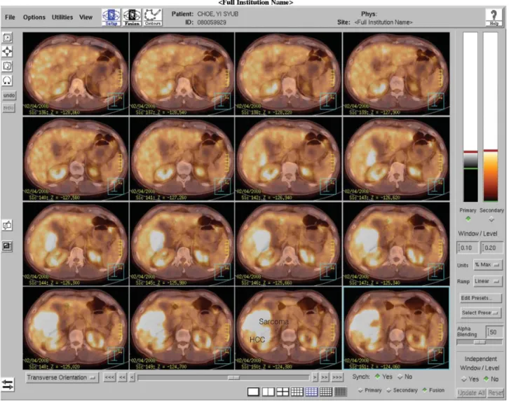
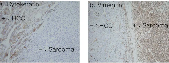
1,2

The present authors experienced a case of a collision tumor that consisted of a HCC and an undifferentiated sarcoma. It was identified from the histopathologic findings of a patient who had a hepatectomy for a mass that had developed in the liver.

To the authors’ knowledge, this is the first case of such a collision tumor, and here we report the case with diagnostic tests, histological findings, and the result of treatment.

A 57-year-old patient who had had periodical

Korean Journal of HBP Surgery

Vol. 13, No 2, June

□ 증 례□

간에 발생한 충돌종양 (간세포암과 미분화성 육종)

Collision tumors represent the coexistence of two adjacent but histologically distinct tumors in an organ. Collision tumors have been identified in various organs, but they are rare in liver. We present a rare case of a 57-year-old man, who was hospitalized for the removal of a liver mass (S5/6 segmentectomy), that was probably a hepatocellular carcinoma. The eventual pathology examination revealed a collision tumor composed of a hepatocellular carcinoma and an undifferentiated sarcoma. Because the tumor recurred 2 months after the operation, reoperation (right hemicolectomy and tumorectomy) was performed. In the 2

nd

month following the second operation we found multiple liver metastases, chest metastasis and abdominal cavity metastasis. To our knowledge this is the first case of a hepatic collision tumor that was composed of a hepatocellular carcinoma and an undifferentiated sarcoma. Herein, we report the case of a hepatic collision tumor and briefly review the literature.

Collision Tumor of the Liver (Hepatocellular Carcinoma and Undifferentiated Sarcoma)

Key Words : Collision tumor, Hepatocellular carcinoma, Sarcoma

Received: 2009. 5. 27.

Accepted: 2009. 6. 19.

부산대학교 의과대학 외과학교실

서 형 일

책임저자

서 형 일

부산시 서구 아미동 1- 10 우 602-739 부산대학교 의과대학 외과학교실 E-mail: [email protected]

※ 2 0 0 8 년 간 담 췌 외 과 추 계 학 회 에 서 구연되었음.

Department of Surgery, Pusan National University College of Medicine, Pusan, Korea

Hyung Il Seo, M.D.

(2)

121

서형일 : 간에 발생한 충돌종양 (간세포암과 미분화성 육종)

examinations (USG+AFP) as a HBV carrier visited the hospital because of a palpable mass. On physical examination, a mass was palpated in the right upper quadrant, and there were no other particular findings.

There were no abnormal findings in hematological and biochemical tests, and alfafetoprotein (AFP) had increased to 1451 ng/ml (normal range; 0-12).

On CT, a 10 cm low-density exophytic mass was observed in the S5/6 segment except some area of S6.

On MRI we observed a mass most of which showed a heterogeneous low signal intensity, and the posterior portion of the mass showed a pattern in which the signal dropped in opposed T1 weighted-images (Fig.

1). On

18

FDG-PET/CT, most of the tumor showed increased metabolism (SUVmax 10.5, Fig. 2) but such

Fig. 1. MRI finding of collision tumor of the liver. Opposed T1 WI showing heterogeneous low signal huge mass (Sarcoma) and signal drop (HCC). This means microscopic fat deposition originated hepatocyte.

Fig. 2. 18FDG-PET/CT showed increasing metabolism of the right lobe (Sarcoma) and no FDG uptake (HCC).

(3)

122 한국간담췌외과학회지 : 제13권 제2호 2009

Discussion

In our case as well, both the undifferentiated sarcoma and the HCC recurred early after the operation. As for undifferentiated sarcomas, there was a report that the survival rate is improved through chemotherapy after surgical excision, but there is no established regimen.

8

Because collision tumors in the liver are very rare, it is hard to diagnose them through postoperative imaging.

As in our case, however, if there are findings from imaging that suggest a collision tumor, it is considered necessary to do a biopsy. In addition, because the prognosis of collision tumors involving a sarcoma and a HCC is expected to be poor, active multimodal treatment is required after excision and closer follow- up is needed.

an increase was not observed in the part where the signal dropped in the MRI. Ultrasonography guided biopsy was performed, and after histological examination the patient was diagnosed with HCC.

On laparotomy, micronodular cirrhotic nodules were observed on the surface of the liver and S5/6 segmentectomy was performed. On the section of the tissue were observed a 5.2cm yellowish multinodular confluent mass and large dark reddish fussy mass lesions with necrotic hemorrhagic features surrounding the mass. The free margin was 1.2cm.

Each tumor was clearly separated by fibrous band- like lesions. On hematoxylin-eosin (HE) staining, HCC lesions showed a trabecular pattern and the mixed form of a clear cell type and a hepatic cell type. Hence, they fell within Edmonson-Steiner grades II-III.

Sarcoma lesions showed oval to round pleomorphic nuclei and took various forms of anaplasia, suggesting t h a t t h e y w e r e u n d i f f e r e n t i a t e d s a r c o m a s.

Immunohistochemical staining confirmed HCC lesions positive for cytokeratin and sarcoma lesions positive for vimentin (Fig. 3a & 3b).

Fifty days after the operation we identified a local recurrence in the abdominal wall, hepatic flexure, and the resection margin of the liver. Re-operation was performed on Day 69 and a 17cm focus of recurrence was removed. Multiple metastatic lesions in the remnant liver were then identified through

ultrasonography. Ethanol injection was applied to each of the 5 lesions, and the recurrence of undifferentiated sarcoma and HCC was confirmed histologically. On a CT exam on Day 8 after the second operation, multiple metastases were detected in both lobes of the remnant liver, and TACE was performed on Day 14. On CT 3 weeks after TACE, we observed, in addition to lipiodol-uptake, multiple liver metastases in the liver, lung and peritoneum.

Fig. 3. Immunohistochemical staining: (a) HCC component stained specifically to cytokeratin; (b) Sarcoma component stained specifically

to vimentin.

(4)

123

서형일 : 간에 발생한 충돌종양 (간세포암과 미분화성 육종)

References

1. Monica TG, Pablo AB, Maria Y, Efren B, Alan L. Tumor of the liver (hepatocellular and high grade neuroendocrine carcinoma): a case report and review of the literature. Virchows Arch 2006;449:376-381.

2. Ishida K, Seki K, Tatsuzawa A, et al. Primary hepatic neuroendocrine carcinoma coexisting with hepatocellular carcinoma in hepatitis C liver cirrhosis: report of a case. Surg Today 2003;33:214-218.

3. Wang XW, Liang P, Li HY. Primary hepatic carcinosarcoma: a case report. Chin Med J 2004;117:1586-1587.

4. Park YN, Park CI. Sarcomatoid hepatocellular carcinoma (in Korean with English abstract). Korean J Hepatol 2006;6:535-538.

5. Trojan J, Schroeder O, Raedle J, Baum RP, Hermann G, Jacobi V, et al. Fluorine-18FDG positron emission tomography for Imaging of hepatocellular carcinoma. Am J Gastroenterol 1999;94:3314-3319.

6. Flope AL, Lyles RH, Sprouse JT, Conrad EU, Eary JF. F-18 fluorodeoxyglucose positron emission tomography as a predictor of pathologic grade and other prognostic variables in bone and soft tissue sarcoma. Clin Cancer Res 2000;4:1279-128.

7. Morimoto H, Takada Y, Akita T, et al. A resected case of collision t u m o r o f h e p a t o c e l l u l a r c a rc i n o m a a n d p r i m a r y l i v e r rhabdomyosarcoma. J Jpn Surg Soc 1986;87(4):456-463.

8. Walker NI, Horn MJ, Strong RW, et al. Undifferentiated (embryonal) sarcoma of the liver. Pathologic findings and long-term survival after complete surgical resection. Cancer 1992;69:52-59.

In patients with liver collision tumors, multimodal treatment including complete surgical resection and systemic chemotherapy is believed to increase the survival rate, but because the incidence of the disease is very low there is no established treatment method.

Collision tumors in the liver are quite rare, and there has been no report on collision tumors of HCC and undifferentiated sarcoma without extrahepatic metastasis. It is important to differentiate this type of tumor from sarcomatoid HCC, and histological examination and the preoperative level of APF can help the differentiation.

3,4

In our patient, there was no transition zone where sarcomatoid cells were observed in typical hepatocellular carcinoma, and tumors were histologically separated by fibrous bands. Because the HCC stained positively for cytokeratin and the sarcoma for vimentin, we could differentiate our case from sarcomatoid HCC, which presents as the intermingled type. Another difference was that while serum AFP is negative or low in sarcomatoid HCC, in our patient AFP had increased to 1451 ng/ml.

Tumors developed in the liver can be localized accurately using ultrasound, CT and MRI, and these methods can be helpful in determining resectability. In t h e p o s t o p e r a t i v e r e v i e w o f o u r c a s e, t h e undifferentiated sarcoma showed a hypovascular and inhomogeneous pattern on CT and the tumor grew rapidly. On MRI, an HCC showing a signal drop (hepatocyte origin) in opposed T1WI imaging was distinguished from the undifferentiated sarcoma showing a heterogeneous low signal, On dynamic T1WI, heterogeneous hypovascular mass lesions were distinguished from hypervascular mass lesions showing signal drops in opposed T1WI. In

18

FDG-PET/

CT as well, an increase in glucose metabolism was observed in the region of the undifferentiated sarcoma but not in the region of the HCC. It has been proposed that glucose metabolism is increased if the

18

FDG- PET/CT of the HCC is low in sensitivity but the tumor has differentiated moderately or has not differentiated, or if the size of the tumor is over 5.0 cm and a markedly elevated AFP level is found.

5

In our case, however, the part of the HCC satisfied all the above conditions, but a false negative finding was observed in

the

18

FDG-PET/CT. It has been reported that the degree of metabolism in the

18

FDG-PET/CT of the sarcoma represents the grade of differentiation.

6

In our case as well, SUVmax was 10.5 and the grade of differentiation was very low, consistent with previous reports.

If tumors in the liver are resectable, then surgical

excision is the basic treatment, but prognosis after the

excision of collision tumors is in general considered

poor. There has been a reported case similar to our

case. According to the report, in that case, the collision

tumor involved a HCC and a rhabdomyosarcoma and

the patient received chemotherapy but died of multiple

metastases of the HCC on day 35 after the operation.

7

수치

Fig. 2. 18FDG-PET/CT showed increasing metabolism of the right lobe (Sarcoma) and no FDG uptake (HCC).
Fig. 3. Immunohistochemical staining: (a) HCC component stained specifically to cytokeratin; (b) Sarcoma component stained specifically  to vimentin.

참조

관련 문서

Weine 14 categorized the root canal systems in single root into four basic types: Type Ⅰ-single canal from the pulp chamber to the apex; Type Ⅱ -two separate canals

In contrast, cells expressing wild-type form or constitutively active form of RapC (GFP-RapC and GFP-RapC G13V ) showed decreased cell area and cell adhesion, whereas rapC

• Generation of different specialized kinds of cells from zygote (fertilized egg) or other precursor cells.. – Generate blood cells, muscle

Type 2 The number of lines degenerate to a point but there is no degeneration of two dimensional phase regions.. Non-regular two-dimensional sections.. Nodal plexi can

Type 2 The number of lines degenerate to a point but there is no degeneration of two dimensional phase regions.. Non-regular two-dimensional sections.. Nodal plexi can

ƒ Monosaccharide may be present in the form of Monosaccharide may be present in the form of a linear or ring structure. ƒ In solution it is in the form of

Type 2 The number of lines degenerate to a point but there is no degeneration of two dimensional phase regions.. Non-regular two-dimensional sections. Nodal plexi can be

- flux of solute mass, that is, the mass of a solute crossing a unit area per unit time in a given direction, is proportional to the gradient of solute concentration