Received March 6, 2015, Revised March 14, 2015, Accepted March 17, 2015 Corresponding author: Sungtae Koo
Division of Meridian and Structural Medicine, School of Korean Medicine, Pusan National University, 49 Busandaehak-ro, Mulgeum, Yangsan 626-870, Korea Tel: +82-51-510-8474, Fax: +82-51-510-8439, E-mail: [email protected]
This work was supported by a 2-Year Research Grant of Pusan National University.
CCThis is an open access article distributed under the terms of the Creative Commons Attribution Non-Commercial License (http://creativecommons.org/licenses/ by-nc/3.0) which permits unrestricted non-commercial use, distribution, and reproduction in any medium, provided the original work is properly cited.
흰쥐 관절염 모델에서 용천 저출력 레이저 자극이 보행행동에 미치는 영향
지병욱ㆍ이성금ㆍ이지은ㆍ구성태
부산대학교 한의학전문대학원 경락구조의학부
Effects of Low Level Laser Treatment Applied to KI 1 on the Gait Behavior in the Rat Model of CFA-Induced Arthritis
Byeong Uk Ji, Chengjin Li, Ji Eun Lee, Sungtae Koo
Division of Meridian and Structural Medicine, School of Korean Medicine, Pusan National University
Objectives : The aim of the study is to investigate the effects of low level laser treatment (LLLT) on the gait behavior in the rat model of arthritis. Methods : Knee arthritis was induced by the injection of 125 μl of Complete Freund`s Adjuvant(CFA) into the right hind knee joint cavity. Arthritic rats were divided 3 groups; arthritis group was used for control(CON), 10 min of laser treated group (LSR10), and 30 min of laser treated group(LSR30). LLLT was applied to KI 1 for 11 times under gaseous anesthesia. We performed several analyses under catwalk test including stand and swing time, duty cycle of paw steps, intensity and print area of steps, and stride length. Results : Stand and duty cycle of paw steps were increased significantly at 12 days after arthritis induction in LSR30 group. Swing time was decreased significantly at 12 days after arthritis induction in LSR10 group. In the analysis of intensity, print area and stride length, however, results did not show statistical significance during the time point of experiments. Conclusions : The data suggest that LLLT on the rat model of CFA induced arthritis showed beneficial effects by increase of stand time and duty cycle of paw steps and decrease of swing time. Therefore, LLLT could be useful option to improve gait discomfort in arthritis patients.
Key words : catwalk test, CFA-induced arthritis, gait analysis, KI 1, low level laser treatment
서 론
관절염은 발생한 이후에 악화와 호전을 반복하며, 증상이나 합 병증이 계속 진행하는 전신적인 질환1-3)으로, 하지에 침범하게 되 면 관절 파괴와 변형으로 보행이상 및 기능장애를 초래한다4). 발에 통증이 있는 경우 보행에 많은 시간과 많은 걸음 수가 필요하게 되며, 삶의 질적인 측면에서는 신체적 및 정신적 저하를 가져와 일
상생활의 기본적인 활동에 제한이 발생한다5). 이러한 관절염의 치 료를 위한 다양한 치료법이 시도되고 있으나 조절이 쉽지 않은 실 정이다. 임상에서의 치료법에는 통증을 감소시켜 주는 재활치료로 물리치료, 운동치료, 작업치료, 보조기 치료 등이 있으며 물리치료 에서는 온열치료, 한랭치료, 전기치료, 레이저 치료 등이 있다6).
레이저는 일상적으로 사용하는 빛과는 달리 매우 국한된 대역폭 을 가지고 있으며 일반 광선과는 다르게 고도의 직진성, 높은 집적
능력, 매우 좁은 면적에 높은 에너지를 전달하는 능력 등을 가지고 있다7). 일반적으로 고출력 레이저는 조사된 조직에 흡수된 에너지 를 열로 전환시켜 세포를 파괴하는데 주로 사용되어 왔고 저출력 레이저는 주로 빛의 파장에 의존하는 생리학적 효과로 이용되어 왔다8). 저출력 레이저는 콜라겐 생성, 섬유아세포 활성 증가, 단백 질 합성 속도 변화와 세포증식의 증가의 생리적 효과를 가지며9) 골 다공증, 근육손상, 관절염과 같은 근골격계의 통증조절을 위해 다양 한 분야에서 널리 사용되고 있다10-12). 임상의 경우 각종 피부질환13), 관절염14), 암15), 심혈관계질환16), 우울증 및 정신신체 장애17) 등 다 양한 분야에서 보고 되었다.
이 연구에서는 관절염 동물 모델에서 저출력 레이저 처치가 보 행에 미치는 영향을 알아보고자 하였다. 보행분석을 위해 사용하는 보행측정장치는 DigiGaitTM System18-20), TreadScanTM21-23), Cat- walk system 등이 있으며 그 중 catwalk system은 자유롭게 움직이 는 설치류에 사용되는 비디오 기반 자동화 보행 분석 시스템이다24). Catwalk system은 신경병증성 통증25), 관절염24,26), 척수손상27), 척추손상28), Huntington’s Disease29), Parkinson’s Disease30), 말 초신경손상31) 등의 모델에서 손상된 보행 기능을 평가하는 도구로 실험에 사용되었다. 관절염 모델에서 catwalk system을 사용한 선 행 연구를 살펴보면 분석 가능한 인자 중에서 stand32), swing32,33), duty cycle10,33), print area7,34), mean intensity3,7,10,34,35)
, stride length32) 같은 인자에서 유의한 변화가 보고되었다.
이 연구에서는 저출력 레이저의 자극이 관절염 모델의 보행에 영향을 미치는지 알아보기 위하여 용천(湧泉; KI 1)에 저출력 레이 저 자극 후 보행측정장치(Catwalk system)를 이용하여 관절염 실 험동물의 보행행동에 미치는 영향을 조사하였다.
대상 및 방법
1. 실험동물
총 24마리 Spargue-Dawley(SD)계 6주령의 수컷 쥐(효창사이 언스, 대구)를 사용하였으며 실험 기간 동안 사육실(온도 22±2oC, 습도 50±5% 유지)에서 물과 먹이를 자유롭게 섭취하도록 하였다.
모든 실험과정은 2008년 제정된 실험동물에 관한 법률이 정한 기 준에 부합하게 수행되었으며, 실험은 부산대학교 동물실험윤리위 원회의 승인(PNU-2014-0604)을 받은 후 수행되었다.
2. 관절염 유발
관절염은 Isoflurane(중외제약, 서울)을 사용하여 가스 마취(3%
농도에서 마취 유도, 1% 농도에서 마취 유지)를 한 상태에서 실험 동물의 오른쪽 무릎 주위의 털을 깎은 후 스크류니들 29G(WJN001,
㈜필텍, 군산)와 1 mL/cc 실린지를 이용하여 오른쪽 무릎 관절강 내에 CFA(Completed Freund`s Adjuvant; SIGMA F5881-10ML, St. Louis, Mo, U.S.A.) 125 μl를 주입하여 유발하였다.
3. 군 분류
실험동물은 군 당 8마리씩 임의로 나누어 관절염 유발한 대조군 (CON)과 가스 마취 상태에서 10분 동안 레이저 처치를 한 군 (LSR10), 30분 동안 레이저 처치를 한 군(LSR30)으로 분류하였다.
4. 레이저 자극
저출력 레이저 자극는 관절염 유발 확인된 후 1일째부터 11일까 지 11회 행동측정 후 매일 레이저 장치를 사용하여 자극하였다.
Isoflurane(중외제약, 서울)을 사용하여 가스 마취(3% 농도에서 마 취 유도, 1% 농도에서 마취 유지)를 한 상태에서 LSR10군과 LSR30 군에 650 nm 파장의 저출력 레이저를 관절염을 유발한 동측(우측) 용천에 각각 10분과 30분 동안 조사하였다. 용천은 해부학적으로 발의 족심부에 위치하고 온바닥쪽발가락신경(common plantar digital n.)을 자극할 수 있는 경혈이기 때문에 반복적인 저출력 레 이저 자극을 위해 선택하였다. 실험동물에서 용천은 발꿈치와 둘째 셋째 발가락 사이를 연결한 선에서 앞으로부터 1/3과 뒤로부터 2/3 가 되는 지점의 오목한 곳에서 취혈하였다.
5. 보행측정검사: Catwalk test
실험동물의 보행변화를 평가하기 위해 관절염 유발 전후로 13일 동안 Catwalk test를 측정하였다. 암실에서 좁고 긴 통로를 자유롭 게 보행하는 동안 다양한 보행패턴을 측정해 주는 장비인 Catwalk XT 9.1 System(Noldus Information Technology; Wageningen, Netherlands)을 사용하여 3회 측정 후 평균값을 사용하였다. 각 매개변수 값 중에서 stand는 시간(second, s)로 표시하였으며, swing은 관절염 유발로 인해 최대로 증가한 시간이 변화하는 양상 을 보여주기 위해 최대변화값(Maximum possible change)으로 표 시하였으며, 최대변화값은 다음과 같은 공식으로 계산하였다.
Maximum possible change(%)=((측정값-관절염 유발 전 정상값)/(CFA 관절염 유발 후 1일째 값-관절염 유발 전
정상값))*100
Duty cycle은 stand 값에 대한 stand 값과 swing 값 합의 비율
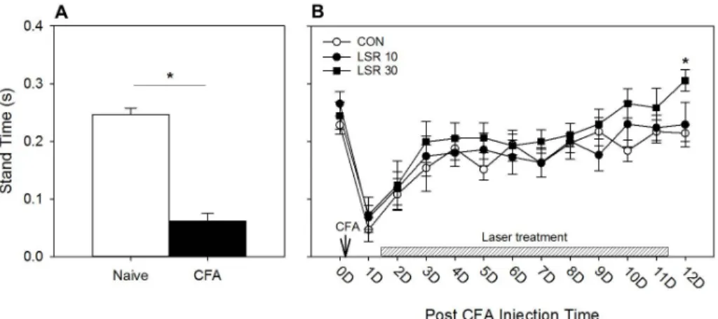
Fig. 1. Effect of Low Level Laser applied to KI 1 on the stand time of the arthritic rat.
Measurements were taken preoperatively and at 1-12 Days post-injection. In the CFA-induced arthritis rat, stand time was decreased significantly at 1 day after arthritis induction(*p<0.05, Paired t-test, Naive vs. CFA)(A). Baseline measurements were performed at 1 day after CFA injection, and then low level laser treatment was given once a day for 11 days. On the 12th day post CFA injection, stand time was increased statistically in LSR 30 group(B). Mean values and S.E.M. were calculated and the results obtained were subjected to repeated measures analysis of variance(One Way ANOVA) with subsequent Bonferroni t-test post hoc comparison of effects by the treatments(*p<0.05, one way ANOVA, CON vs. LSR30 Group)(B).
(%)로 표시되는 매개변수로서 다음과 같은 공식으로 계산하였다.
Duty cycle(%)=((Stand+Swing)/Stand)*100
Print area, Mean intensity, Stride length는 관절염을 유발하 지 않은 쪽 측정값(왼쪽 뒷발; LH)에 대한 관절염을 유발한 쪽 측정 값(오른쪽 뒷발; RH)의 비율로 표시하였다.
Relative ratio(%)=RH/LH*100
6. 통계
Catwalk test 측정 결과는 평균±표준오차(S.E.M.)로 표시하였 고 통계분석은 Sigma Plot 12를 사용하였다. 관절염 유발로 인한 각 매개변수의 변화는 paired t-test로 전후 비교하였고, 레이저 처 치 후 각 군간의 비교는 one way ANOVA를 시행하고 사후검정은 Bonferroni t-test를 시행하였다. 통계적 유의 수준은 p<0.05로 하였다.
결 과
1. 저출력 레이저 자극이 stand 변화에 미치는 영향 Stand 시간은 관절염 유발 전 naïve 상태일 때 0.2461±
0.0117(s)에서 관절염 유발 후 0.0617±0.0134(s)로 감소하였다
(n=24, Fig. 1A). 레이저 처치 전인 관절염 유발 후 1일째에 CON군 0.0468±0.0209(s), LSR10군 0.0680±0.0206(s), LSR30군 0.0727±0.0307(s) 이었으며, 레이저 11회 처치 후인 관절염 유발 후 12일에 CON군 0.2142±0.0142(s), LSR10군 0.2292±0.0385(s), LSR30군 0.3056±0.0185(s)로 나타났다. 관절염 유발 12일째 LSR10군은 CON군과 LSR30군간의 통계적 유의성이 나타나지 않 았으나 LSR30군은 CON군과 비교해서 통계적으로 유의하게 증가 하였다(Fig. 1B).
2. 저출력 레이저 자극이 swing 변화에 미치는 영향 Swing 시간은 관절염 유발 전 naïve 상태일 때와 비교하여 관절 염 유발 후 약 2.5배(252.7±31.1) 증가하였다(n=24, Fig. 2A).
Swing 값이 최대로 증가하는 관절염 유발 1일째 값을 기준으로 레이저 처치 후 나타나는 변화를 최대변화값으로 측정하였다. 관절 염 유발 후 12일째 CON군은 29.4±1.8(%), LSR10군은 16.9±
2.5(%), LSR30군은 17.3±4.7(%)로 나타났다. 관절염 유발 12일째 LSR10군은 CON군과 비교하여 통계적으로 유의한 차이가 있었으 나, LSR30군에서는 통계적 유의성이 나타나지 않았다(Fig. 2B).
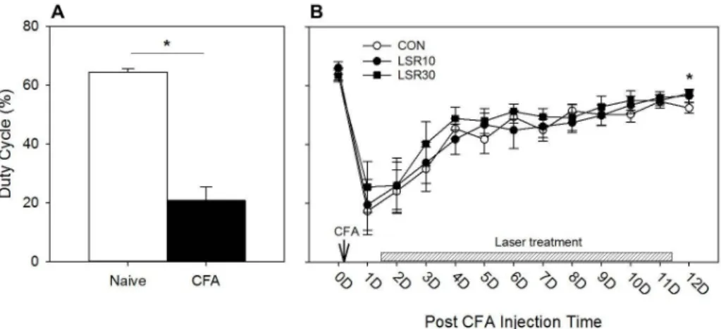
3. 저출력 레이저 자극이 duty cycle 변화에 미치는 영향 Duty cycle은 관절염 유발 전 naïve 상태일 때 64.3±1.1(%)에 서 관절염 유발 후 20.7±4.7(%)로 감소하였다(n=24, Fig. 3A). 레 이저 처치 전인 관절염 유발 후 1일째 CON군은 17.3±8.0(%), LSR10군은 19.4±8.6(%), LSR30군은 25.5±8.6(%) 이었으며, 11
Fig. 2. Effect of Low Level Laser applied to KI 1 on the swing time of the arthritic rat.
Measurements were taken preoperatively and at 1-12 Days post-injection. In the CFA-induced arthritis rat, swing time was increased significantly at 1 day after arthritis induction(*p<0.05, Paired t-test, Naive vs. CFA)(A). Baseline measurements were performed at 1 day after CFA injection, and then low level laser treatment was given once a day for 11 days. On the 12th day post CFA injection, swing time was decreased statistically in LSR 10 group(B). Mean values and S.E.M. were calculated and the results obtained were subjected to repeated measures analysis of variance(One Way ANOVA) with subsequent Bonferroni t-test post hoc comparison of effects by the treatments(*p<0.05, one way ANOVA, CON vs. LSR10 Group)(B).
Fig. 3. Effect of Low Level Laser applied to KI 1 on the duty cycle of the arthritic rat.
Measurements were taken preoperatively and at 1-12 Days post-injection. In the CFA-induced arthritis rat, duty cycle was decreased significantly at 1 day after arthritis induction(*p<0.05, Paired t-test, Naive vs. CFA)(A). Baseline measurements were performed at 1 day after CFA injection, and then low level laser treatment was given once a day for 11 days. On the 12th day post CFA injection, duty cycle was increased statistically in LSR 30 group(B). Mean values and S.E.M. were calculated and the results obtained were subjected to repeated measures analysis of variance(One Way ANOVA) with subsequent Bonferroni t-test post hoc comparison of effects by the treatments(*p<0.05, one way ANOVA, CON vs. LSR30 Group)(B).
회 레이저 처치를 받은 후인 관절염 유발 후 12일째 CON군은 52.3±1.7(%), LSR10군은 56.5±2.1(%), LSR30군은 57.3±0.9(%) 로 나타났다. 관절염 유발 12일째 LSR10군은 다른 두 군 사이에 통계적으로 유의한 차이가 없었지만 LSR30군은 CON군과 비교하 여 통계적으로 유의하게 증가하였다(Fig. 3B).
4. 저출력 레이저 자극이 print area 변화에 미치는 영향 Print area는 관절염 유발 전 naïve 상태일 때에는 좌우 뒷발 간에 차이가 없었지만(100.4±3.4) 관절염 유발 후 관절염으로 인 한 통증 때문에 좌측에 비해 현저하게 감소(17.9±4.1)하였다 (n=24, Fig. 4A). 레이저 처치 전인 관절염 유발 후 1일째 CON군은 13.1±7.3(%), LSR10군은 17.0±7.8(%), LSR30군은 18.4±
6.0(%) 이었으며, 11회 레이저 처치 후인 관절염 유발 후 12일째
Fig. 4. Effect of Low Level Laser applied to KI 1 on the print area of the arthritic rat.
Measurements were taken preoperatively and at 1-12 Days post-injection. In the CFA-induced arthritis rat, the print area was decreased significantly at 1 day after arthritis induction(*p<0.05, Paired t-test, Naive vs. CFA)(A). Baseline measurements were performed at 1 day after CFA injection, and then low level laser treatment was given once a day for 11 days. Mean values and S.E.M. were calculated.
There were no significant differences between groups during all time period of experiments(B).
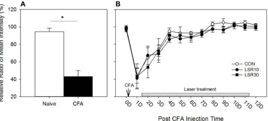
Fig. 5. Effect of Low Level Laser applied to KI 1 on the mean intensity of the arthritic rat.
Measurements were taken preoperatively and at 1-12 Days post-injection. In the CFA-induced arthritis rat, the mean intensity was decreased significantly at 1 day after arthritis induction(*p<0.05, Paired t-test, Naive vs. CFA)(A). Baseline measurements were performed at 1 day after CFA injection, and then low level laser treatment was given once a day for 11 days. Mean values and S.E.M. were calculated. There were no significant differences between groups during all time period of experiments(B).
CON군은 65.8±9.5(%), LSR10군은 64.6±4.8(%), LSR 30군은 62.9±8.3(%)으로 나타났다. Print area는 모든 측정기간 동안 세 군간에 통계적으로 유의한 차이는 없었다(Fig. 4B).
5. 저출력 레이저 자극이 mean intensity 변화에 미치 는 영향
Mean intensity는 관절염 유발 전 naïve 상태일 때에는 좌우 뒷발 간에 통계적으로 유의한 차이가 없었지만(94.3±4.1) 관절염 유발 후 딛을 때 발생하는 통증을 줄이려는 경향이 있어 왼발에 비해서 통증이 있는 오른발 딛는 강도는 절반 이하로 현저하게 감
소(42.9±6.9)하였다(n=24, Fig. 5A). 레이저 처치 전인 관절염 유 발 후 1일째 mean intensity는 CON군은 44.2±8.7(%), LSR10군 은 41.1±11.2(%), LSR30군은 43.3±14.3(%) 이었으며, 관절염 유 발 후 4일째 CON군 95.1±3.6(%)로 거의 정상값으로 회복하였다.
Mean intensity에서는 모든 측정기간 동안 세 군간에 통계적으로 유의한 차이는 없었다(Fig. 5B).
6. 저출력 레이저 자극이 stride length 변화에 미치는 영향
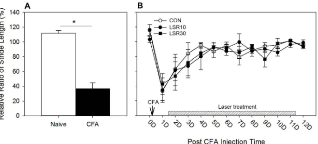
Stride length은 관절염 유발 전 naïve 상태일 때에는 좌우 뒷발
Fig. 6. Effect of Low Level Laser applied to KI 1 on the stride length of the arthritic rat.
Measurements were taken preoperatively and at 1-12 Days post-injection. In the CFA-induced arthritis rat, the stride length was decreased significantly at 1 day after arthritis induction(*p<0.05, Paired t-test, Naive vs. CFA)(A). Baseline measurements were performed at 1 day after CFA injection, and then low level laser treatment was given once a day for 11 days. Mean values and S.E.M. were calculated. There were no significant differences between groups during all time period of experiments(B).
간에 통계적으로 유의한 차이가 없었지만(111.6±3.7) 관절염 유발 후 관절염이 있는 우측 뒷발 보폭이 통계적으로 유의하게 감소 (36.7±8.0)하였다(n=24, Fig. 6A). 레이저 처지 전인 관절염 유발 후 1일째 stride length는 CON군은 33.0±12.7(%), LSR10군은 34.3±16.4(%), LSR30군은 42.6±14.2(%)이었으며, 관절염 유발 후 4일째 CON군의 경우 95.3±3.4(%)로 거의 좌우 차이가 없는 정상값으로 회복하였다. Stride length 항목에서는 모든 측정기간 동안 세 군간에 통계적으로 유의한 차이를 관찰할 수 없었다(Fig.
6B).
고 찰
이 연구에서는 관절염 동물의 보행행동을 관찰하기 위하여 관절 염 실험동물 모델로서 일반적으로 많이 사용하는 CFA를 관절강에 주입하여 유도한 관절염 모델을 사용하였다. 관절염 유발로 인해 보행에 장애가 있는 실험동물의 용천에 저출력 레이저를 반복적으 로 조사한 후 catwalk system을 사용하여 보행행동 변화를 알아보 았다. Catwalk system 보행 측정 매개변수 중 선행연구에서 유의 한 변화가 보고된 바 있는 인자인 stand, swing, duty cycle, print area, mean intensity, stride length를 분석하였다. CFA로 관절염 을 유발 한 1일째에는 6개 변수 모두가 통계적으로 유의한 변화가 나타났으며, 반복적인 레이저 처치 후에는 관절염 유발 후 12일째 에 stand와 duty cycle은 증가하고 swing은 감소하였다. 그러나 나머지 세 매개변수인 print area, mean intensity, stride length
에서는 통계적으로 유의한 변화가 측정되지 않았다. 또한 LSR10군 과 LSR30군 사이에서 유의한 차이는 나타나지 않았다.
이와 같은 결과는 관절염이 유발된 직후에는 염증으로 인한 통 증이 가장 심하기 때문에 통증이 있는 쪽 발을 딛고 있는 시간 (stand)이 감소하고, 내 딛는데 걸리는 시간(swing)은 증가하며, 바 닥에 닫는 면적(print area)과 강도(mena intensity), 보폭(stride length)은 감소한다는 것을 의미한다. 관절염 동물의 용천에 매일 1회씩 11일 동안 저출력 레이저를 처치한 결과 통증이 있는 발을 딛고 있는 시간이 유의하게 증가하였으며, 내 딛는 시간은 유의하 게 빨라졌는데 이는 관절염으로 인한 보행행동이 개선된 결과로 해석할 수 있다.
Catwalk system을 활용한 선행연구를 살펴보면 carrageenan 으로 유발된 관절염 모델에서 급성 염증성 통증성에 대한 행동변화 를 분석한 연구36)에서는 intensity, duty cycle, base of support of the hind limbs, print area, maximum area, stride length, step cycle, swing speed 등의 매개변수 중 intensity, duty cycle, print area에서 유의한 행동변화를 보였다.
이 실험에서는 catwalk test 매개변수 중 print area, mean intensity, stride length에서는 통계적으로 유의한 차이가 나타나 지 않았다. 이러한 결과가 나타난 이유는 실험에 사용한 CFA 유발 관절염모델의 경우 아픈 쪽 발의 딛는 면적, 강도, 보폭 등이 정상 값으로 회복이 빨라 반복적인 처치에 따른 효과를 관찰하기에 적당 하지 않은 문제가 있으며 실험 디자인 상의 제한된 레이저 자극 혈위 및 혈위 수, 레이저 자극시간, catwalk 측정 시간 및 기간이 영향을 미쳤을 것으로 사료된다.
저출력 레이저 자극 혈위는 관절염에 흔히 사용하는 족삼리37)에 자극했다면 다른 효과가 없었던 매개변수에서도 유의한 차이가 있 을 수 있지만 이 연구에서는 만성퇴행성 관절염 환자에 수시로 경 혈에 레이저 자극을 하여 보행에 도움을 줄 수 있는 기능성 신발과 같은 장치에 응용이 가능 하도록 발바닥에 있는 경혈인 용천을 선 택하였다. 또한 이 연구에서는 용천 한 곳만 선택하여 자극을 했지 만 양구(ST34), 족삼리(ST36), 슬안(EX-LE5), 양릉천(GB34), 음릉 천(SP9) 등 혈위에 동시에 레이저 자극을 했다면 다른 분석 인자에 서도 유의한 결과가 나타날 수 있기 때문에 추가적인 연구가 필요 한 부분이다.
기존 연구결과에 의하면 저출력 레이저 자극시간은 화농성 슬관 절염을 일으킨 흰쥐에 He-Ne 레이저를 4분과 10분 시행한 결과 레이저 치료는 관절염에 효과가 있는 것으로 알려져 있어38), 이 연 구에서는 저출력 레이저의 자극시간에 따른 보행 변화의 반응을 보기 위해 레이저 10분 군과 레이저 30분 군으로 나눠 각각 시행하 였다. 그러나 레이저 10분 군과 레이저30분 군간의 차이는 확인 되지 않았다. 레이저 자극 시간에 따른 용량 반응(dose-response) 관계 연구 또한 향후 추가 연구가 필요할 것으로 사료된다.
이 연구에서는 catwalk 측정시간은 반복적인 처치에 따른 누적 효과를 보기 위하여 관절염 유발 후 12일 동안 레이저 자극 후 다음 날 자극 전(24시간째)에 행동 측정을 하였다. 이 연구에서는 실험설 계상 레이저 자극 후 단시간에 나타나는 효과는 측정하지 않았지 만, 단시간에 나타나는 효과도 의미가 있을 것이기 때문에 30분에 서 수시간 이내에 나타나는 단기적 효과에 대한 확인이 필요 할 것이다. 아울러 이번 실험에서는 CFA로 유발한 관절염 모델을 사 용하여 몇 가지 인자에서 관절염 유발 후 12일째에 통계적으로 유 의한 차이가 나타났으나 조금 더 장기간 관찰이 가능한 iodoace- tate로 유발하는 퇴행성 관절염 모델 같은 실험모델을 사용하여 12 일보다 장기적인 누적 효과를 확인하면 장기간 사용했을 때 효과가 더 증가되는지 알아볼 수 있을 것이다.
결 론
용천에 조사한 저출력 레이저 자극이 보행에 미치는 영향을 알 아보기 위하여 CFA 유발 관절염 흰쥐모델에서 catwalk을 이용하 여 stand, swing, duty cycle, print area, mean intensity, stride length의 매개변수를 측정 분석했다.
분석 결과 관절염 유발 후 모든 매개변수가 통계적으로 유의하 게 변화하였으며, 저출력 레이저를 11회 조사한 후인 관절염 유발
후 12일째 LSR30 군과 CON군에서 stand와 duty cycle이 유의하 게 증가하였으며, LSR10 군과 CON군에서 swing이 유의하게 감소 하였다. 반면에 저출력 레이저 자극이 print area, mean intensity, stride length의 변화에는 통계적으로 유의한 차이를 보이지 않았다.
이상 본 연구 결과를 종합해 보면 stand 및 swing, duty cycle의 변화에 저출력 레이저 조사가 유의한 변화를 미치는 반면, print area, mean intensity, stride length에서는 유의한 변화가 없었다.
관절염으로 인해 감소한 딛는 시간(stand)을 유의하게 증가시켜 주 고, 늘어난 내 딛는데 걸리는 시간(swing)은 유의하게 줄여주는 효 과가 있기 때문에 관절염으로 인한 보행장애에 용천에 조사한 저출 력 레이저 자극은 퇴행성 관절염 환자의 보행을 개선시키는 효과가 있을 것으로 예상된다. 이러한 효과는 추후 사람을 대상으로 하는 연구를 통해 확인이 필요할 것이다.
감사의 글
This work was supported by a 2-Year Research Grant of Pusan National University.
References
1. Jaakkola JI, Mann RA. A review of rheumatoid arthritis affecting the foot and ankle. Foot & Ankle International. 2004 ; 25 : 866-74.
2. Michelson J, Easley M, Wigley FM, Hellmann D. Foot and ankle problems in rheumatoid arthritis. Foot & Ankle International.
1994 ; 15 : 608-13.
3. Semple R, Turner DE, Helliwell PS, Woodburn J. Regionalised centre of pressure analysis in patients with rheumatoid ar- thritis. Clin Biomech(Bristol, Avon). 2007 ; 22 : 127-9.
4. Backhouse MR, Pickles DA, Mathieson HR, Edgson L, Emery P, Helliwell PS, et al. Diurnal variation of gait in patients with rheumatoid arthritis: The DIVIGN study. Clinical Biome- chanics. 2014 ; 29 : 811-4.
5. Benvenuti F, Ferrucci L, Guralnik JM, Gangemi S, Baroni A. Foot pain and disability in older persons: an epidemiologic survey.
Journal of the American Geriatrics Society. 1995 ; 43 : 479-84.
6. Oh KY. Rehabilitation Interventions for the Patient with
Rheumatoid Arthritis. J of Soonchunhyang Medical Science.
2008 ; 14 : 71-82.
7. Whittaker P. Laser acupuncture: past, present, and future.
Lasers in Medical Science. 2004 ; 19 : 69-80.
8. Park MH, Rho MH, Kim JY. The Effect of Swimming and Low Power Laser on the Healing of the Freund`s Complete Adjuvant Induced Arthritis in Rat. The J of Korean Academy of Physical Therapist. 2006 ; 13 : 7-20.
9. Na SY. Effect of Low level laser on pain and regeneration of car- tilage in a model of osteoarthritis rat. Department of Physical Therapy Graduate Schoolo Dong Shin University. 2012 ; Ph. D.
Dissertation: 65-85.
10. Kang H, Son T, Lee A, Youn I, Seo DH, Kim HS, et al. The effects of a minimally invasive laser needle system on complete Fre- und's adjuvant-induced arthritis. Lasers in Medical Science.
2014 ; 29 : 1599-606.
11. Patrocinio-Silva TL, de Souza AM, Goulart RL, Pegorari CF, Oliveira JR, Fernandes K, et al. The effects of low-level laser ir- radiation on bone tissue in diabetic rats. Lasers in Medical Science. 2014 ; 29 : 1357-64.
12. Chang MS. The effects of laser and ultrasound treatment on re- generation nerve and muscle on injured peripheral nerve of rats medel. Department of Physical Therapy Graduate School of DongShin University. 2005 ; Ph. D. Dissertation: 68.
13. Efendiev AI, Tolstykh PI, Dadashev AI, Azimov SA. Increasing the scar strength after preventive skin irradiation with low-in- tensity laser. Klinicheskaia Khirurgiia. 1992 : 23-5.
14. Ozdemir F, Birtane M, Kokino S. The clinical efficacy of low-power laser therapy on pain and function in cervical osteoarthritis. Clinical Rheumatology. 2001 ; 20 : 181-4.
15. Schaffer M, Sroka R, Fuchs C, Schrader-Reichardt U, Schaffer PM, Busch M, et al. Biomodulative effects induced by 805 nm la- ser light irradiation of normal and tumor cells. Journal of Photochemistry and Photobiology B, Biology. 1997 ; 40 : 253-7.
16. Ad N, Oron U. Impact of low level laser irradiation on infarct size in the rat following myocardial infarction. International Journal of Cardiology. 2001 ; 80 : 109-16.
17. Kartelishev AV, Kolupaev GP, Chebotkov AA. Low-intensity magnetic-laser therapy in the combined treatment of alco- holics with neurotic disorders. Voenno-Meditsinskii Zhurnal.
2000 ; 321 : 38-43.
18. Cops EJ, Sashindranath M, Daglas M, Short KM, da Fonseca Pereira C, Pang TY, et al. Tissue-type plasminogen activator is an extracellular mediator of Purkinje cell damage and altered gait. Experimental Neurology. 2013 ; 249 : 8-19.
19. Eftaxiopoulou T, Macdonald W, Britzman D, Bull AM. Gait com- pensations in rats after a temporary nerve palsy quantified using temporo-spatial and kinematic parameters. Journal of Neuro- science Methods. 2014 ; 232 : 16-23.
20. Hansen ST, Pulst SM. Response to ethanol induced ataxia be- tween C57BL/6J and 129X1/SvJ mouse strains using a treadmill based assay. Pharmacology, Biochemistry, and Behavior. 2013 ; 103 : 582-8.
21. Dorman CW, Krug HE, Frizelle SP, Funkenbusch S, Mahowald ML. A comparison of DigiGait and TreadScan imaging systems:
assessment of pain using gait analysis in murine monoarthritis.
J Pain Res. 2014 ; 7 : 25-35.
22. Kuypers NJ, James KT, Enzmann GU, Magnuson DS, Whitte- more SR. Functional consequences of ethidium bromide de- myelination of the mouse ventral spinal cord. Experimental Neurology. 2013 ; 247 : 615-22.
23. Myers SA, DeVries WH, Gruenthal MJ, Andres KR, Hagg T, Whittemore SR. Sildenafil improves epicenter vascular perfu- sion but not hindlimb functional recovery after contusive spinal cord injury in mice. Journal of Neurotrauma. 2012 ; 29 : 528-38.
24. Masocha W, Parvathy SS. Assessment of weight bearing changes and pharmacological antinociception in mice with LPS-in- duced monoarthritis using the Catwalk gait analysis system. Life Sciences. 2009 ; 85 : 462-9.
25. Huehnchen P, Boehmerle W, Endres M. Assessment of paclitax- el induced sensory polyneuropathy with "Catwalk" automated gait analysis in mice. PloS One. 2013 ; 8 : e76772.
26. Ferreira-Gomes J, Adaes S, Castro-Lopes JM. Assessment of movement-evoked pain in osteoarthritis by the knee-bend and CatWalk tests: a clinically relevant study. The Journal of Pain : Official Journal of the American Pain Society. 2008 ; 9 : 945-54.
27. Forgione N, Karadimas SK, Foltz WD, Satkunendrarajah K, Lip A, Fehlings MG. Bilateral contusion-compression model of in- complete traumatic cervical spinal cord injury. Journal of Neu- Rotrauma. 2014 ; 31 : 1776-88.
28. Miyagi M, Ishikawa T, Kamoda H, Suzuki M, Sakuma Y, Orita S, et al. Assessment of Pain Behavior in a Rat Model of Interverte- bral Disc Injury Using the Catwalk Gait Analysis System. Spine.
2013 ; 38 : 1459-65.
29. Abada YS, Nguyen HP, Schreiber R, Ellenbroek B. Assessment of motor function, sensory motor gating and recognition memory in a novel BACHD transgenic rat model for huntington disease.
PloS One. 2013 ; 8 : e68584.
30. Wang XH, Lu G, Hu X, Tsang KS, Kwong WH, Wu FX, et al.
Quantitative assessment of gait and neurochemical correlation in a classical murine model of Parkinson's disease. BMC Neu- roscience. 2012 ; 13 : 142.
31. Chen YJ, Cheng FC, Sheu ML, Su HL, Chen CJ, Sheehan J, et al.
Detection of subtle neurological alterations by the Catwalk XT gait analysis system. Journal of Neuroengineering and Rehabi- litation. 2014 ; 11 : 62.
32. Masocha SSPaW. Gait analysis of C57BL/6 mice with complete Freund's adjuvant-induced arthritis using the CatWalk system.
Parvathy and Masocha BMC Musculoskeletal Disorders. 2013 ; 14 : 1471-2474.
33. Ferland CE, Laverty S, Beaudry F, Vachon P. Gait analysis and pain response of two rodent models of osteoarthritis. Pharma-
cology, Biochemistry, and Behavior. 2011 ; 97 : 603-10.
34. Angeby-Moller K, Berge OG, Hamers FP. Using the CatWalk method to assess weight-bearing and pain behaviour in walk- ing rats with ankle joint monoarthritis induced by carrageenan:
effects of morphine and rofecoxib. Journal of Neuroscience Methods. 2008 ; 174 : 1-9.
35. Ferreira-Gomes J, Adães S, Mendonça M, Castro-Lopes JM.
Analgesic effects of lidocaine, morphine and diclofenac on movement-induced nociception, as assessed by the Knee-Bend and CatWalk tests in a rat model of osteoarthritis. Pharmacology Biochemistry and Behavior. 2012 ; 101 : 617-24.
36. Gabriel AF, Marcus MA, Honig WM, Walenkamp GH, Joosten EA.
The CatWalk method: a detailed analysis of behavioral changes after acute inflammatory pain in the rat. Journal of Neurosci- ence Methods. 2007 ; 163 : 9-16.
37. Manyanga T, Froese M, Zarychanski R, Abou-Setta A, Friesen C, Tennenhouse M, et al. Pain management with acupuncture in osteoarthritis: a systematic review and meta-analysis. BMC Complementary and Alternative Medicine. 2014 ; 14 : 312.
38. Kim JY, No MH, Ko JB. The Effect of He-Ne IR Laser Therapy on The Healing of Acute Septic Knee Arthritis in Rat. Korean J Biomed Lab Sci 2000 ; 6 : 223-8.