Abstract
Prosthodontic treatment using implants has many advantages in comparison with conventional treatment. However, it is reported that there are several complications associated with implants. They are divided into mechanical, biological, and esthetic aspects in prosthodontics. To overcome them, there have been numerous attempts such as a connection type of abutment-fixture, microthread, crestal module design, and abutment profile. Recently, one of the methods involves the development of a 1-piece implant. A 1-piece implant has many advantages in comparison with previous 2-piece implant. It is free of mechanical complications such as screw looseness, screw fracture, and fixture fracture. Also, in a biological aspect, absence of microgap, micromovement, and dis/reconnection of abutment leads to the stable maintenance of soft and hard tissue. However, 1-piece implants have limited indications. Selection of abutment is very strict and correction of the path is difficult after the installation of the fixture. Also, bone quality and primary stability are very important factors in 1-piece implants because it is based on immediate provisionalization. Although there are not many kinds of available 1-piece implants, one of the most well-known 1-piece implants is NobelDirect® (Nobel Biocare). However, clinical results of NobelDirect® are controversial and improvement is necessary. In most studies, it is reported that long term studies and improvements of implant design are required. Therefore, this research focuses on the advantages, design, clinical application and practical result of 1-piece implants.
Key Words: dental implants, 1-piece implant, one-piece implant, review
일체형 임플란트
최희곤, 손용하, 권주현, 김선재, 한종현 연세대학교 치과대학 강남세브란스 치과병원 보철과
1-Piece Implant
Hee-Gon Choi, Yong-Ha Son, Joo-Hyun Kwon, Sunjai Kim, Chong-Hyun Han
Department of Prosthodontics, Gangnam Severance Dental Hospital, College of Dentistry, Yonsei University, Seoul, Korea
Reprint requests: Chong-Hyun Han
Department of Prosthodontics, Gangnam Severance Dental Hospital, College of Dentistry, Yonsei University, 211 Eonju-ro, Gangnam-gu, Seoul 135-720, Korea Tel: 82-2-2019-1340, Fax: 82-2-2019-1347
E-mail: [email protected]
Received for publication: September 5, 2012 Accepted for publication: September 18, 2012
교신저자: 한종현
(135-720) 서울시 강남구 언주로 211
연세대학교 치과대학 강남세브란스 치과병원 보철과 Tel: 82-2-2019-1340, Fax: 82-2-2019-1347 E-mail: [email protected]
원고접수일: 2012년 9월 5일 게재확정일: 2012년 9월 18일
Copyright © 2012. The Korean Academy of Oral & Maxillofacial Implantology
This is an Open Access article distributed under the terms of the Creative Commons Attribution Non-Commercial License (http://creativecommons.org/licenses/by-nc/3.0/) which permits
merged)과 비매립형(non-submerged) 간의 차이가 없었 고 비매립형(non-submerged) 방식의 이체형 임플란트 에 비해 일체형 임플란트(1-piece implant)가 기계적, 생 물학적 이점이 있는 것으로 밝혀졌다7. 일체형 임플란트 는 임시 임플란트(transitional implant)로 예전부터 사용 되었고8 최근에는 영구 임플란트(definitive implant)로서 의 연구가 활발하다. Finne 등9은 Nobel Biocare사의 일 체형 임플란트를 이용한 3년간의 전향적 연구에서 안정 적인 경조직, 연조직의 보존과 높은 임플란트 생존율을 보고하였다. 이에 앞서 Hahn10은 Nobel Biocare사의 일 체형 임플란트를 이용한 3년간의 연구에서 안정적인 경 조직의 보존과 높은 누적 생존율을 보였으며 즉시 임시 보철물 수복 시에 이점을 보인다고 하였다. Sohn 등11은 Biohorizon사의 3.0 mm 직경의 일체형 임플란트를 사용 한 후향적 연구에서 3년간 0.53 mm의 적은 변연골 소실 을 보고하였다. 하지만 일체형 임플란트를 이용한 연구 에서 좋지 않은 결과를 보인 경우도 있었다. Ostman 등12 은 Nobel Biocare사의 일체형 임플란트를 이용한 전향적 연구에서 이체형 임플란트에 비해 많은 변연골 소실을 보였고 임플란트 디자인, 식립 깊이, rough surface의 점 막 내 노출, 식립 당일 preparation 등이 영향을 미쳤다 고 보고하였다. 또한 Zembić 등13은 NobelDirect® 3.0 (Nobel Biocare)을 사용한 즉시 부하 연구에서 전반적인 생존율은 높았지만 다수의 임플란트가 많은 양의 변연골 소실을 보여 주의를 할 필요가 있다고 보고하였다.
일체형 임플란트를 이용한 연구들에서 장기 연구의 필 요성과 임플란트 디자인의 개선을 주장하고 있다. 따라 서 본 연구에서는 일체형 임플란트의 이점, 디자인, 임상 적 적용, 실제 임상 결과 등에 대하여 소개하고자 한다.
I 서론
P.I.Brånemark에 의해 osseointegration 이라는 개념이 처음 도입된 후 임플란 트의 발전은 치의학 전반에 걸쳐 많은 변화를 가져왔다. 특히 보철학 분야에서 임플란트를 이 용한 치료는 장기간의 임상 연구 및 높은 성공률을 보이 며 기존의 고정성 국소의치, 가철성 국소의치, 총의치에 의한 치아 수복과 비교하여 많은 장점을 가진다. 바야흐 로 전치부, 구치부의 단일 결손부 수복, 부분 무치악, 완 전 무치악 환자의 보철 수복에 있어서 임플란트가 치료 계획 전반에 영향을 미치는 시대가 되었다1.
하지만 임플란트를 이용한 수복 치료가 발전하고 보편 화되는 과정에서 여러 합병증이 보고되고 있다. 보철적 인 측면에서 합병증은 생물학적(biological), 기계적 (mechanical), 심미적(esthetic)인 것으로 나뉜다. 생물학 적 합병증은 임플란트와 보철물 주위 조직에 영향을 미 치는 것으로 변연골 소실, 임플란트 주위염, 임플란트 주 위 점막염, 임플란트 고정체와 지대주의 계면에서 생기 는 누공, 연조직의 과증식, 임플란트 소실 등이 있다2. 기 계적인 합병증은 임플란트 고정체, 지대주, 상부 구조의 기계적인 결함을 포함한다. 주된 항목은 veneering material이나 금속 구조물의 파절, 유지력 상실, 나사 풀 림과 파절, 고정체의 파절 등이다2-6. 마지막으로 심미적 인 합병증은 보철물과 주위 조직이 관련되어 보철물의 외형에 영향을 미치는 것을 의미한다.
위와 같은 합병증을 극복하고자 고정체와 지대주의 연 결 방식, 미세나사선, 임플란트 경부 디자인, 지대주의 구조와 형상 등에서 많은 시도와 발전이 있었다. 기존의 임플란트는 매립형(submerge) 방식의 이체형 임플란트 (2-piece implant)였다. 하지만 연구 결과 매립형(sub-
II 일체형 임플란트의 이점
1. 생물학적 측면(biological aspect)
1) 미세간극과 미세동요(microgap and micromovement)
일반적으로 임플란트 보철과 관련된 합병증은 위에서 언급한 바와 같이 생물학적, 기계적, 심미적인 측면으로 나뉜다. 이 중에서 이체형 임플란트와 관련된 생물학적 합병증은 미세간극(microgap), 미세동요(micromove- ment)로 인하여 발생되는 marginal bone loss가 있다. 일 반적인 임플란트 주위 변연골은 기능 후 1년 동안 1.5~2.0 mm의 골소실을 야기하는 것으로 알려져 있다.
이러한 골소실은 첫 번째 나사산까지 발생한 후 안정되 어 이후로는 적은 양의 골소실이 일어난다. 임플란트 식 립 후 초기에 발생하는 변연골 소실을 설명하기 위하여 여러 연구들이 진행되었다. 연구들에 의하면 변연골 소 실은 여러 가지 요소들이 복합적으로 작용하는 것으로 보인다. 보철 후 발생하는 변연골 흡수는 임플란트에 가 해지는 부하에 의하여 응력이 주로 치조정 부위에 집중 하는 것과 관련이 있을 수 있다. 임플란트 변연골 소실의 또 다른 원인으로 임플란트와 지대주의 연결부(implant- abutment junction, IAJ)의 위치가 거론되며 이는 치조골 의 흡수와 밀접하게 연관이 있음이 밝혀졌다14,15. 동물실 험에서 임플란트에 healing abutment를 연결하면 IAJ에 서 하방으로 1.5~2.0 mm 정도의 골소실이 발생하였다.
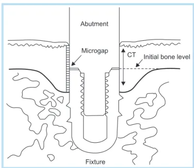
다른 연구들에서도 임플란트가 구강 환경에 노출되면 임 플란트와 지대주 사이에 존재하는 미세한 간극 내로 세 균들의 침투가 발생하여 인접 조직내 염증 반응으로 골 소실이 발생하는 것으로 보고하였다(Fig. 1)16,17. 이체형 임플란트를 조직학적으로 관찰하였을 때, IAJ 주변조직 에는 염증세포에 의한 침윤대(abutment inflammatory
cell infiltrate)가 발생하며, 하방으로 염증세포의 침윤이 없는 1 mm 정도의 건전한 결합조직에 의하여 치조정과 분리되어 있다18. 이는 임플란트 표면에 건전한 결합조직 부착을 통하여 생물학적인 점막 방어막을 형성하여 외부 환경으로부터 치조골을 보호하기 위한 생물학적인 반응 이며, 임플란트 주위에도 생물학적 폭경(biologic width) 이 존재하고 있음을 보여준다19,20. Hermann 등21은 치조 정보다 1~2 mm 상방에 미세간극(microgap)이 위치하도 록 식립하고 미세간극 size와 미세간극의 laser welding 이 crestal bone loss에 미치는 영향을 분석하였다. 그 결 과 미세간극의 size는 변연골 흡수 정도에 영향을 주지 않았고 미세간극의 laser-welding은 미세동요를 없애 변 연골 유지에 기여하는 것이 밝혀졌다. 이로 보아 미세동 요도 crestal bone loss에 영향을 미치는 것을 알 수 있다.
일체형 임플란트는 미세간극, 미세동요가 없다는 측면에 서 안정적인 변연골 유지에 유리할 것이라 생각된다.
Fig. 1. Implant-abutment junction (microgap).
Hee-Gon Choi et al. : 1-Piece Implant. Implantology 2012
2) 지대주의 탈착과 재부착(disconnection, reconnection of abutment)
Abrahamsson 등22은 동물실험에서 지대주의 착탈을 반복한 결과 실험군이 대조군보다 유의적으로 높은 변연 골 흡수량을 보였다. 지대주 착탈 시 결합조직(connec- tive tissue)이 disruption되면서 wound로 인식이 되고 접합 상피세포는 결합조직의 wound 부위로 하방 이주 (down growth)하게 되어 결과적으로는 생물학적 폭경을 확보하기 위하여 변연골 흡수가 일어나기 때문이다.
Rodríguez 등23은 platform-switched/non platform- swtiched implant에 abutment를 착탈한 횟수에 따라 변 연골 흡수량을 연구한 동물 실험에서 모든 이체형 임플 란트에서 healing 기간 동안 일정량의 변연골 흡수를 보 이며 흡수량은 non platform-switched implant에서 더 컸고 지대주를 착탈한 횟수가 증가할수록 변연골 소실량 이 더 커진다는 것을 보고하였다. 일체형 임플란트는 healing 기간과 보철물 수복 과정에서 지대주의 착탈이 없기 때문에 임플란트 주위 조직들의 손상이 없어 경조 직과 연조직 유지에 도움을 줄 것으로 생각된다.
2. 기계적 측면(mechanical aspect)
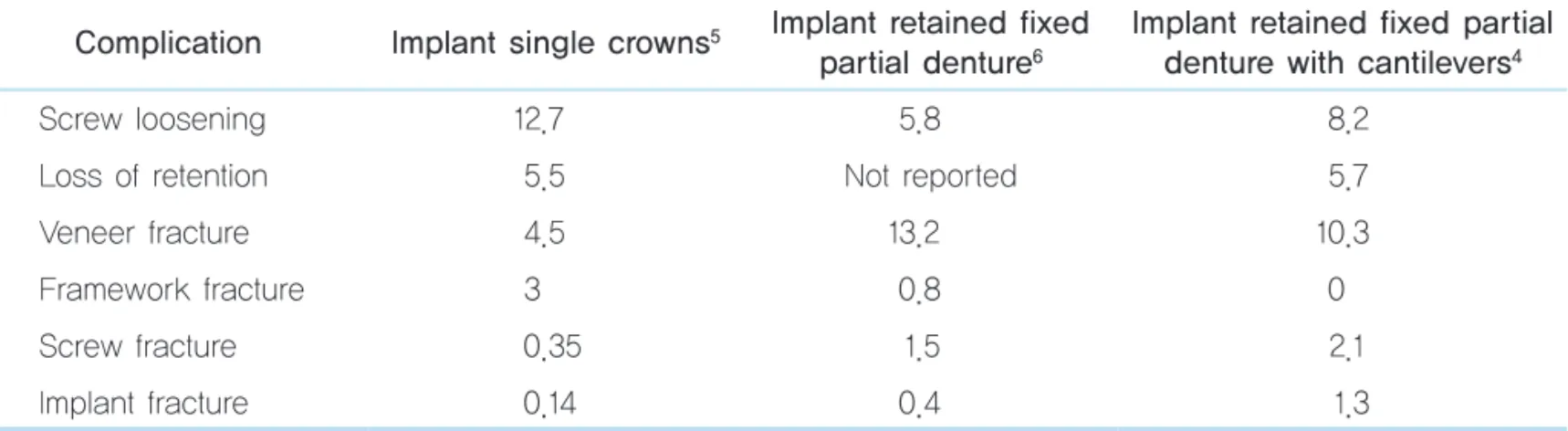
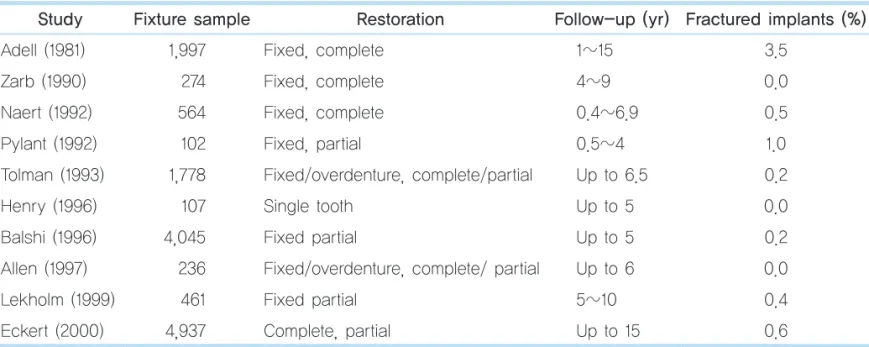
Jung 등5, Aglietta 등4, Pjetursson 등6에 의하면 임플 란트 보철과 관련하여 빈번하게 발생되는 기계적 합병증 은 Table 1과 같다. 이 중에서도 임플란트 조임 나사의 풀 림은 연구에 따라 다양하며 3.8~27%에 이른다. 여기에 나사의 파절 또한 0.09~5.1%에 이르며 이를 합산하면 약 30%대에 육박한다. 임플란트의 고정체-지대주 연결 방 식이나 사용된 지대주의 종류 등 여러 가지 조건에 따라 차이가 있지만 간과할 수 없는 정도이다. 빈도가 높지는 않지만 고정체의 파절(implant fracture)도 일어날 수 있 다(Table 2). 이는 특히 내측 연결형 임플란트에서 더욱 높은 빈도로 발생할 수 있으며 원인으로는 생역학적 과 부하, 임플란트 디자인과 재료의 결함, 보철물의 부적절 한 장착, 고정체 내측벽의 취약함 등이 있다. 특히 잔존 치조제의 폭이 충분치 않은 경우 직경이 작은(narrow- diameter) 임플란트를 식립하게 되는데 이러한 경우에는 고정체의 내측벽이 얇아지므로 파절이 일어날 가능성이 높아진다.
일체형 임플란트의 경우는 고정체와 지대주 간의 연결 부위나 나사가 없으므로 여러 기계적 합병증으로부터 자 유롭다.
Table 1. Mechanical complication of implant retained fixed prosthesis (%) Complication Implant single crowns5 Implant retained fixed
partial denture6
Implant retained fixed partial denture with cantilevers4
Screw loosening 12.7 5.8 8.2
Loss of retention 5.5 Not reported 5.7
Veneer fracture 4.5 13.2 10.3
Framework fracture 3 0.8 0
Screw fracture 0.35 1.5 2.1
Implant fracture 0.14 0.4 1.3
Hee-Gon Choi et al. : 1-Piece Implant. Implantology 2012
일체형 임플란트의 디자인
III (design of 1-piece implant)
현재 상용화되어 있는 일체형 임플란트는 많지 않으며 직경을 기준으로 regular, wide diameter와 narrow diameter로 나눌 수 있다. Lazzara25와 Ivanoff 등26은 다 른 조건들이 충족되는 한 길이가 길고 직경이 굵은 임플 란트가 좋으며, 3.75~4.0 mm 직경의 임플란트는 stan- dard implant, 그 이하는 narrow implant로 분류하였다.
Narrow implant는 임플란트 직경에 비하여 교합면이 넓 어져 cantilever를 보이며 과부하(overload)의 가능성도 높다. 무치악부의 근원심 거리가 큰 경우 emergence profile을 확보하기 어렵고 black triangle로 인한 식편압 입(food impaction) 등의 문제도 생길 수 있다. Petrie와 Williams27은 유한요소 분석법을 이용한 응력 분산 연구 에서 regular, wide diameter implant에 비해 최대 응력 값이 높았고 이는 변연골 소실의 원인이 될 수 있다고 하 였다. Narrow implant는 이 같은 단점들 때문에 하악 전
치부와 같이 잔존골 폭이 제한적인 곳에만 식립할 것을 추천하고 있다.
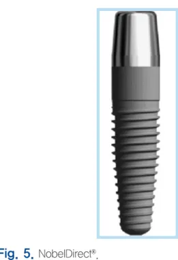
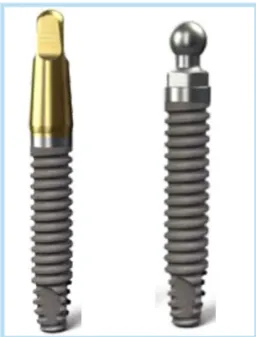
현재 시판되고 있는 일체형 임플란트는 NobelDirect® 와 NobelPerfect® 1-piece (Nobel biocare)가 가장 대표적 이다. 이 중 NobelPerfect® 1-piece는 더 이상 생산이 안 되고 NobelDirect® groovy와 NobelDirect® posterior, NobelDirect® 3.0 (Figs. 2~4)이 시판되고 있다. 이 중 NobelDirect® 3.0은 narrow implant이고 NobelDirect® groovy와 NobelDirect® posterior는 regular, wide diam- eter implant이다. NobelDirect®는 tapered body이며 anodizing oxidation 방식의 TiUniteTM surface이다. 초기 에는 rough surface의 bone part와 smooth surface를 가 진 치은관통부로 구분이 되었지만(Fig. 5) 현재는 치은관 통부가 rough 또는 smooth surface이다.
Biohorizon사는 one-piece 3.0® (구 Maximus®)이라는 3.0 mm 직경의 narrow one-piece implant (Fig. 6)를 생 산하고 있다. Resorbable blasting texturing과 hydroxy- apatite (HA) coating surface가 있다. 지대주의 종류는 straight abutment와 o-ring abutment가 있다.
Table 2. Incidence of implant fixture fracture24
Study Fixture sample Restoration Follow-up (yr) Fractured implants (%)
Adell (1981) 1,997 Fixed, complete 1~15 3.5
Zarb (1990) 274 Fixed, complete 4~9 0.0
Naert (1992) 564 Fixed, complete 0.4~6.9 0.5
Pylant (1992) 102 Fixed, partial 0.5~4 1.0
Tolman (1993) 1,778 Fixed/overdenture, complete/partial Up to 6.5 0.2
Henry (1996) 107 Single tooth Up to 5 0.0
Balshi (1996) 4,045 Fixed partial Up to 5 0.2 Allen (1997) 236 Fixed/overdenture, complete/ partial Up to 6 0.0
Lekholm (1999) 461 Fixed partial 5~10 0.4
Eckert (2000) 4,937 Complete, partial Up to 15 0.6
Hee-Gon Choi et al. : 1-Piece Implant. Implantology 2012
Zimmer Dental사에는 3.0, 4.0, 4.7 mm 직경의 일체 형 임플란트가 있다(Fig. 7). 표면은 HA grit blasted and mild acid etching한 MTXTM surface이며 지대주로는 straight, angled abutment가 있다.
또한 Biodenta사의 titanium oxide surface의 일체형
임플란트, Tatum사의 titanium oxide surface의 일체형 임플란트 등이 있다. Titanium이 아닌 일체형 임플란트 는 CeraRoot® (Oral Iceberg)가 있으며 이는 zirconium oxide에 acid-etching을 시행한 ICE surfaceTM이다(Fig.
8).
Fig. 2. NobelDirect® groovy.
Hee-Gon Choi et al. : 1-Piece Implant. Implantology 2012
Fig. 3. NobelDirect® posterior.
Hee-Gon Choi et al. : 1-Piece Implant. Implantology 2012
Fig. 4. NobelDirect® 3.0.
Hee-Gon Choi et al. : 1-Piece Implant. Implantology 2012
Fig. 5. NobelDirect®.
Hee-Gon Choi et al. : 1-Piece Implant. Implantology 2012
국내에서 시판되고 있는 일체형 임플란트로는 Onebody® (Warantec)(Fig. 9)와 Slimline® (Dentium) (Fig. 10)이 있다. Warantec사의 Onebody®는 grit- blasted and acid-etched surface이고 지대주로는 microgrooved, straight abutment가 있으며 concave
profile의 narrow neck design이다. 직경은 4.3, 5.3 mm 가 있으며 microthread, rounded square thread를 가진 tapered body form이다.
Dentium사의 Slimline®은 grit-blasted and acid- etched surface를 가지며 2.0, 2.5, 3.0, 3.5 mm 직경인 Fig. 6. One-piece 3.0® (Biohorizon).
Hee-Gon Choi et al. : 1-Piece Implant. Implantology 2012
Fig. 7. One-piece implant of Zimmer Dental.
Hee-Gon Choi et al. : 1-Piece Implant. Implantology 2012
Fig. 8. Ceraroot® (Oral Iceberg).
Hee-Gon Choi et al. : 1-Piece Implant. Implantology 2012
Fig. 9. Onebody® (Warantec).
Hee-Gon Choi et al. : 1-Piece Implant. Implantology 2012
narrow implant들이다. 지대주는 straight abutment, angled abutment, o-ring abutment가 있다.
여러 회사에서 시판되고 있는 임체형 임플란트는 다양 한 표면, 지대주, 디자인을 가지고 있다. Fixture 재질은 titanium, zirconium이 있고 표면은 대부분이 rough surface이다. 직경을 기준으로 narrow, regular, wide diameter가 있고 임플란트 디자인 측면에서는 micro- thread의 존재 유무로 나눌 수 있다. 지대주는 straight, angled, o-ring abutment가 있으며 straight 또는 con- cave profile을 가진다.
IV 기존의 연구(previous study)
1. 실험 연구(experimental study)
Hermann 등15,21은 일체형 임플란트와 이체형 임플란트 를 비교하였을 때 이체형 임플란트가 더 많은 변연골 흡 수량을 보였으며 이는 미세간극과 미세동요가 원인이라
고 보고하였다. Implant의 표면은 경조직의 유지와 많은 관련이 있으며 현재는 rough surface implant가 대부분 이다. 하지만 abutment surface characterizaion은 연구 가 많이 진행되어 있지 않다. Glauser 등28은 acid-etched surface, anodized oxidation surface, machined surface 의 일체형 임플란트 주위 연조직 반응을 살펴보았다. 그 결과 acid-etched surface와 anodized oxidation surface 에서 더 긴 connective tissue attachement를 보였고 더 짧은 epithelial downgrowth를 보였다. 하지만 rough surface (grit blased-etched surface), machined sur- face, microgrooved surface에서 fibroblast의 adhesion을 조사한 Furuhashi 등29의 연구에서는 machined surface 와 microgrooved surface에서 더 많은 fibroblast cell 개 수와 높은 adhesion strength를 보였다. 다른 연구에서 도 microgrooved surface에서 더 높은 결합조직 부착 (connective tissue attachment)을 보였다. 일반적으로 rough surface가 연조직과 접한 경우 치태의 축적을 우 려하지만 Wennerberg 등30은 abutment surface의 roughness가 임상적으로 치태의 축적과 염증 반응을 야 기하지 않는다고 보고한 바 있다. 아직까지 확실한 결론 이 없는 상태이다.
Kim 등31은 동물 실험에서 치은관통부가 각각 나팔 (flared), 오목(concave), 일직선(straight) 형태를 갖는 3 가지 일체형 임플란트를 비교 분석한 결과, 오목한 abutment profile의 일체형 임플란트가 결합조직 부착 (connective tissue attachment)의 양이 가장 많았으며 변 연골 소실은 가장 적다고 보고하였다.
Chun 등32은 미세나사선을 가진 외측 연결형, 내측 연 결형, 일체형 임플란트의 유한요소 분석에서 내측 연결 형의 임플란트가 가장 낮은 최대 응력값을 보였다. 일체 형 임플란트는 내측 연결형에 비해선 높은 최대 응력값 을 보였지만 외측 연결형보다는 좋은 결과를 보였다고 보고하였다. 미세나사선이 bone-to-implant contac Fig. 10. Slimline® (Dentium).
Hee-Gon Choi et al. : 1-Piece Implant. Implantology 2012
ratio을 증가시키고 변연골 유지에 도움을 준다는 것은 이미 이전의 연구에서 밝혀진 바 있다33,34.
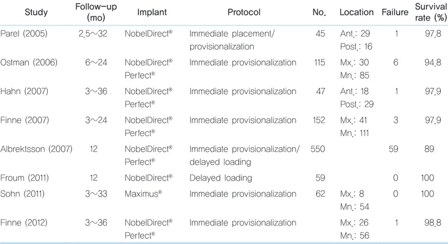
2. 임상 연구(clinical study)
문헌상에 고찰된 일체형 임플란트는 대부분 Nobel Biocare사의 NobelDirect®이다(Table 3). Parel과 Schow35 는 45개의 NobelDirect®를 식립하여 32개월 동안 경과 관 찰한 결과 1개의 immediate provisionalization 임플란트 만 실패하였으며 97.8%의 성공률을 보고하였다. Hahn10 은 47개의 NobelDirect®, NobelPerfect® 1-piece를 식립 하여 36개월 동안 경과 관찰하여 1개의 임플란트만 실패 하였으며 97.9%의 누적 생존율을 보고하였다. Bone level은 1년째 -0.78 mm, 2년째에 -0.26 mm, 3년째에 -0.54 mm를 보였다.
Finne 등36,37이 발표한 첫 번째 연구에서는 82개의 Nobel Biocare사의 NobelDirect®, NobelPerfect® 1-piece 를 식립 후 2년간의 관찰에서 98.8%의 누적 생존율을 보 였고 2년째 -0.58 mm의 안정적인 bone level을 보였다.
두 번째 연구에서는 152개의 일체형 임플란트를 식립하 여 97.9%의 누적 생존율과 1 mm 이하의 골흡수를 보였 다. 또한 최근 논문에서 Finne 등9은 마찬가지로 NobelDirect®, NobelPerfect® 1-piece 임플란트를 56명의 환자에게 82개 식립하였고, 3년까지 경과 관찰한 결과 안정적인 경조직, 연조직의 유지와 98.8%의 누적 생존율 을 보고하였다.
Sohn 등11은 36명의 환자에게 62개의 one-piece 3.0® (Biohorizon)을 식립하여 100%의 생존율을 보고하였고 1 년째 평균 골흡수량은 0.53 mm였다.
Table 3. Literature review of 1-piece implant Study Follow-up
(mo) Implant Protocol No. Location Failure Survival rate (%) Parel (2005) 2.5~32 NobelDirect® Immediate placement/
provisionalization
45 Ant.: 29 Post.: 16
1 97.8
Ostman (2006) 6~24 NobelDirect® Perfect®
Immediate provisionalization 115 Mx.: 30 Mn.: 85
6 94.8
Hahn (2007) 3~36 NobelDirect® Perfect®
Immediate provisionalization 47 Ant.: 18 Post.: 29
1 97.9
Finne (2007) 3~24 NobelDirect® Perfect®
Immediate provisionalization 152 Mx.: 41 Mn.: 111
3 97.9
Albrektsson (2007) 12 NobelDirect® Perfect®
Immediate provisionalization/
delayed loading
550 59 89
Froum (2011) 12 NobelDirect® Delayed loading 59 0 100 Sohn (2011) 3~33 Maximus® Immediate provisionalization 62 Mx.: 8
Mn.: 54
0 100
Finne (2012) 3~36 NobelDirect® Perfect®
Immediate provisionalization Mx.: 26 Mn.: 56
1 98.8
Ant.: anterior, Post.: posterior, Mx.: maxilla, Mn.: mandible.
Hee-Gon Choi et al. : 1-Piece Implant. Implantology 2012
Ostman 등12은 115개의 NobelDirect®와 NobelPerfect® 1-piece와 380개의 2-piece implant (Nobel Biocare)를 최소 1년 이상 경과 관찰하여 비교한 결과 평균 골흡수량 은 1-piece implant가 2.1 mm, 2-piece implant가 0.8 mm였다. Albrektsson 등38이 제시한 success grade에 따 르면 1-piece implant는 success grade I에 해당하는 임 플란트가 46.1%, success grade II implant가 72.2%였다.
그에 반하여 2-piece implant는 success grade I이 85.5%, grade II가 91.6%였으며, 1-piece implant는 식립 시 신중하게 선택해야 하는 디자인이라고 하였다.
Albrektsson 등39은 multi-center에서 식립한 550개의 NobelDirect®를 경과 관찰하여 90.1%의 survival rate를 보고하였다. 이 중 58개의 임플란트는 delayed loading을 시행하였고 1개의 임플란트만 실패하여 98.3%의 생존율 을 보였으며 immediate loading을 가한 492개의 임플란 트는 58개가 실패하여 88.2%의 생존율을 보였다. 이 결 과로 보아 immediate loading은 신중하게 결정해야 함을 알 수 있다.
Sennerby 등40은 117개의 NobelDirect®를 분석한 결과 success grade I은 41%, success grade II는 58%를 보였 다. 평균 10.2개월의 관찰 기간 동안 2.4 mm의 골흡수를 보였다.
Zembić 등13에 의하면 47명의 환자에서 식립한 57개의 NobelDirect® 3.0 implant를 1년간 경과 관찰한 결과 98%
의 생존율을 보였다. 하지만 success grade의 관점에서 볼 때 전체 임플란트의 18%에서 3 mm가 넘는 골흡수량 이 관찰되는 등 좋지 않은 결과를 보였다. 대부분의 논문 에서 골흡수에 영향을 미치는 요인은 임플란트 디자인, 식립 깊이, rough surface가 점막과 접한 부분, 즉시 부 하 등이라고 분석하였다.
V 임상 적용(clinical application)
1. 임상 적용 원칙(principles of clinical application)
일체형 임플란트의 가장 큰 장점이자 단점은 지대주와 고정체가 붙어있다는 것이다. 위에서 언급한 미세간극이 없으며, 나사 풀림, 파절이 없고 고정체의 파절이 적다는 등의 장점을 가지고 있지만, 지대주의 선택이 제한적이 며 일단 식립한 후에는 지대주의 경로 수정이 어렵다는 단점이 있다. 그러므로 수술시에는 surgical guide를 사 용할 것을 권장하고 있다(Fig. 11)35.
전산화 단층 촬영(computed tomography [CT])과 진단 모형 분석 등의 방법으로 정확한 임플란트의 위치를 미 리 계획한다면 flapless surgery도 생각해 볼 수 있다. 일 체형 임플란트의 식립 시 flap, flapless surgery 간 변연
Fig. 11. Use of surgical guide prefabricated in the Lab (NobelDirect®)35.
Hee-Gon Choi et al. : 1-Piece Implant. Implantology 2012
골 흡수량은 통계적으로 유의하지 않았으나41 flapless surgery가 연조직 보존에는 긍정적으로 작용할 수 있다.
수술시 고려해야 할 중요한 점 중의 또 한 가지는 초기 고정(primary stability)이다. 일체형 임플란트가 imme- diate provisionalization을 전제로 하고 있기 때문에 일 반적으로 최소 30~35 Ncm torque 이상의 초기고정을 권장하고 있다9,10,35. 초기 고정(primary stability)은 식립 시와 초기 치유기(healing phase)에서 bone과 implant의 계면에서 발생할 수 있는 micromotion으로 인하여 골유 착이 실패할 수 있다는 관점에서 매우 중요하다. 이러한 초기 고정(primary stability)에 영향을 주는 인자로는 bone quality, density와 implant design, surgical tech- nique 등이 있다. 특히나 type III, IV bone에 식립 시 under-drilling을 통하여 식립 토크(insertion torque)가 높게 나올 수 있지만 진정한 의미의 초기 고정이 아니며 골질이 가장 중요함을 잊지 말아야 한다.
문헌마다 약간의 차이는 있지만 대부분의 경우 술 후 48시간 내에 immediate provisionalization을 시행한다.
Cochran 등42에 의하면 immediate provisionalization, 즉 non-functional loading은 술 후 48시간 이내에 임시 수 복물을 장착하지만 이 때 대합치와 교합 접촉을 하지 않 는 것을 의미한다. Immediate loading은 술 후 48시간 이 내에 대합치와 교합 접촉이 되는 임시 수복물을 장착하 는 것을 뜻한다. 대부분의 문헌에서 권장하는 것은 교합 접촉이 되지 않거나 light centric contact이며 교합접촉
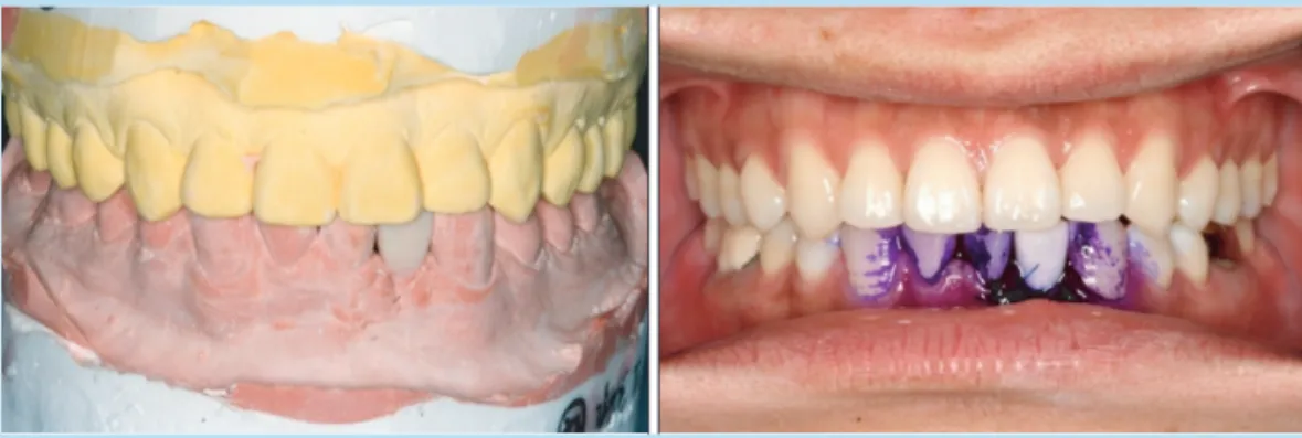
이 되지 않는 것이 빈도가 높았다9,10,35,41. Provisional restoration은 보통 acrylic resin을 이용하며 미리 제작한 crown shell을 이용할 수도 있으며, chair side에서 직접 제작할 수도 있고 인상 채득 후 기공실에서 제작할 수도 있다(Fig. 12).
술 후 provisional restoration을 제작하기 위해 지대주 를 삭제하게 되는데 이 때 고온의 열이 발생하게 되므로 주의해야 한다. Gabay 등43은 임플란트 식립 후 47oC 이 상의 열이 가해질 경우 bone formation에 영향을 미치게 되며 골유착이 실패할 수도 있기 때문에 water-spray irrigation이 꼭 필요하며 irrigation하지 않은 group에서 는 골과 접하고 있는 상부 thread에서 최고 60oC 이상의 열이 발생한다고 보고하였다. 최소 분당 15 ml 이상의 irrigation이 필요하며 주수량에 대해선 dose-dependent manner를 따른다.
2. 임상 적용 결과(results of clinical application)
본과에서 2009년 7월부터 2011년 12월까지 Onebody® (Warantec) implant를 식립한 환자들에 대하여 방사선 사진 촬영, 인상 채득, 임상 사진 촬영 등의 방법을 통하 여 후향적인 연구를 실시하였다. 총 34명의 환자에게 43 개의 임플란트를 식립하였고 이 중 방사선학적 평가는 보철물 장착을 시행하여 기능 부하가 가해진 후 6개월 이 상된 환자를 대상으로 하였다. 단, 생존율에 대한 평가는
Fig. 12. Provisional restoration by indirect technique.
Hee-Gon Choi et al. : 1-Piece Implant. Implantology 2012
보철물 장착 전이나 보철물 장착 후 6개월이 안 된 환자 도 대상으로 하였다. 임플란트 식립 후 마지막 내원일까 지의 기간은 24개월이었다. 포함된 환자는 남자가 13명 여자가 21명이며 상악에 식립된 것이 30개, 하악이 13개 였다. 수술 후 1주, 보철물 장착 시, 장착 후 3개월, 6개 월, 1년, 2년에 check-up을 시행하였다.
1개의 임플란트가 실패하였으며 survival rate는 97.7%
였다. 상악 좌측 중절치 부위에 즉시 식립하였고 식립 당 일 immediate provisional restoration을 장착하였다. 식 립 1개월 후 임플란트가 흔들린다는 주소로 내원하였고 골유착 실패로 고정체를 제거하였다.
방사선 촬영은 구내 디지털 치근단 방사선 사진 촬영기 인 Planmeca Intra® (Planmeca, Hesinki, Finland)를 이 용하여 63 kVp, 8 mA의 조건으로 촬영하여 computed dental radiography 센서인 Schick® (Schick Technologies, Long Island City, NY, USA)를 통하여 영상정보를 얻었 다. 얻은 영상정보는 gateway 프로그램인 Dentigate® (INFINITT Technology, Seoul, Korea)를 통하여 JPEG 파일로 변환하였다. 각 사진은 UTHSCSA Image tool for
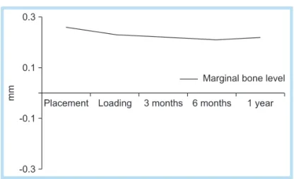
Windows version 3.0 (University of Texas Health Science Center at San Antonio, TX, USA)을 이용하여 소수점 둘째 자리까지 mm 단위로 bone level을 측정하였 다(Fig. 13). 모든 방사선 사진의 확대율이 동일하지 않으 므로 각 측정 시마다 이미 알고 있는 수치를 이용하여 calibration 과정을 거쳐 정확한 거리를 측정하였다. 변연 골 흡수량은 임플란트 최상방 shoulder 부위부터 bone crest 최상부까지의 거리를 기준으로 근심, 원심 두 군데 에서 측정하였으며 모든 경우에서 2회 측정하여 그 평균 값을 최종 데이터로 결정하였다(Fig. 14). 각 기간별 bone level과 골흡수량은 다음과 같다(Table 4).
통계는 SPSS 12.0 (SPSS Inc., Chicago, IL, USA) 프로 그램을 이용하였으며 식립 후 기간에 따른 평균 bone level change를 Kruskal-Wallis test를 이용하여 분석한 결과 각 기간에 따른 bone level의 유의 차는 없었다. 이 로 보아 식립 후부터 보철물 기능 1년까지 골흡수량은 그 정도가 미미하여 임상적으로 영향을 주지 않는 범주에 있다고 볼 수 있다(Fig. 15).
Fig. 13. Reference point of measurement.
Hee-Gon Choi et al. : 1-Piece Implant. Implantology 2012
Fig. 14. Marginal bone level change of Onebody® implant (mm).
Hee-Gon Choi et al. : 1-Piece Implant. Implantology 2012
V 총괄 및 고찰
일체형 임플란트는 이전부터 여러 가지 용도로 사용되 었다. 대표적으로 임시 임플란트는 시술 후 골유착이 완 성되기 전까지 임시 임플란트를 이용하여 즉시부하를 시 행함으로써 치유 기간 동안 환자에게 심미적, 심리적 안 정감을 부여하고 임플란트에 가해지는 미세동요나 부하 를 줄여주는 데 사용되며8,44,45 영구 임플란트의 surgical guide로도 사용할 수 있다. 교정 치료에 사용되는 mini screw 또한 일체형 임플란트의 한 종류로 볼 수 있다. 하 지만 definitive prosthesis를 위한 임플란트는 비교적 최 근에 연구와 개발이 진행되고 있다. 현재 가장 널리 알려 져 있으며 임상 연구가 진행되어 있는 일체형 임플란트 는 NobelDirect®이다. 하지만 NobelDirect®의 임상 연구 결과는 논란이 분분하다. 위에서 언급한 것처럼 Finne
등9,36,37은 안정적인 경조직, 연조직의 보존을 보고하였지
만 Ostman 등12은 광범위한 골흡수를 보고하는 등 아직 까지 고려가 필요한 임플란트라고 하였다. 이러한 연구 들에서 골흡수에 영향을 미치는 요소로는 즉시 부하, 임 플란트 디자인, 표면 처리, 식립 깊이, rough surface의 점막 내 노출, 지대치 삭제 등이 거론되고 있다. 다른 종 류의 일체형 임플란트는 아직 임상 연구가 많지 않다.
유한요소 분석을 통한 응력 분산 연구나 실제 임상 연 구 결과에서 보여지듯이 이체형 임플란트와 일체형 임플 란트와는 분명한 차이가 존재한다. 하지만 기존의 연구 에서 일체형 임플란트가 임상적으로 영향을 미칠 정도로 높은 최대응력을 보이지는 않았으며 2-piece implant와 의 비교, 분석에서도 임플란트 디자인이 각각 동일하지 는 않았다. 본원에서 시행한 Onebody®의 임상 연구와 Kim 등31의 연구에서 유추해 보면 일체형 임플란트의 디 자인은 이체형 임플란트와 같은 선상에 있는 것으로 보 인다. 경조직 보존에 있어서 Abrahamsson과 Berglundh33는 이체형 임플란트에서 미세나사선이 있는 임플란트가 골흡수량은 적고 골-임플란트 접촉률은 높 Fig. 15. Radiographic evaluation.
Hee-Gon Choi et al. : 1-Piece Implant. Implantology 2012
Table 4. Marginal bone level change of Onebody® implant (mm)
Placement Loading 3 months 6 months 1 year Marginal bone level 0.26 (0.36) 0.23 (0.32) 0.22 (0.24) 0.21 (0.23) 0.22 (0.19)
Marginal bone loss -0.03 0.04 -0.05 -0.04
Values are presented as mean (standard deviation).
Hee-Gon Choi et al. : 1-Piece Implant. Implantology 2012
다고 보고하였으며 이는 일체형 임플란트에도 적용이 될 것으로 보인다. 연조직 측면에서 볼 때는 임플란트 주위 결합조직이 상피조직의 치근단 측 이주를 방지하는 효과 를 가진다고 하였으므로46 임플란트 주위에 생물학적 폭 경의 크기가 같더라도 결합조직 부착 길이가 큰 경우가 작은 경우보다 유리하다. Kim 등31에 의하면 3가지 형태 의 치은관통부 중에서 concave한 profile, 즉 narrow neck을 가질 때 결합조직의 부착량이 가장 많았다. 표면 처리에 있어서도 초기의 machined surface나 titanium plasma spray (TPS) surface보다 최근의 blasted &
etched suface와 같은 rough surface가 좋은 결과를 보일 것이다. 즉 일체형 임플란트에서도 2-piece implant와 마찬가지로 microthread, narrow neck, rough surface 등은 경조직, 연조직 보존에 도움을 줄 것으로 생각된다.
Implant fixture의 surface characterization은 위에서 언급한 것처럼 어느 정도 연구가 많이 진행된 상태이나 abutment의 surface와 soft tissue의 연관성은 아직 명확 하게 밝혀지지 않았다. Previous study에서 살펴본 것처 럼 machined surface나 microgrooved surface가 rough surface보다 fibroblast adhesion에 더 좋은 결과를 보였 지만 실제 in vivo study에서는 machined surface에서 epithelial cell이 더 많은 하방 이주를 보였다28. 또한 abutment surface의 roughness가 plaque acculmulation 이나 염증반응을 야기하지 않았다는 임상 연구도 있었으 므로30 아직까지는 더욱 연구가 필요할 것이라 생각한다.
일체형 임플란트의 식립 시에는 세심한 주의가 필요하 다. 식립 후 보철적 수정이 어려우므로 surgical guide가 필요하다. 특히나 flapless surgery를 계획하고 있다면 x-ray뿐만 아니라 진단 모형 등을 이용하여 정확하게 식 립하는 것이 중요하다. NobelDirect®의 경우 임플란트를 너무 깊게 식립하면 rough surface와 접해 있는 부분의 bone loss가 일어날 수 있으며 너무 얕게 식립할 시에는 rough surface의 색조가 치은을 관통하여 변색이 일어날
수가 있다. 그러므로 치은의 두께와 잔존골의 양, 교합고 경을 정확히 파악해야 한다.
식립 시 초기 고정(primary stability)은 immediate provisionalization, loading을 전제로 하는 일체형 임플 란트에서 매우 중요하다. 초기 고정은 식립 시 주위 bone 과의 mechanical engagement와 관련이 있으며 임플란트 의 장기간 성공률에 큰 영향을 미친다. 이는 초기 고정이 healing 과정에서 일어나는 micromotion을 방지하기 때 문이다. 계속적인 50~150 μm의 micromotion은 골-임플 란트 접촉 계면에서 fibrous tissue를 만들어 골유착을 방 해한다. 초기 고정에 영향을 미치는 factor는 임플란트의 디자인, 임플란트의 표면 처리, surgical technique, bone quality와 density 등이며 이 중 bone quality와 density가 가장 중요하다47. 골질이 좋지 않은 경우 under-drilling 을 시행하여 식립 토크(insertion torque)를 높이는 방법 도 있지만 식립 토크가 primary stability를 완전히 대변 하지는 못하므로 무엇보다도 식립 부위의 골질이 중요하 다는 것을 명심해야 한다. Song 등48은 임플란트 식립 후 cone beam CT를 이용하여 식립 부위를 단층 촬영하고 primary stability를 implant stability quotient (ISQ) value를 이용하여 측정하였다. 그 결과 bone quality, density가 초기 고정과 높은 상관성이 있는 것으로 밝혀 졌다.
Immediate provisionalization, loading은 전체적인 치 료기간의 단축, 심미적, 심리적 안정감, 가철성 보철물의 불필요성, 연조직의 profile 확립, 수술 횟수의 감소 등의 이점이 있다. 일체형 임플란트의 경우 immediate provi- sionalization에 유리한 조건을 가지고 있으며 지대주를 착탈하는 번거로움을 피할 수가 있다. 교합 설정에 관한 의견은 조금씩 다르나 대부분의 연구에서 대합치와의 비 교합접촉, 특히 excursive movement시 비접촉이 필수적 이라고 주장하고 있다. Hahn49은 임상적 경험에 비추어 봤을 때 모든 방향의 excursive movement시 최소 0.5
mm씩 clearance를 가져야 하며 특히 3.0 mm 이하의 narrow diameter implant를 사용할 시에는 1 mm까지 clearance를 가져야 한다고 보고하였다.
Immediate provisionalization, loading을 전제로 하기 때문에 식립 당일에 지대주 삭제 등의 술식을 행하는 경 우가 많다. 지대치 삭제는 그 자체만으로 골에 비가역적 인 손상을 입힐 수 있고 임플란트 골유착의 실패도 야기 할 수 있다고 하였다. Gabay 등43은 최소 15 ml/min의 irrigation이 필요하다고 하였으며 삭제가 멈춘 후에도 계속적인 물분사를 하는 것이 급속 냉각을 하는데 도움 이 된다고 하였다. 또한 Tawse-Smith 등50은 metal par- ticle에 의해 peri-implantitis가 야기될 수 있다고 하였으 므로 지대치 삭제 시 발생하는 titanium particle의 침습 을 방지하기 위해 rubber dam을 사용해야 한다.
VII 결론
현재까지 여러 연구들을 분석, 고찰해 본 결과 일체형 임플란트는 계속적인 연구가 필요하다고 볼 수 있다. 기 계적, 생물학적 이점이 있으며 안정적인 경조직, 연조직 보존을 할 수 있는 잠재력이 있지만 널리 사용되지 못하 고 있으며 결과가 좋지 않은 연구도 있다. 하지만 다음 사항들에 대해 이해하고 적절히 임상에 적용한다면 높은 성공률을 기대할 수 있을 것이다.
1. 일체형 임플란트는 나사 풀림과 파절, 지대주와 고 정체의 파절과 같은 기계적 합병증이 적다.
2. 일체형 임플란트는 미세간극(microgap), 미세동요 (micromovement), 지대주의 탈착과 재부착 등이 없으므 로 생물학적 합병증이 적다.
3. 일체형 임플란트 디자인의 원칙은 기존의 임플란트 와 동일하다.
4. 일체형 임플란트는 적응증이 제한적이며 철저한 술 전 준비가 필요하다.
5. Immediate provisionalization 시에 bone quality와 초기 고정(primary stability)이 중요하다.
6. Immediate provisionalization 시에 non-occlusal contact을 권장한다.
참고문헌
1. Albrektsson T, Dahl E, Enbom L, et al. Osseointegrated oral implants. A Swedish multicenter study of 8139 consecutively inserted Nobelpharma implants. J Periodontol. 1988; 59: 287-296.
2. Berglundh T, Persson L, Klinge B. A systematic review of the incidence of biological and technical complications in implant dentistry reported in prospective longitudinal studies of at least 5 years. J Clin Periodontol.
2002; 29 Suppl 3: 197-212.
3. Becker W, Becker BE, Alsuwyed A, et al. Long-term evaluation of 282 implants in maxillary and mandibular molar positions: a prospective study. J Periodontol. 1999; 70: 896-901.
4. Aglietta M, Siciliano VI, Zwahlen M, et al. A systematic review of the survival and complication rates of implant supported fixed dental prostheses with cantilever extensions after an observation period of at least 5 years. Clin Oral Implants Res. 2009; 20: 441-451.
5. Jung RE, Pjetursson BE, Glauser R, et al. A systematic review of the 5-year survival and complication rates of implant-supported single crowns. Clin Oral Implants Res. 2008; 19: 119-130.
6. Pjetursson BE, Tan K, Lang NP, et al. A systematic review of the survival and complication rates of fixed partial dentures (FPDs) after an observation period of at least 5 years. Clin Oral Implants Res. 2004; 15:
667-676.
7. Hermann JS, Buser D, Schenk RK, et al. Biologic width around one- and two-piece titanium implants. Clin Oral Implants Res. 2001; 12: 559- 571.
8. Zubery Y, Bichacho N, Moses O, et al. Immediate loading of modular transitional implants: a histologic and histomorphometric study in dogs.
Int J Periodontics Restorative Dent. 1999; 19: 343-353.
9. Finne K, Rompen E, Toljanic J. Three-year prospective multicenter
study evaluating marginal bone levels and soft tissue health around a one-piece implant system. Int J Oral Maxillofac Implants. 2012; 27:
458-466.
10. Hahn JA. Clinical and radiographic evaluation of one-piece implants used for immediate function. J Oral Implantol. 2007; 33: 152-155.
11. Sohn DS, Bae MS, Heo JU, et al. Retrospective multicenter analysis of immediate provisionalization using one-piece narrow-diameter (3.0- mm) implants. Int J Oral Maxillofac Implants. 2011; 26: 163-168.
12. Ostman PO, Hellman M, Albrektsson T, et al. Direct loading of Nobel Direct and Nobel Perfect one-piece implants: a 1-year prospective clinical and radiographic study. Clin Oral Implants Res. 2007; 18: 409- 418.
13. Zembić A, Johannesen LH, Schou S, et al. Immediately restored one- piece single-tooth implants with reduced diameter: one-year results of a multi-center study. Clin Oral Implants Res. 2012; 23: 49-54.
14. Cochran DL, Hermann JS, Schenk RK, et al. Biologic width around titanium implants. A histometric analysis of the implanto-gingival junction around unloaded and loaded nonsubmerged implants in the canine mandible. J Periodontol. 1997; 68: 186-198.
15. Hermann JS, Buser D, Schenk RK, et al. Crestal bone changes around titanium implants. A histometric evaluation of unloaded non-submerged and submerged implants in the canine mandible. J Periodontol. 2000;
71: 1412-1424.
16. Persson LG, Lekholm U, Leonhardt A, et al. Bacterial colonization on internal surfaces of Brånemark system implant components. Clin Oral Implants Res. 1996; 7: 90-95.
17. Piattelli A, Vrespa G, Petrone G, et al. Role of the microgap between implant and abutment: a retrospective histologic evaluation in monkeys.
J Periodontol. 2003; 74: 346-352.
18. Ericsson I, Persson LG, Berglundh T, et al. Different types of inflammatory reactions in peri-implant soft tissues. J Clin Periodontol.
1995; 22: 255-261.
19. Berglundh T, Lindhe J. Dimension of the periimplant mucosa.
Biological width revisited. J Clin Periodontol. 1996; 23: 971-973.
20. Abrahamsson I, Berglundh T, Wennström J, et al. The peri-implant hard and soft tissues at different implant systems. A comparative study in the dog. Clin Oral Implants Res. 1996; 7: 212-219.
21. Hermann JS, Schoolfield JD, Schenk RK, et al. Influence of the size of the microgap on crestal bone changes around titanium implants. A histometric evaluation of unloaded non-submerged implants in the canine mandible. J Periodontol. 2001; 72: 1372-1383.
22. Abrahamsson I, Berglundh T, Lindhe J. The mucosal barrier following
abutment dis/reconnection. An experimental study in dogs. J Clin Periodontol. 1997; 24: 568-572.
23. Rodríguez X, Vela X, Méndez V, et al. The effect of abutment dis/
reconnections on peri-implant bone resorption: a radiologic study of platform-switched and non-platform-switched implants placed in animals. Clin Oral Implants Res. 2011. [Epub ahead of print]
24. Virdee P, Bishop K. A review of the aetiology and management of fractured dental implants and a case report. Br Dent J. 2007; 203: 461- 466.
25. Lazzara RJ. Criteria for implant selection: surgical and prosthetic considerations. Pract Periodontics Aesthet Dent. 1994; 6: 55-62.
26. Ivanoff CJ, Gröndahl K, Sennerby L, et al. Influence of variations in implant diameters: a 3- to 5-year retrospective clinical report. Int J Oral Maxillofac Implants. 1999; 14: 173-180.
27. Petrie CS, Williams JL. Comparative evaluation of implant designs:
influence of diameter, length, and taper on strains in the alveolar crest. A three-dimensional finite-element analysis. Clin Oral Implants Res. 2005;
16: 486-494.
28. Glauser R, Schüpbach P, Gottlow J, et al. Periimplant soft tissue barrier at experimental one-piece mini-implants with different surface topography in humans: a light-microscopic overview and histometric analysis. Clin Implant Dent Relat Res. 2005; 7 Suppl 1: S44-51.
29. Furuhashi A, Ayukawa Y, Atsuta I, et al. The difference of fibroblast behavior on titanium substrata with different surface characteristics.
Odontology. 2012; 100: 199-205.
30. Wennerberg A, Sennerby L, Kultje C, et al. Some soft tissue characteristics at implant abutments with different surface topography. A study in humans. J Clin Periodontol. 2003; 30: 88-94.
31. Kim S, Oh KC, Han DH, et al. Influence of transmucosal designs of three one-piece implant systems on early tissue responses: a histometric study in beagle dogs. Int J Oral Maxillofac Implants. 2010; 25: 309-314.
32. Chun HJ, Shin HS, Han CH, et al. Influence of implant abutment type on stress distribution in bone under various loading conditions using finite element analysis. Int J Oral Maxillofac Implants. 2006; 21: 195- 202.
33. Abrahamsson I, Berglundh T. Tissue characteristics at microthreaded implants: an experimental study in dogs. Clin Implant Dent Relat Res.
2006; 8: 107-113.
34. Lee SY, Piao CM, Koak JY, et al. A 3-year prospective radiographic evaluation of marginal bone level around different implant systems. J Oral Rehabil. 2010; 37: 538-544.
35. Parel SM, Schow SR. Early clinical experience with a new one-piece
implant system in single tooth sites. J Oral Maxillofac Surg. 2005; 63(9 Suppl 2): 2-10.
36. Finne K, Rompen E, Toljanic J. Prospective multicenter study of marginal bone level and soft tissue health of a one-piece implant after two years. J Prosthet Dent. 2007; 97(6 Suppl): S79-85.
37. Finne K, Rompen E, Toljanic J. Clinical evaluation of a prospective multicenter study on 1-piece implants. Part 1: marginal bone level evaluation after 1 year of follow-up. Int J Oral Maxillofac Implants.
2007; 22: 226-234.
38. Albrektsson T, Zarb G, Worthington P, et al. The long-term efficacy of currently used dental implants: a review and proposed criteria of success. Int J Oral Maxillofac Implants. 1986; 1: 11-25.
39. Albrektsson T, Gottlow J, Meirelles L, et al. Survival of NobelDirect implants: an analysis of 550 consecutively placed implants at 18 different clinical centers. Clin Implant Dent Relat Res. 2007; 9: 65-70.
40. Sennerby L, Rocci A, Becker W, et al. Short-term clinical results of Nobel Direct implants: a retrospective multicentre analysis. Clin Oral Implants Res. 2008; 19: 219-226.
41. Froum SJ, Cho SC, Elian N, et al. Survival rate of one-piece dental implants placed with a flapless or flap protocol--a randomized, controlled study: 12-month results. Int J Periodontics Restorative Dent.
2011; 31: 591-601.
42. Cochran DL, Morton D, Weber HP. Consensus statements and recommended clinical procedures regarding loading protocols for endosseous dental implants. Int J Oral Maxillofac Implants. 2004; 19
Suppl: 109-113.
43. Gabay E, Cohen O, Machtei EE. Heat production during prosthetic preparation of a one-piece dental implant. Int J Oral Maxillofac Implants. 2010; 25: 1131-1136.
44. Babbush CA. Titanium plasma spray screw implant system for reconstruction of the edentulous mandible. Dent Clin North Am. 1986;
30: 117-131.
45. Froum S, Emtiaz S, Bloom MJ, et al. The use of transitional implants for immediate fixed temporary prostheses in cases of implant restorations.
Pract Periodontics Aesthet Dent. 1998; 10: 737-746.
46. Chehroudi B, Gould TR, Brunette DM. The role of connective tissue in inhibiting epithelial downgrowth on titanium-coated percutaneous implants. J Biomed Mater Res. 1992; 26: 493-515.
47. O'Sullivan D, Sennerby L, Meredith N. Measurements comparing the initial stability of five designs of dental implants: a human cadaver study.
Clin Implant Dent Relat Res. 2000; 2: 85-92.
48. Song YD, Jun SH, Kwon JJ. Correlation between bone quality evaluated by cone-beam computerized tomography and implant primary stability.
Int J Oral Maxillofac Implants. 2009; 24: 59-64.
49. Hahn J. One-piece root-form implants: a return to simplicity. J Oral Implantol. 2005; 31: 77-84.
50. Tawse-Smith A, Ma S, Siddiqi A, et al. Titanium particles in the peri- implant tissues: surface analysis and histological response. Clin Adv Periodontic. 2012; 2: 232-238.