Copyright © 2021. Anatomy & Cell Biology
Introduction
Waterpipe and its alternative names like goza, hookah, narghile, and shisha is a single or multi-stemmed instrument
involved in inhalation of tobacco smoking after passing through tap water (Fig. 1A) [1]. World health organization listed waterpipe tobacco smoking as a public health problem that needs fast solutions to regulate its prevalence worldwide by implementing tobacco control measures [2]. The reasons behind that, are due to high concentrations of toxicants pres- ent in charcoal and tobacco that have an adverse impact on human health [3]. These toxicants include nicotine, tar, car- bon monoxide, volatile organic compounds, heavy metals, polycyclic aromatic hydrocarbons, and heterocyclic aromatic amines (Fig. 2) [4-6]. These compounds were reported to
Corresponding author:
Alia Khwaldeh
Department of Medical Laboratory Sciences, Faculty of Pharmacy, Jadara University, Irbid 21110, Jordan
E-mail: [email protected]
Ameliorative effects of curcumin and caffeic acid against short term exposure of waterpipe tobacco smoking on lung, heart and kidney in mice
Alia Khwaldeh
1, Ali Abu Siyam
1, Ahmed Alzbeede
2,3, Mohammad Farajallah
2, Ziad Shraideh
2, Darwish Badran
41Department of Medical Laboratory Sciences, Faculty of Pharmacy, Jadara University, Irbid, 2Department of Biological Sciences, School of Science, The University of Jordan, Amman, Jordan, 3Department of Medical Laboratory Science, College of Science, Komar University of Science and Technology, Sulaymaniyah, Iraq, 4Department of Anatomy and Histology, School of Medicine, The University of Jordan, Amman, Jordan
Abstract: This study aims to evaluate the chemopreventive activity of two antioxidants (curcumin [CUM] and caffeic acid [CAF]), focusing on how these antioxidants could reduce cytotoxicity induced by short term secondhand exposure of waterpipe tobacco smoking. Forty-eight adult male BALB/c albino mice were equally divided into four groups. Antioxidants were delivered intraperitoneally, and the exposure to waterpipe smoking (WPS) was performed using a smoking machine.
This experiment lasts for 14 consecutive days. Serum were collected from mice before dissection to quantify the activity of some liver enzymes, kidney function tests and proinflammatory cytokines. Lung, heart, and kidney were isolated and processed for light microscopy technique. Parallel treatment of CUM or CAF along with exposure to WPS showed less inflammation, less vacuolized, and more inflated alveoli, less deteriorations in cortex part of kidney, and less disintegration of cardiac myofibers in comparison to waterpipe only. Besides, CUM and CAF significantly reduced the activity of aspartate aminotransferase and proinflammatory cytokines. CUM and CAF were found to have anti-inflammatory and ameliorative effects against the cytotoxicity induced by exposure to waterpipe tobacco smoking, and CUM showed better chemopreventive activity than CAF.
Key words: Antioxidants, Biochemical parameters, Histopathology, Hookah, Proinflammatory cytokines
Received July 25, 2020; 1st Revised September 12, 2020; 2nd Revised November 7, 2020; Accepted November 9, 2020
induce the oxidative stress status in humans, increase the ex- pression of carcinogenic genes, and disruption of the physi- ological activity of internal organs, especially those of the respiratory and cardiovascular systems [3, 7]. The amount of nicotine present in unflavored tobacco is ten times more than flavored tobacco (maassal) as shown by gas chroma- tography, and it was suggested that the reason behind lower nicotine content in flavored maassal is the addition of honey, glycerin and other flavors that contributed in minimizing
the tobacco content of maassal [8]. The secondhand smokers could retain up to 80% of nicotine, and the same amount of exhaled carbon monoxide in comparison with the active wa- terpipe smokers [9]. Another meta-analysis study illustrated that waterpipe smokers could induce the tumor biomark- ers in cigarette smokers. Also, waterpipe smoking (WPS) is highly suspected in inducing head and neck, esophagus, and lung cancer [10]. Consumption of antioxidants is one of the top strategies that contribute to attenuate the oxidative stress
Head
Body
Bowl
Water Hose Mouthpiece Tobacco
Coal
A
B
C
Fig. 1. The main components of this study (WPS, CUM, and CAF); (A) shows a typical waterpipe with a brief illustration of its parts. Chemical structure of CUM (B) and CAF (C) with three-dimensional conformer represented in ball and stick model. CAF, caffeic acid; CUM, curcumin;
WPS, waterpipe smoking. Adapted from Bhatnagar et al. Circulation 2019;139:e917-36, according to the Creative Commons license PubChem [1].
H
N
N
(Nicotine) (Carbon monoxide) (Nitric oxide) (Catechol Tar ) OH
OH
OH
OH CH3
H C3 O
CH3 H
O
H H
O
CH2
H C3 H
O H C3
C O N O
(Pyrene)
(Benzo[a]pyrene)
(Fluoranthene)
(Benzo[a]anthracene) (Chrysene) (Hydroquinone Tar )
(Benzo[g,h,i]perylene) (Indeno[1,2,3-cd]pyrene)
H C3 O
H
(Acetaldehyde)
(Propionaldehyde) (Methacrolein) (Formaldehyde) (Acetone)
Fig. 2. List of toxicant contents gene- rated from tobacco-based waterpipe smoking, according to Shihadeh et al.
[5]. Adapted from Shihadeh et al. Tob Control 2015;24(Suppl 1):i22-30 [5].
status induced by the toxicants. Antioxidants are chemical compounds extracted from natural sources, especially me- dicinal plants. These compounds can react with the reactive oxygenic and nitrogenic species to prevent these free radicals from damaging the cellular component of different tissues [11, 12].
Curcumin (CUM) is a phytopolyphenol pigment isolated from the plant Curcuma longa, commonly known as turmeric, with a variety of pharmacologic properties. CUM has molecu- lar formula of C21H20O6 and molecular weight of 368.4 g/mol (Fig. 1B) [1]. This molecule has antioxidant capacity by pre- venting the formation of reactive oxygen species, possesses anti-inflammatory properties as a result of inhibition of cy- clooxygenases and other enzymes involved in inflammation [13]. Also, CUM has anti-carcinogenic activity by inhibiting proliferation of tumor cells, suppression of chemically in- duced carcinogenesis and tumor growth in animal models of cancer [14]. CUM inhibits toxin-mediated stress responses via its anti-inflammatory and antioxidant properties in addi- tion to inducing the expression of cytoprotective proteins [15].
CUM neutralizes nitric oxide, provides continuity to anti- oxidant enzymes such as catalase and superoxide dismutase, and lowers lipid peroxidation, to protect tissues and organs from oxidative damage caused by nicotine [16].
Caffeic acid (CAF [3,4-Dihydroxycinnamic acid]) is an organic compound found naturally in plants such as argan oil, oats, wheat, rice, olive oil, and narrow-leaved purple coneflower. It has molecular formula of C9H8O4 and molecu- lar weight of 180.16 g/mol (Fig. 1C) [1] and exhibit potent an- tioxidant activity [17]. CAF showed antioxidant and chemo preventive properties against toxic chemical models in vivo and in vitro [18]. In vivo, CAF showed enhancement of repro- ductive functions in male rats by increasing the concentra- tion of testicular zinc, glycogen, cholesterol, and hydroxys- teroid dehydrogenases, increasing superoxide dismutase and total thiol in the epididymis and stimulating the secretion of testosterone and follicle-stimulating hormone [19]. Also, CAF attenuates titanium dioxide nanoparticles toxicity by decreasing DNA damage and possesses renal and hepatopro- tective functions [20]. In vitro, CAF reduces reactive oxygen species level, attenuates lipid peroxidation, elevates cellular glutathione content, and increases cellular viability in cul- tured hepatocytes induced by hydrogen peroxide toxicity [21, 22]. CAF showed anti-cancer properties by inhibiting the migration of breast cancer cell line (MCF7) and reducing its cellular viability [23]. In addition, CAF has antimicrobial ac-
tivity by promoting intracellular potassium leakage, changes physiochemical properties of surface membrane which con- sequently lead to cell death [24]. CAF showed antiviral prop- erties by inhabiting replication and propagation of hepatitis C virus [25].
In this study, we aim to evaluate the cytoprotective ef- fect of CUM and CAF against second-hand smoking model induced by waterpipe tobacco smoking using a modified smoking machine. The experimental exposure lasts for two consecutive weeks in parallel with CUM and CAF adminis- tration. Immunochemical inflammatory biomarker includes interleukin-1 beta (IL-1β), interleukin-6 (IL-6), and tumor necrosis factor-alpha (TNF-α), and biochemical tests for liver and kidney functions include alanine aminotransferase (ALT), aspartate aminotransferase (AST), lactate dehydroge- nase (LDH), creatinine, and blood urea nitrogen (BUN) were investigated. Five microns stained sections from the heart, kidney, and lung were examined under the light microscope to evaluate the histopathological changes.
Materials and Methods
Experimental animalsAdult male albino BALB/c mice (eight weeks old, weight 22±3 g) were purchased from the animal care unit at Jordan University of Science and Technology, Irbid, Jordan. Mice were acclimatized in room temperature with 12 hours light/
dark period for one week prior starting of the experiment.
Mice were lived in semi-transparent plastic cages; each cage contains six or seven mice fed with standard chew and ad libitum for drinking water. The experiment was performed in Animal households in the Department of Biological Sci- ences at The University of Jordan, Amman, Jordan. The code of practice for the housing and care of animals used in scien- tific procedures was observed. All applicable guidelines for the care and use of animals at The University of Jordan were followed. All experimental protocols were approved by the Graduate Studies and Research Committee of the School of Science at the University of Jordan, Amman, Jordan.
Fifty-two adult male (8 weeks old) BALB/c albino mice were divided randomly into four different groups; each con- tains thirteen mice divided as following: Group 1 (Control) was intraperitoneally (IP) injected with vehicle solution and exposed to fresh air; Group 2 (WPS) was IP injected with vehicle solution and exposed to waterpipe tobacco smoking;
Group 3 (WPS+CUM) was IP injected with 0.2 ml (40 mg/
kg/day) of CUM prior to the exposure of waterpipe tobacco smoking; Group 4 (WPS+CAF) was IP injected with 0.2 ml (40 mg/kg/day) of CAF prior to the exposure of waterpipe tobacco smoking.
Preparation of antioxidants
CUM and CAF were purchased from (Sigma-Aldrich, China) with high purity. Stock solutions of these chemicals were prepared according to manufacturer instruction using ethanol as dissolving agent. Working solutions were pre- pared by using phosphate-buffered saline (pH 7.2, 0.1 M) as vehicle in the same day of exposure to ensure the significant bioactivity of the prepared antioxidants, with making con- siderations that the experimental dose was 40 mg/kg/day for both CUM and CAF according to [26, 27]. These solutions were packed in 1 ml disposable insulin syringes.
Tobacco honeyed-maassal and waterpipe preparation Tobacco Honeyed-Maassal was purchased from available local markets (Two Apples Flavoured Molasses, Mazaya, Jor- dan). Waterpipe instrument is composed of four main parts:
Head bowl, body with neck, glass vase and hose with mouth- piece (Fig. 1A) [1]. Five grams of maassal was uploaded to waterpipe head and the vase was half filled with tape water.
The mouthpiece was cleaned with 75% alcohol every day. For burning tobacco, one fast lighting disk charoal 40 mm was burned upon a perforated aliminium foil covering the water- pipe head.
Smoking exposure
Each group of mice exposed to waterpipe tobacco smoking was placed inside transparent plexiglass box (0.6×0.5×0.1 m3) act- ing as inhalation chamber to perform the smoking process using a modified smoking machine described by Shraideh et al. [28]. After placing mice inside the inhalation chamber, a vacuum pump withdraws smoke from burned waterpipe tobacco inside the chamber to provide a smoky environment (each group was exposed individually to prevent mixing).
Smoking procedure was 15 minutes in duration with 15 smoking intervals (10 seconds WPS puff+50 seconds stop puffing with adequate ventilation).
Blood collection and serum separation
The chew and ad libitum were removed from the cages in the night of the 14th day of the exposure. Next morning, fasting blood was collected from mice by introducing non-
heparinized capillary into the retro orbital venous sinus.
Blood droplets flew into the Eppendorf tube, left in room temperature at dark condition for one hour, then centrifu- gated at 3,800 rpm for 10 minutes. The resulting pale-yellow supernatant was the designated serum. The serum was col- lected carefully by micropipette without up-taking the coag- ulated red blood cells and transferred it into new disposable- labeled Eppendorf tube.
Liver and kidney biochemical tests
Three common tests were performed to evaluate liver function include (ALT, AST, and LDH) with two tests for kidney function tests (creatinine and blood urea nitrogen BUN) using serum samples. ALT and AST kits were pur- chased from (Teco Diagnostics, Annaheim, CA, USA), cre- atinine and BUN (BioSystems, Barcelona, Spain). These tests were operated using UV/VIS single beam spectrophotometer (EMC-11D-V; EMCLAB instruments, Duisburg, Germany).
LDH test was operated through automated clinical chemistry analyzer (HumaStar 600, Human Diagnostics, Wiesbaden, Germany).
Histological samples
After blood collection, the mice were euthanized by cervi- cal dislocation. Heart, kidney, and lungs were isolated from mice, washed with phosphate buffered saline, and fixed in 10% formal saline. Tissue specimens were dehydrated with ascending concentrations of ethanol, cleared with xylene, and impregnated in paraffin wax using automated processor (Citadel 2000 Tissue Processor; Thermo Scientific Shandon, Loughborough, UK). These specimens were embedded in paraffin wax, sectioned with 5 µm thickness using manual rotary microtome (Leica RM2125RT, Leica Biosystems, Wet- zlar, Germany), stained using the hematoxylin and eosin Y (BioGnost, Zagreb, Croatia), mounted in DPX. Stained sec- tions were microphotographed by (Leica inverted light mi- croscopy, Leica Microsystems, Wetzlar, Germany) installed with colored digital camera (Leica EC3; Leica Microsystems, Heerburg, Switzerland) and monitored manually by com- puter software (Leica Application Suite LAS EZ version 3.4.0, Leica Microsystems).
Immunochemical tests for proinflammatory cytokines Three ELISA kits were purchased from (Abcam, UK) to measure the levels of IL-1β, IL-6, and TNF-α in mice serum.
All procedures were performed according to the instructions
written in manual sheets. All plates were read using micro- plate reader (RT-2100C, Rayto, Shenzhen, China).
Statistical analysis
The results were inserted in (GraphPad Prism version 7.0.0; GraphPad Software Inc., San Diego, CA, USA) for statistical analysis and designing the bar graphs. All data
for the treatment groups for each test were analyzed by one-way ANOVA followed by the post comparison test us- ing Tukey post-hoc, considering results with P-value <0.05,
<0.01, <0.001 as significant, highly significant and extreme significant respectively. The graphs were represented as mean±standard error of the mean.
Contro l
WPS
WPS+CUM WPS+CAF 80
60
40
20
ALT(U/l)
0
A
Treatment groups NS
NS NS
NS NS
Contro l
WPS
WPS+CUM WPS+CAF 150
100
(U/l)AST 50
0
B
Treatment groups
NS NS
*** **
***
Contro l
WPS
WPS+CUM WPS+CAF 5,000
4,000 3,000 2,000 1,000
LDH(U/l)
0
C
Treatment groups NS
* NS
* NS
Contro l
WPS
WPS+CUM WPS+CAF 125
100 75 50 (mol/l) Creatinine 25
0
D
Treatment groups NS
NS NS
NS NS
Contro l
WPS
WPS+CUM WPS+CAF 30
20
10
BUN(mg/dl)
0
E
Treatment groups NS
NS NS
NS NS
Contro l
WPS
WPS+CUM WPS+CAF 125
100 75 50 25
IL-1(pg/ml)
0
F
Treatment groups NS
***
***
*** ***
Contro l
WPS
WPS+CUM WPS+CAF 100
80 60 40 20
IL-6(pg/ml)
0
G
Treatment groups NS
***
**
*** ***
Contro l
WPS
WPS+CUM WPS+CAF 2,500
2,000 1,500 1,000 (pg/ml)TNF- 500
0
H
Treatment groups NS
***
***
***
***
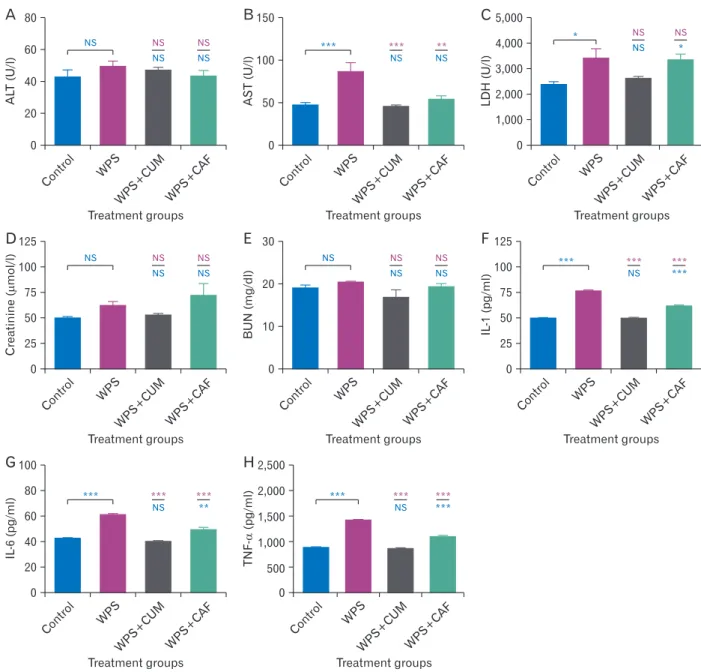
Fig. 3. Bar charts for illustration level of different liver function, kidney function, and proinflammatory cytokines in serum samples of albino mice; (A) for the concentration of ALT; (B) for the concentration of AST; (C) for the concentration of LDH; (D) for quantifying the level of creatinine; (E) for the level of BUN; (F) shows the level for IL-1β; (G) shows the level for IL-6, and (H) shows the level for TNF-α. Asterisks (*,
**, ***) indicates for (P<0.05, P<0.01, P<0.001) respectively, and (NS) indicates for no significant result when P≥0.05. Significance against the control group written in blue color, whereas against WPS group written in red. WPS, WPS+CUM, and WPS+CAF respectively. ALT, alanine aminotransferase; AST, aspartate aminotransferase; BUN, blood urea nitrogen; CAF, caffeic acid; CUM, curcumin; IL-1β, interleukin-1 beta;
IL-6, interleukin-6; LDH, lactate dehydrogenase; NS, not significant; TNF-α, tumor necrosis factor-alpha; WPS, waterpipe smoking.
Results
Liver and kidney function tests
The results for ALT enzyme did not show any significant differences among the examined groups at P<0.05 (Fig. 3A).
The results for AST enzyme were (47±3.2 U/l, 86±10.3 U/
l, 44±2.8 U/l, 53±4.6 U/l) for Control, WPS, WPS+CUM and WPS+CAF respectively (Fig. 3B). WPS group showed extreme significant result against Control group at P<0.001.
WPS+CUM group did not show a significant change against Control group but had extreme significant result against WPS group at P<0.001. WPS+CAF group did not show a significant change against Control group but had highly sig- nificant result against WPS group at P=0.003.
The results for LDH enzyme were 2,366±119 U/l, 3,408±386 U/l, 2,609±89 U/l, 3,325±250 U/l for Control, WPS, WPS+CUM and WPS+CAF respectively (Fig. 3C).
WPS group showed significant difference against Control group at P=0.023. WPS+CUM group did not show signifi- cant differences against both the Control and WPS groups at P<0.05. WPS+CAF showed a significant difference against Control group at P=0.042 and did not show a significant dif- ference against WPS group at P<0.05 (Fig. 3C).
The results for two kidney function tests (creatinine and BUN) were 49.5±2.15 µmol/l, 61.5±4.47 µmol/l, 52.3±1.86
µmol/l, 71.5±11.8 µmol/l for Control, WPS, WPS+CUM and WPS+CAF groups respectively in creatinine test (Fig.
3D), and 11.9±0.77 mg/dl, 20.3±0.17 mg/dl, 16.7±1.89 mg/
dl, 19.2±0.79 mg/dl for Control, WPS, WPS+CUM and WPS+CAF groups respectively in BUN test (Fig. 3E). The statistical results did not show significant differences among the groups in both creatinine and BUN tests.
Immunochemical tests for proinflammatory cytokine biomarkers
The results for IL-1β marker were 49.2±0.91 pg/ml, 76.0±1.07 pg/ml, 49.1±0.92 pg/ml, 60.9±1.63 pg/ml for Con- trol, WPS, WPS+CUM, and WPS+CAF respectively (Fig. 3F).
WPS group showed extreme significant difference against Control group at P<0.001. WPS+CUM group did not show significant difference against Control group but had ex- treme significant difference against WPS group at P<0.001.
WPS+CAF group showed extreme significant differences against both the Control and WPS groups at P<0.001 (Fig. 3F).
The results for IL-6 marker were 42.1±0.81 pg/ml, 60.3±1.51 pg/ml, 40.2±0.85 pg/ml, 49.1±1.90 pg/ml for Con- trol, WPS, WPS+CUM, and WPS+CAF respectively (Fig.
3G). WPS group showed extreme significant difference against Control group at P<0.001. WPS+CUM group did not show significant difference against Control group but
50 m 50 m
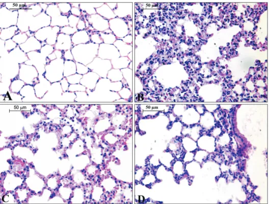
Fig. 4. Thin sections of 5 µm for the lung of different experimental groups in this study; (A) represents the control group; (B) for WPS; (C) for CUM+WPS; (D) for CAF+WPS.
CAF, caffeic acid; CUM, curcumin;
WPS, waterpipe smoking. Sections were stained with H&E, magnification,
×400. Scale bars=50 µm, n=13 (A–D).
had extreme significant difference against WPS group at P<0.001. WPS+CAF group showed highly significant differ- ence against Control group at P=0.004 and extreme signifi- cant difference against WPS group at P<0.001 (Fig. 3G).
The results for TNF-α marker were 881±14 pg/ml, 1,413±22 pg/ml, 859±13 pg/ml, 1,086±39 pg/ml for Control, WPS, WPS+CUM, and WPS+CAF respectively (Fig. 3H).
WPS group showed extreme significant difference against Control group at P<0.001. WPS+CUM group did not show significant difference against Control group but had ex- treme significant difference against WPS group at P<0.001.
WPS+CAF group showed extreme significant differences against both the Control and WPS groups at P<0.001 (Fig.
3H).
Comparative histopathological description for the selected organs in this study
The control group sections showed regular spaces of al- veolar sacs, alveolar septa were integral, alveolar walls were well-inflated and blood vessels had normal endothelial cells without dilations in smooth muscles (Fig. 4A). WPS group showed thickening of alveolar walls, alveolar spaces were collapsed and not properly inflated, sort of nuclear conden- sation and cellular vacuolization, bleeding of RBCs and in- filtration of inflammatory cells (Fig. 4B). WPS+CUM group
showed less infiltration of inflammatory cells, alveoli were better inflated and less thickening in comparison to WPS group, but bleeding and hemorrhage were still observed (Fig.
4C). WPS+CAF group showed well inflated alveoli, less al- veolar vacuolization, less hemorrhage and bleeding, but has significant infiltration of inflammatory cells (Fig. 4D).
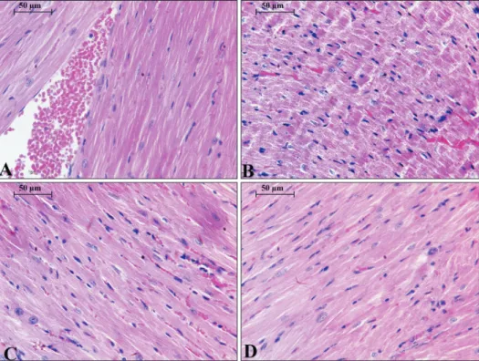
The control group showed well developed cardiac muscle fibers without deterioration or inflammation (Fig. 5A). WPS group showed partial degradation and disintegration of car- diac myofiber, nuclei of cardiomyocytes displayed necrotic features and partial vacuolization in the sarcoplasm, mild myofibrillar spaces and mild blood congestion in the inter- stitial fluid without significant infiltration of inflammatory cells (Fig. 5B). WPS+CUM group showed normal cardiomy- ocytes with euchromatic nuclei, compact myofibrils and ob- vious intercalated discs (Fig. 5C). WPS+CAF group showed normal of structure for cardiomyocytes with mild myofibril spaces without inflammation or hemorrhage (Fig. 5D).
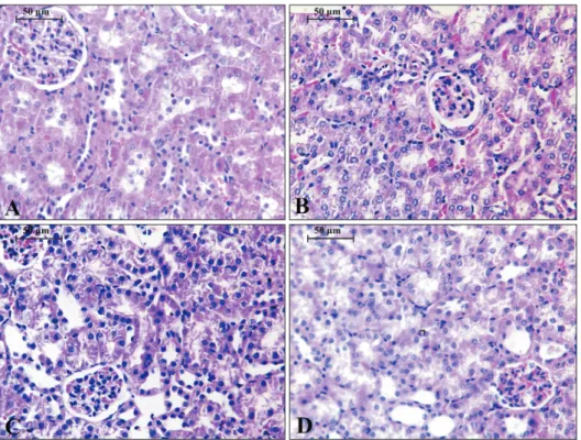
Microscopic examination of the kidney revealed the nor- mal structure of kidney cortex was free from any abnormali- ties (Fig. 6A). WPS group showed deteriorations in kidney cortex and dilation of collecting ducts, the epithelial cells with some necrotic nuclei containing condensed chromatin, epithelial vacuolization, and infiltration of inflammatory cells (Fig. 6B). WPS+CUM group showed blood conges-
Fig. 5. Thin sections of 5 µm for the heart of different experimental groups in this study; (A) represents the control group; (B) for WPS; (C) for CUM+WPS; (D) for CAF+WPS.
CAF, caffeic acid; CUM, curcumin;
WPS, waterpipe smoking. Sections were stained with H&E, magnification,
×400. Scale bars=50 µm, n=13 (A–D).
tion, partial recovery of epithelium and glomerulus less vacuolization, significant decrease in number of counted necrotic nuclei with condensed chromatin (Fig. 6C). Kidney in WPS+CAF group showed slight deterioration in proximal not distilled convoluted tubules, no signs of vacuolization or necrotic nuclei in cells of cortex part of the kidney (Fig. 6D).
Discussion
There are common social views in middle eastern coun- tries that waterpipe tobacco smoking is less harmful than cigarette smoking, especially among youth and teenagers.
This study showed that short term exposure to waterpipe tobacco smoking could elevate liver enzymes, induce inflam- matory cells, proinflammatory cytokines, and deteriorate the histological architecture of lung, heart, and kidney in albino mice without significant increasing in levels of creatinine and blood urea nitrogen. These results were congruent with previous findings that showed waterpipe tobacco smoking elevate total white blood cells count, significantly increase TNF-α and IL-6 in bronchoalveolar fluid lavage [29]. In ad- dition, waterpipe tobacco smoking decreases the enzymatic activity of some endogenous free radical scavengers like superoxide dismutase, catalase, and glutathione peroxidase, and increases lipid peroxidation in mice [30]. Also, the influx
of inflammatory cells in the peribronchial and interstitial spaces consisting predominantly of neutrophil polymorphs with increasing in the levels of 8-isoprostane, nuclear factor kappa-B, DNA migration, and significantly alters the physi- ological airway resistance in conducting portion of the lung in mice exposed to waterpipe tobacco smoking [31]. The im- pact of WPS in lung tissue of rat showed many morphologi- cal changes including induce the bleeding of red blood cell, thickening of alveolar walls, Furthermore, the inflammatory cells were increased [29]. In addition, short term exposure of WPS could induce vacuolization with mild glomerular degeneration, mild degeneration of endothelial cells of glom- erulus capillaries, and mild ultrastructural changes in proxi- mal convoluted tubules in mice kidney without significant increase in amount of creatinine clearance and proteins in urine [32]. Also, WPS showed histopathological alternations and ultrastructural changes in cardiomyocyte organelles in rat ventricular tissue [33].
Our findings showed that CUM had attenuated the cyto- toxicity induced by waterpipe tobacco smoking by retaining the inflation of alveoli and decrease the infiltration of in- flammatory cells in the lung, displayed normal cardiomyo- cytes with compact myofibrils and evident intercalated discs in heart, reduced the level of AST and LDH enzymes, but did not prevent the deteriorations of nephrons in the kidney.
Fig. 6. Thin sections of 5 µm for the kidney of different experimental groups in this study; (A) represents the control group; (B) for WPS; (C) for CUM+
WPS; (D) for CAF+WPS. CAF, caffeic acid; CUM, curcumin; WPS, waterpipe smoking. Sections were stained with H&E, magnification, ×400. Scale bars=
50 µm, n=13 (A–D).
Previous studies stated that CUM has chemopreventive effect for human urothelial cells and human bladder carcinoma cells that induced by chronic exposure of cigarette smok- ing [34]. Also, CUM can abrogate the expression of genes of cyclin D1, matrix metalloproteinase-9, and act as COX-2 inhibitor in lung normal and carcinogenic cell lines exposed to cigarette smoking [35]. CUM could increase the transcrip- tion of antioxidant genes in primary alveolar macrophage, decrease the infiltration of inflammatory cells in bronchoal- veolar lavage fluid, and reduce the enlargement of air space in lung of mice exposed to long-term of cigarette smoking [36].
CAF showed a chemopreventive action against alveolar thickening induced by WPS, reduces necrosis and vacuoliza- tion in cardiomyocytes, protects the typical architecture of the kidney to a reasonable extent, significantly decrease AST level in serum, but not LDH.
It was previously stated that CAF exhibits hepatoprotec- tive activity through inhibiting the enzymatic activity of ALT and AST in rodents induced by the sub-lethal dose of paracetamol [37]. It was previously illustrated that CAF has potent inhibition on microsomal P450 cytochrome enzymes in human liver by acting as noncompetitive inhibitor to CYP1A2 enzyme and competitive inhibitor to CYP2C9 and CYP2D6 [38]. CAF has less ability than CUM in inhibiting 12-O-tetradecanoylphorbol-13-acetate-induced inflamma- tion and promotion of skin tumors [39]. Moreover, CAF has more potent to inhibit lipid peroxidation than CUM [39].
Inflammation is major to the body's immunity against infection action. Besides, it is likewise a basic part of ordi- nary tissue repair. Tobacco smoking has been appeared to modify the host reaction and, in this way, alters the pro- gression and result of irritation. The specific components of tobacco smoking prompting irritation are not obviously comprehended. Not with standing, it has been proposed that smoking influences various inflammatory process through its impact on immune inflammatory cells, causing an im- munosuppressant state and cytokine secretion [40]. Several investigations revealed that tobacco smoke presentation is related with expanded inflammation. It has been discovered that the degree of serum inflammatory marker, for example, TNF-α is altogether higher in smokers contrasted with non- smokers [41, 42]. The present research gave experimental evidence that exposure to WPS induces systemic inflamma- tion and oxidative stress. Critically, we demonstrated that treatment with the nourishment added substance CUM and
caffeic corrosive reduced the systemic inflammation [43, 44].
Nutritional compounds that show anti-inflammatory and antioxidant agent impacts have explicit applications in fore- stalling oxidative stress related injury [45].
We have as of late exhibited that exposure to WPS for 5 days and one month induced inflammation and oxidative stress in some studied organs [46, 47]. Therefore, in this study, the levels of inflammatory markers, for example, IL- 1β, IL-6 and TNF-α, were estimated in serum of water pipe smoking with and without of CUM and CAF and we found that CUM and CAF attenuate the increase in serum con- centrations of IL-1β, IL-6 and TNF-α caused by a two weeks acute secondhand exposure to WPS, suggesting the systemic anti-inflammatory effects of CUM and CAF. Besides, CUM showed better results than CAF in term of anti-inflammato- ry effect.
In conclusion, this study illustrated that both CUM and CAF had anti-inflammatory effect against the elevated levels of proinflammatory cytokines induced by exposure to wa- terpipe tobacco smoking. In addition, both antioxidants had ameliorative effects against the histopathological alternations induced by acute secondhand exposure of waterpipe tobacco smoking.
ORCID
Alia Khwaldeh: https://orcid.org/0000-0002-7492-4906 Ali Abu Siyam: https://orcid.org/0000-0002-6672-6184 Ahmed Alzbeede: https://orcid.org/0000-0002-1009-5440 Mohammad Farajallah:
https://orcid.org/0000-0002-1858-5348
Ziad Shraideh: https://orcid.org/0000-0002-3194-3728 Darwish Badran: https://orcid.org/0000-0002-8117-1762
Author Contributions
Conceptualization: AK, AAl. Data acquisition: AK, AAb, AAl, MF. Data analysis or interpretation: AAl, AK, ZS, DB.
Drafting of the manuscript: AK, AAb, AAl. Critical revision of the manuscript: MF, ZS, DB. Approval of the final version of the manuscript: all authors.
Conflicts of Interest
No potential conflict of interest relevant to this article was reported.
Acknowledgements
We like to thank the Department of Biological Sciences at The University of Jordan for the technical facilitation to ac- complish this work.
References
1. Bhatnagar A, Maziak W, Eissenberg T, Ward KD, Thurston G, King BA, Sutfin EL, Cobb CO, Griffiths M, Goldstein LB, Rezk-Hanna M. Water pipe (hookah) smoking and cardiovas- cular disease risk: a scientific statement From the American Heart Association. Circulation 2019;139:e917-36.
2. WHO Study Group on Tobacco Product Regulation (TobReg).
Waterpipe tobacco smoking: health effects, research needs and recommended actions for regulators. 2nd ed. Geneva: WHO;
2015.
3. Jukema JB, Bagnasco DE, Jukema RA. Waterpipe smoking:
not necessarily less hazardous than cigarette smoking: pos- sible consequences for (cardiovascular) disease. Neth Heart J 2014;22:91-9.
4. Kaplan B, Sussan T, Rule A, Moon K, Grau-Perez M, Olmedo P, Chen R, Carkoglu A, Levshin V, Wang L, Watson C, Blount B, Calafat AM, Jarrett J, Caldwell K, Wang Y, Breysse P, Strick- land P, Cohen J, Biswal S, Navas-Acien A. Waterpipe tobacco smoke: characterization of toxicants and exposure biomarkers in a cross-sectional study of waterpipe employees. Environ Int 2019;127:495-502.
5. Shihadeh A, Schubert J, Klaiany J, El Sabban M, Luch A, Saliba NA. Toxicant content, physical properties and biological activi- ty of waterpipe tobacco smoke and its tobacco-free alternatives.
Tob Control 2015;24(Suppl 1):i22-30.
6. Pryor WA, Stone K, Zang LY, Bermúdez E. Fractionation of aqueous cigarette tar extracts: fractions that contain the tar radical cause DNA damage. Chem Res Toxicol 1998;11:441-8.
7. Alsaad AM, Al-Arifi MN, Maayah ZH, Attafi IM, Alanazi FE, Belali OM, Alhoshani A, Asiri YA, Korashy HM. Genotoxic impact of long-term cigarette and waterpipe smoking on DNA damage and oxidative stress in healthy subjects. Toxicol Mech Methods 2019;29:119-27.
8. Hadidi KA, Mohammed FI. Nicotine content in tobacco used in hubble-bubble smoking. Saudi Med J 2004;25:912-7.
9. Chaouachi K. Hookah (Shisha, Narghile) smoking and en- vironmental tobacco smoke (ETS). A critical review of the relevant literature and the public health consequences. Int J Environ Res Public Health 2009;6:798-843.
10. Mamtani R, Cheema S, Sheikh J, Al Mulla A, Lowenfels A, Maisonneuve P. Cancer risk in waterpipe smokers: a meta- analysis. Int J Public Health 2017;62:73-83.
11. Zhang YJ, Gan RY, Li S, Zhou Y, Li AN, Xu DP, Li HB. Anti- oxidant phytochemicals for the prevention and treatment of chronic diseases. Molecules 2015;20:21138-56.
12. Tan BL, Norhaizan ME, Liew WP, Sulaiman Rahman H. Anti- oxidant and oxidative stress: a mutual interplay in age-related diseases. Front Pharmacol 2018;9:1162.
13. Bengmark S. Curcumin, an atoxic antioxidant and natural NFkappaB, cyclooxygenase-2, lipooxygenase, and inducible ni- tric oxide synthase inhibitor: a shield against acute and chronic diseases. JPEN J Parenter Enteral Nutr 2006;30:45-51.
14. Oyagbemi AA, Saba AB, Ibraheem AO. Curcumin: from food spice to cancer prevention. Asian Pac J Cancer Prev 2009;10:963-7.
15. Bisht K, Wagner KH, Bulmer AC. Curcumin, resveratrol and flavonoids as anti-inflammatory, cyto- and DNA-protective dietary compounds. Toxicology 2010;278:88-100.
16. Kalpana C, Sudheer AR, Rajasekharan KN, Menon VP. Com- parative effects of curcumin and its synthetic analogue on tis- sue lipid peroxidation and antioxidant status during nicotine- induced toxicity. Singapore Med J 2007;48:124-30.
17. Stojković D, Petrović J, Soković M, Glamočlija J, Kukić- Marković J, Petrović S. In situ antioxidant and antimicrobial activities of naturally occurring caffeic acid, p-coumaric acid and rutin, using food systems. J Sci Food Agric 2013;93:3205-8.
18. Khan FA, Maalik A, Murtaza G. Inhibitory mechanism against oxidative stress of caffeic acid. J Food Drug Anal 2016;24:695- 702.
19. Akomolafe SF, Akinyemi AJ, Oboh G, Oyeleye SI, Ajayi OB, Omonisi AE, Owolabi FL, Atoyebi DA, Ige FO, Atoki VA. Co- administration of caffeine and caffeic acid alters some key enzymes linked with reproductive function in male rats. An- drologia 2018;50:e12839.
20. Dekanski D, Spremo-Potparević B, Bajić V, Živković L, Topalović D, Sredojević DN, Lazić V, Nedeljković JM. Acute toxicity study in mice of orally administrated TiO2 nanopar- ticles functionalized with caffeic acid. Food Chem Toxicol 2018;115:42-8.
21. Li Y, Chen LJ, Jiang F, Yang Y, Wang XX, Zhang Z, Li Z, Li L.
Caffeic acid improves cell viability and protects against DNA damage: involvement of reactive oxygen species and extracel- lular signal-regulated kinase. Braz J Med Biol Res 2015;48:502- 8.
22. Ahn CB, Je JY, Kim YS, Park SJ, Kim BI. Induction of Nrf2- mediated phase II detoxifying/antioxidant enzymes in vitro by chitosan-caffeic acid against hydrogen peroxide-induced hepatotoxicity through JNK/ERK pathway. Mol Cell Biochem 2017;424:79-86.
23. Kabała-Dzik A, Rzepecka-Stojko A, Kubina R, Wojtyczka RD, Buszman E, Stojko J. Caffeic acid versus caffeic acid phenethyl ester in the treatment of breast cancer MCF-7 cells: migration rate inhibition. Integr Cancer Ther 2018;17:1247-59.
24. Andrade M, Benfeito S, Soares P, Magalhães e Silva D, Loureiro J, Borges A, Borges F, Simões M. Fine-tuning of the hydro- phobicity of caffeic acid: studies on the antimicrobial activity against Staphylococcus aureus and Escherichia coli. RSC Adv 2015;5:53915-25.
25. Tanida I, Shirasago Y, Suzuki R, Abe R, Wakita T, Hanada K,
Fukasawa M. Inhibitory effects of caffeic acid, a coffee-related organic acid, on the propagation of hepatitis C virus. Jpn J In- fect Dis 2015;68:268-75.
26. Zhu Q, Sun Y, Yun X, Ou Y, Zhang W, Li JX. Antinociceptive effects of curcumin in a rat model of postoperative pain. Sci Rep 2014;4:4932.
27. Salem AM, Ragheb AS, Hegazy MGA, Matboli M, Eissa S. Caf- feic acid modulates miR-636 expression in diabetic nephropa- thy rats. Indian J Clin Biochem 2019;34:296-303.
28. Shraideh ZA, Awaida W, Najjar H, Musleh M. A modified smoking machine for monitoring the effect of tobacco smoke on albino rats. Jordan J Biol Sci 2011;4:109-12.
29. Khabour OF, Alzoubi KH, Bani-Ahmad M, Dodin A, Eis- senberg T, Shihadeh A. Acute exposure to waterpipe tobacco smoke induces changes in the oxidative and inflammatory markers in mouse lung. Inhal Toxicol 2012;24:667-75.
30. Al-Sawalha NA, Alzoubi KH, Khabour OF, Alyacoub W, Almahmood Y. Effect of waterpipe tobacco smoke exposure during lactation on learning and memory of offspring rats: role of oxidative stress. Life Sci 2019;227:58-63.
31. Nemmar A, Al-Salam S, Beegam S, Yuvaraju P, Ali BH. Water- pipe smoke exposure triggers lung injury and functional de- cline in mice: protective effect of Gum Arabic. Oxid Med Cell Longev 2019;2019:8526083.
32. Nemmar A, Beegam S, Yuvaraju P, Yasin J, Ali BH, Adeghate E. Nose-only water-pipe smoke exposure in mice elicits renal histopathological alterations, inflammation, oxidative stress, DNA damage, and apoptosis. Front Physiol 2020;11:46.
33. Al-Awaida W, Najjar H, Shraideh Z. Structural characteriza- tion of rat ventricular tissue exposed to the smoke of two types of waterpipe. Iran J Basic Med Sci 2015;18:942-9.
34. Liang Z, Lu L, Mao J, Li X, Qian H, Xu W. Curcumin reversed chronic tobacco smoke exposure induced urocystic EMT and acquisition of cancer stem cells properties via Wnt/β-catenin.
Cell Death Dis 2017;8:e3066.
35. Shishodia S, Majumdar S, Banerjee S, Aggarwal BB. Ursolic acid inhibits nuclear factor-kappaB activation induced by car- cinogenic agents through suppression of IkappaBalpha kinase and p65 phosphorylation: correlation with down-regulation of cyclooxygenase 2, matrix metalloproteinase 9, and cyclin D1.
Cancer Res 2003;63:4375-83.
36. Suzuki M, Betsuyaku T, Ito Y, Nagai K, Odajima N, Moriyama C, Nasuhara Y, Nishimura M. Curcumin attenuates elastase-
and cigarette smoke-induced pulmonary emphysema in mice.
Am J Physiol Lung Cell Mol Physiol 2009;296:L614-23.
37. Janbaz KH, Saeed SA, Gilani AH. Studies on the protective ef- fects of caffeic acid and quercetin on chemical-induced hepato- toxicity in rodents. Phytomedicine 2004;11:424-30.
38. Rastogi H, Jana S. Evaluation of inhibitory effects of caffeic acid and quercetin on human liver cytochrome p450 activities.
Phytother Res 2014;28:1873-8.
39. Anand P, Thomas SG, Kunnumakkara AB, Sundaram C, Hari- kumar KB, Sung B, Tharakan ST, Misra K, Priyadarsini IK, Ra- jasekharan KN, Aggarwal BB. Biological activities of curcumin and its analogues (Congeners) made by man and Mother Na- ture. Biochem Pharmacol 2008;76:1590-611.
40. Gonçalves RB, Coletta RD, Silvério KG, Benevides L, Casati MZ, da Silva JS, Nociti FH Jr. Impact of smoking on inflam- mation: overview of molecular mechanisms. Inflamm Res 2011;60:409-24.
41. Ghazali WS, Romli AC, Mohamed M. Effects of honey supple- mentation on inflammatory markers among chronic smokers:
a randomized controlled trial. BMC Complement Altern Med 2017;17:175.
42. Petrescu F, Voican SC, Silosi I. Tumor necrosis factor-alpha serum levels in healthy smokers and nonsmokers. Int J Chron Obstruct Pulmon Dis 2010;5:217-22.
43. Mercantepe T, Unal D, Tümkaya L, Yazici ZA. Protective ef- fects of amifostine, curcumin and caffeic acid phenethyl ester against cisplatin-induced testis tissue damage in rats. Exp Ther Med 2018;15:3404-12.
44. Yang WS, Jeong D, Yi YS, Park JG, Seo H, Moh SH, Hong S, Cho JY. IRAK1/4-targeted anti-inflammatory action of caffeic acid. Mediators Inflamm 2013;2013:518183.
45. Hussain T, Tan B, Yin Y, Blachier F, Tossou MC, Rahu N. Oxi- dative stress and inflammation: what polyphenols can do for us? Oxid Med Cell Longev 2016;2016:7432797.
46. Nemmar A, Al Hemeiri A, Al Hammadi N, Yuvaraju P, Bee- gam S, Yasin J, Elwasila M, Ali BH, Adeghate E. Early pulmo- nary events of nose-only water pipe (shisha) smoking exposure in mice. Physiol Rep 2015;3:e12258.
47. Nemmar A, Yuvaraju P, Beegam S, John A, Raza H, Ali BH.
Cardiovascular effects of nose-only water-pipe smoking expo- sure in mice. Am J Physiol Heart Circ Physiol 2013;305:H740- 6.