Korean J Clin Microbiol Vol. 15, No. 4, December, 2012 http://dx.doi.org/10.5145/KJCM.2012.15.4.147
Lung Abscess and Bacteremia Caused by Neisseria flavescens and Streptococcus sanguis in Patient with Idiopathic
Hypereosinophilic Syndrome
Ju Hyun Kong1, Sung Hyun Shin1, Su Eun Park1, Hee Ju Park1, Jongyoun Yi2, Shine Young Kim3, Seung Kook Son1
Departments of
1Pediatrics,
2Laboratory Medicine, Pusan National University School of Medicine, Yangsan,
3
Department of Laboratory Medicine, Pusan National University Hospital, Busan, Korea
Neisseria flavescens has been rarely reported as a pathogen in the literature. We experienced a case of N. flavescens bacteremia and lung abscess co-in- fected with Streptococcus sanguis in patient with idi- opathic hypereosinophilic syndrome. A 15-year-old boy was diagnosed with idiopathic hypereosinophilic syn- drome complicated with pulmonary thromboembolism.
He was given systemic steroids and thrombolytics.
After 8 weeks of therapy, a lung abscess appeared on the plain chest radiograph. We treated him with empirical antibiotics and carried out surgical drainage.
Two types of microorganisms were cultured from both blood and pus samples, obtained in the first
day of hospitalization. Pus was aspirated from the lung abscess with an aseptic technique. Neisseria species and S. sanguis were identified using tradi- tional methods. To confirm the identity of the Neisseria species, we conducted further testing using 16S ribo- somal ribonucleic acid sequencing whereupon N. fla- vescens was identified. This is the first case report of pulmonary infection caused by N. flavescens. We suggest that N. flavescens may act as a pathogen.
(Korean J Clin Microbiol 2012;15:147-150)
Key Words: Neisseria flavescens, Sepsis, 16S ribosomal RNA sequencing
Received 20 September, 2012, Revised 15 October, 2012 Accepted 17 October, 2012
Correspondence: Seung Kook Son, Department of Pediatrics, Pusan National University Children's Hospital, Pusan National University School of Medicine, 20, Geumo-ro, Mulgeum-eup, Yangsan 626- 770, Korea. (Tel) 82-55-360-2180, (Fax) 82-55-360-2181, (E-mail) [email protected]
147 INTRODUCTION
Neisseria flavescens is one of the non-meningococcal and non-gonococcal Neisseria species that has infrequently caused meningitis, endocarditis, and sepsis [1]. Therefore they are dis- tinguished from meningococcus or gonococcus and so called non-pathogenic Neisseria.
Although N. flavescens can colonize as a normal flora in the upper respiratory tract, an infection of the lower respiratory tract was never reported. However, we experienced a case of a lung abscess caused by N. flavescens with consequential bacteremia.
CASE REPORT
A 15-year-old boy was transferred to our center with com- plaints of cough, sputum, and progressive dyspnea for 4 days.
The plain chest radiograph showed pneumonic infiltration with pleural effusion in both lungs. The chest CT scan revealed patch consolidation on both lower lobes with pleural effusion and the thromboembolus within the left pulmonary artery. Thromboem- boli were also detected in the portal vein and the left common iliac vein on the diagnostic work-up. Laboratory examinations showed a white blood cell count of 23,530/μL, segmental neu- trophil of 56%, lymphocyte of 9.9%, eosinophil of 26.3%
(6,630/μL), hemoglobin of 13.7 g/dL, platelet count of 77,000/μL, C-reactive protein of 11.4 mg/dL (reference range <0.5 mg/dL) and eosinophil cation protein >200 μg/L (reference range,
≤19 μg/L). He was diagnosed as idiopathic hypereosinophilic syndrome (IHES) complicated with pulmonary thromboembo- lism [2]. He received systemic steroid therapy for IHES, throm- bolytics and anti-coagulation therapy.
Hypereosinophilia was improved immediately after medication.
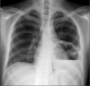
Methylprednisolone was taken in doses of 2 mg/kg/day for 4 days and then prednisolone was used in doses of 1 mg/kg/day for a month. After 4 weeks of tapering periods, he had inter- mittent fever and cough over 2 days. The plain chest radiograph
148
Korean J Clin Microbiol 2012;15(4):147-150Fig. 1. Chest plain radiograph showing lung abscess.
showed a shadow of a cavity with an air-fluid level (Fig. 1). He was diagnosed with lung abscess and hospitalized for antibiotics therapy and surgical drainage. We selected the antibiotics nafcil- lin and cefotaxime empirically.
Bacterial cultures of blood, urine, sputum, and pus drained from the abscess were carried out before antibiotics therapy.
Two species of bacteria were cultivated from the blood and the pus obtained at the first day of hospitalization. These two were identified by conventional methods and the Vitek 2 system (BioMérieux, inc., Marcy-l'Etoile, France). One (gram-positive cocci) was identified as Streptococcus sanguis, and the other (gram-negative diplococci) as Neisseria species using Vitek 2 NH Identification Card. To confirm the identity of this isolate, 16S rRNA sequencing analysis was performed. InstaGene Matrix (Bio-Rad Laboratories, Hercules, CA, USA) was used to extract the bacterial genomic DNA, and the first 500 base pairs on the 5' end of the 16S rRNA gene were amplified and se- quenced using MicroSeq 500 16S rDNA Bacterial Identification PCR and Sequencing Kits (Applied Biosystems, Foster City, CA, USA). The sequencing product was analyzed on a 3130 Genetic Analyzer (Applied Biosystems) according to the manu- facturer’s instructions. The resulting sequence was compared with sequences stored in GenBank (http://www.ncbi.nlm.nih.
gov/). A GenBank BLAST search revealed that the 16S rRNA gene sequence of the isolate was 100% homologous with that of N. flavescens (GenBank Accession no. GU417548.1). This showed percent identity with >0.8% separation from other spe- cies that were listed up in the search (Neisseria gonorrhea, Neisseria weaveri, Neisseria subflava, etc.), and the isolate was
confirmed to be N. flavescens [3].
After the antibiotics therapy, the clinical symptoms were improved. According to the results of the bacterial culture, we continued to use cefotaxime except for nafcillin. The sensitivity test of antibiotics was not performed because there was no pro- tocol about the proper sensitivity test for N. flavescens. Subse- quently the patient was discharged on the 37th day of hospita- lization. On the second month of the follow-up he was in good condition without recurrence.
DISCUSSION
Neisseria is the genus characteristic of gram-negative diplo- cocci. There are two well-known pathogenic organisms in the genus Neisseria, N. gonorrhea and N. meningitidis. These two organisms characterized the specific disease entity, respectively.
N. meningitidis usually colonize in the nasopharynx of humans and sometimes invades into the bloodstream. Moreover, when invaded, it may cause bacteremia, meningitis, and severe me- ningococcemia. N. gonorrhea infects the genitourinary tract mu- cosa and rarely causes disseminated infection. While non-patho- genic Neisseria are commensal organisms living in the upper respiratory tract, and relatively few cases of infection were re- ported compared to pathogenic Neisseria.
Like other non-pathogenic Neisseria, N. flavescens has been rarely reported as a causative microorganism. The first case re- port of significant infection by this organism was of 14 cases of meningitis, which were a single epidemic in Chicago in 1928 [4]. Subsequently four other cases of meningitis were reported in 1957 [4]. The septicemia by this organism was reported first in 1968 [4]. It was a 20-year-old woman and her symptoms were discovered 7 days after dental surgery. And in 1984, more cases of meningitis and septicemia by N. flavescens were re- ported, which consisted of 15 cases of meningitis accompanied by septicemia and one case of septicemia [5]. After that, the first case of infectious endocarditis by N. flavescens was noted and reported in 1987 [6]. Infectious endocarditis was also ob- served in the case of a patient with narcotic addiction and re- ported in 1990 [7]. A case of septic shock was reported in 1990 which occurred in a pediatric patient [8]. Therefore, our report is one of the rare cases of the infection caused by N. flavescens especially in a pediatric patient, and also the first case report of the lung abscess, which is one of its resultant pulmonary infections.
N. meningitidis causes septicemia, sepsis, and meningitis by
Ju Hyun Kong, et al. : Lung Abscess and Bacteremia Caused by Neisseria flavescens
149
invading into the blood stream [9]. In the present case, N. fla- vescens was found both in the abscess and blood. We predicted that N. flavescens caused localized pulmonary infection and then invaded into the bloodstream through the site.
In this context, the present case demonstrates several charac- teristic points. First, while, N. flavescens are commensal organ- isms, yet less virulent than the other pathogenic Neisseria spe- cies, it can potentially infect its host.
Second, N. flavescens not only cause sepsis, meningitis or in- fective endocarditis but it also locally infects the respiratory system. This would be more likely happened in patients who have underlying pulmonary problems or diseases. As with this case, the lung abscess occurred at the site, where pulmonary embolism and combined pneumonia was previously noticed.
There are articles supporting this suggestion. One study revealed that the lower respiratory tract was non-sterile in patients with chronic obstructive pulmonary disease (COPD) and the in- creased number of microbial loads was related to acute ex- acerbation of COPD [10]. Another study showed common spe- cies identified in the lower respiratory tract of COPD patients, which included of Neisseria [11]. In comparison, the low respi- ratory tract of healthy subjects was relatively sterile [12].
Another study compared patients who had contracted cicatricial laryngeal stenosis after surgery with healthy subjects. The pa- tients showed a significantly greater occurrence of staphylococci, Neisseria and Candida in the pharyngeal mucosa. What is more, they had a higher rate of dysbacteriosis, which was charac- terized by a predominance of N. flavescens, Staphylococcus aur- eus, alpha-hemolytic streptococcus, Staphylococcus epidermidis and Candida species [13].
Third, his illness could be influenced by immune suppression following the steroid therapy. The immune deficiency allows an extraordinary infection by a less virulent organism. Abscesses complicated in steroid therapy occur infrequently and have been reported as a case report, but it may be one of the important factors causing the infection.
In interpretating of the organism as a causative micro- organism, it can be argued whether the identified organism is a
contaminant or not. In the present case, we believe that N. fla- vescens was a pathogen co-infected with S. sanguis, because same organisms were cultivated from both blood and pus simultaneously. The specimens were collected by an aseptic technique and the pus was attained by an aspiration through the chest wall not through the airway. The fact that N. flavescens was not a common colonizer of the skin also supports the con- tention that this microorganism was not a contaminant.
REFERENCES
1. Feder HM Jr and Garibaldi RA. The significance of nongonococcal, nonmeningococcal Neisseria isolates from blood cultures. Rev Infect Dis 1984;6:181-8.
2. Roufosse FE, Goldman M, Cogan E. Hypereosinophilic syndromes.
Orphanet J Rare Dis 2007;2:37.
3. Clinical and Laboratory Standards Institute. Interpretive criteria for identification of bacteria and fungi by DNA target sequencing:
Approved guideline. CLSI Document MM18-A. Wayne, PA; Clinical and Laboratory Standards Institute, 2008.
4. Wertlake PT and Williams TW Jr. Septicaemia caused by Neisseria flavescens. J Clin Pathol 1968;21:437-9.
5. Coovadia YM. Neisseria flavescens septicaemia with meningitis. A case report. S Afr Med J 1984;66:308-9.
6. Sinave CP and Ratzan KR. Infective endocarditis caused by Neisseria flavescens. Am J Med 1987;82:163-4.
7. Szabo S, Lieberman JP, Lue YA. Unusual pathogens in narcotic- ssociated endocarditis. Rev Infect Dis 1990;12:412-5.
8. Quintero Otero S, Rubio Quiñones F, Hernández Gonzalez A, Díaz Portillo J, García Martos P, Pantoja Rosso S. Septic shock caused by Neisseria flavescens. An Esp Pediatr 1990;33:64-5.
9. Melican K and Dumenil G. Vascular colonization by Neisseria meningitidis. Curr Opin Microbiol 2012;15:50-6.
10. Rosell A, Monsó E, Soler N, Torres F, Angrill J, Riise G, et al.
Microbiologic determinants of exacerbation in chronic obstructive pulmonary disease. Arch Intern Med 2005;165:891-7.
11. Cabrera-Rubio R, Garcia-Núñez M, Setó L, Antó JM, Moya A, Monsó E, et al. Microbiome diversity in the bronchial tracts of patients with chronic obstructive pulmonary disease. J Clin Microbiol 2012;50:3562-8.
12. Thorpe JE, Baughman RP, Frame PT, Wesseler TA, Staneck JL.
Bronchoalveolar lavage for diagnosing acute bacterial pneumonia.
J Infect Dis 1987;155:855-61.
13. Kovalyk AP and Govda AV. Characteristics of microflora of laryngeal mucosa in healthy subjects and patients with cicatrical stenosis of the larynx. Vestn Otorinolaringol 2010:17-20.
150
Korean J Clin Microbiol 2012;15(4):147-150=국문초록=
특발성 호산구증가증 환자에서 발생한
Neisseria flavescens
와Streptococcus sanguis
중복감염에 의한 폐농양과 균혈증 1예부산대학교 의학전문대학원 1소아과학교실, 2진단검사의학교실, 3부산대학교병원 진단검사의학과
공주현1, 신성현1, 박수은1, 박희주1, 이종윤2, 김신영3, 손승국1
Neisseria flavescens는 지금까지 감염원으로 보고된 예가 드문 균종이다. 저자들은 특발성 호산구 증가증으로 진단된 환 자에서 Streptococcus sanguis와 중복 감염된 N. flavescens 균혈증과 폐농양 감염 증례를 경험하였다. 환자는 폐혈전색전증 이 병발된 특발성 호산구증가증으로 진단된 15세 남아로 전신 스테로이드 치료 및 혈전용해제 치료를 받았다. 총 8주간 의 스테로이드 치료 중에 폐농양이 발생하여 경험적 항생제 치료 및 배농을 시행하였다. 입원 1일에 시행한 혈액 배양 검사와 폐농양 천자를 통해 얻은 농 배양 검사에서 모두 동일한 두개의 균종이 배양되었다. 일반적인 균동정법에 의해 두 균종은 Neisseria species와 S. sanguis로 확인되었고, Neisseria species의 정확한 동정을 위해 16S ribosomal ribonucleic acid (16S rRNA) 염기서열분석을 시행하였고, N. flavescens로 확인되었다. 본 증례는 N. flavescens에 의한 폐감염에 대한 최초의 보고로서 N. flavescens가 병원균으로 작용할 수 있다는 것을 제시하여 준다. [대한임상미생물학회지 2012;
15:147-150]
교신저자 : 손승국, 626-770, 양산시 물금읍 금오로 20
부산대학교 의학전문대학원 부산대학교 어린이병원 소아청소년과 Tel: 055-360-2180, Fax: 055-360-2181
E-mail: [email protected]