D I A B E T E S & M E T A B O L I S M J O U R N A L D I A B E T E S & M E T A B O L I S M J O U R N A L
This is an Open Access article distributed under the terms of the Creative Commons Attribution Non-Commercial License (http://creativecommons.org/licenses/by-nc/4.0/) which permits unrestricted non-commercial use, distribution, and reproduction in any medium, provided the original work is properly cited.
Evogliptin, a Dipeptidyl Peptidase-4 Inhibitor, Attenuates Renal Fibrosis Caused by Unilateral Ureteral Obstruction in Mice
Mi-Jin Kim1,*, Na-young Kim1,*, Yun-A Jung1, Seunghyeong Lee2,3, Gwon-Soo Jung4, Jung-Guk Kim1, In-Kyu Lee1, Sungwoo Lee4, Yeon-Kyung Choi1, Keun-Gyu Park1
1Division of Endocrinology and Metabolism, Department of Internal Medicine, School of Medicine, Kyungpook National University, Daegu,
2Department of Biomedical Science, Graduate School, Kyungpook National University, Daegu,
3BK21 Plus KNU Biomedical Convergence Program, Kyungpook National University, Daegu,
4New Drug Development Center, Daegu-Gyeongbuk Medical Innovation Foundation, Daegu, Korea
Renal fibrosis is considered to be the final common outcome of chronic kidney disease. Dipeptidyl peptidase-4 (DPP-4) inhibi- tors have demonstrated protective effects against diabetic kidney disease. However, the anti-fibrotic effect of evogliptin, a DPP-4 inhibitor, has not been studied. Here, we report the beneficial effects of evogliptin on unilateral ureteral obstruction (UUO)-in- duced renal fibrosis in mice. Evogliptin attenuated UUO-induced renal atrophy and tubulointerstitial fibrosis. Immunohisto- chemistry and Western blotting demonstrated that evogliptin treatment inhibits pro-fibrotic gene expressions and extracellular matrix production. In vitro findings showed that the beneficial effects of evogliptin on renal fibrosis are mediated by inhibition of the transforming growth factor-β/Smad3 signaling pathway. The present study demonstrates that evogliptin is protective against UUO-induced renal fibrosis, suggesting that its clinical applications could extend to the treatment of kidney disease of non-dia- betic origin.
Keywords: Dipeptidyl-peptidase IV inhibitors; Kidney failure, chronic; Transforming growth factor beta
Corresponding authors: Keun-Gyu Park https://orcid.org/0000-0002-8403-1298 Division of Endocrinology and Metabolism, Department of Internal Medicine, School of Medicine, Kyungpook National University, 130 Dongdeok-ro, Jung-gu, Daegu 41944, Korea E-mail: [email protected]
Yeon-Kyung Choi https://orcid.org/0000-0003-0996-6437
Division of Endocrinology and Metabolism, Department of Internal Medicine, School of Medicine, Kyungpook National University, 130 Dongdeok-ro, Jung-gu, Daegu 41944, Korea E-mail: [email protected]
INTRODUCTION
Diabetic kidney disease (DKD) is a leading cause of chronic kidney disease (CKD), and a considerable number of cases of CKD progress to end-stage renal disease (ESRD) [1]. Renal fi- brosis is a major pathologic finding of ESRD and is character- ized by extracellular matrix (ECM) accumulation induced by activation of the transforming growth factor-β (TGF-β)/Smad signaling pathway. It is generally accepted that TGF-β/Smad signaling induces the expression of pro-fibrotic genes, such as
α-smooth muscle actin (α-SMA), plasminogen activator in- hibitor-1 (PAI-1), and type 1 collagen [2]. For this reason, inhi- bition of TGF-β/Smad signaling is considered a promising therapeutic strategy to delay the development of renal fibrosis [3].
Dipeptidyl peptidase-4 (DPP-4) inhibitors reduce blood glu- cose levels by decreasing the degradation of glucagon-like pep- tide-1 [4]. In addition to their glucose-lowing effect, previous studies have revealed the anti-fibrotic effect of DPP-4 inhibi- tors in various organ systems, including heart, lung, liver, and https://doi.org/10.4093/dmj.2018.0271
pISSN 2233-6079 · eISSN 2233-6087
kidney [5-8].
Evogliptin is a novel DPP-4 inhibitor and has a low potential for interaction with other drugs [9]. However, few reports have described the effects of evogliptin on renal tubulointerstitial fi- brosis. Thus, we investigated whether evogliptin has a protec- tive effect against renal fibrosis following unilateral ureteral obstruction (UUO) in mice, a well-characterized animal mod- el of renal fibrosis. We also evaluated whether evogliptin inhib- its the TGF-β/Smad3 signaling pathway in vitro.
METHODS
To generate UUO-induced renal fibrosis, the left ureter of C57BL/6J mice was doubly ligated, as previously described [10]. To investigate the effect of evogliptin at early and late time points, mice were into two groups; those destined to be sacri- ficed on day 7 and those destined to be sacrificed on day 11 af- ter UUO. For each group, mice were randomly divided into three subgroups: Sham operation+vehicle (n=6), UUO+vehi- cle (n=6), and UUO+evogliptin (n=6). Evogliptin at a dose of 300 mg/kg (Dong-A ST Co., Ltd., Seoul, Korea) [11] or vehicle was orally administered from 3 days before UUO and on each day until they were scarified at 7 or 11 days after UUO. All pro- cedures were performed in accordance with the guidelines specified by the Committee on Laboratory Animal Ethics, Kyungpook National University (KNU 2017-0010, Daegu, Ko- rea) [12]. Other experimental methods are described in Sup- plementary Materials.
RESULTS
Evogliptin attenuates UUO-induced renal fibrosis and inhibits pro-fibrotic gene expression
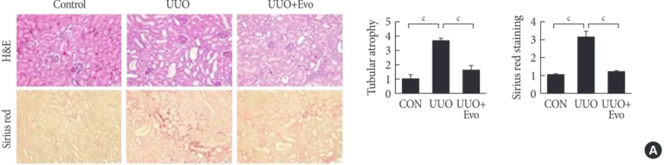
First, we investigated whether evogliptin ameliorates UUO-in- duced renal tubulointerstitial fibrosis by performing hematox- ylin and eosin (H&E) and Sirius red staining. On day 11 after UUO, UUO induces prominent renal tubular atrophy and tu- bulointerstitial fibrosis. However, evogliptin-treated kidneys showed significant attenuation of both these pathological fea- tures (Fig. 1A). To evaluate the mechanism by which evo- gliptin attenuates UUO-induced renal fibrosis and ECM accu- mulation, we examined the effects of evogliptin on TGF-β/
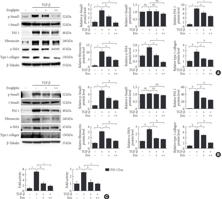
Smad3 signaling, which is a key mediator of renal fibrosis caused by UUO. Immunohistochemistry revealed that for phosphorylated Smad3, PAI-1, fibronectin, α-SMA, and type I collagen were markedly increased in the UUO kidneys, where- as these effects were ameliorated by evogliptin (Fig. 1B). These inhibitory effects of evogliptin on pro-fibrotic gene expression and ECM production were further evaluated by Western blot analysis. Consistent with the result of immunohistochemistry, the protein levels of phosphorylated Smad3, PAI-1, fibronec- tin, α-SMA, and type I collagen were lower in evogliptin-treat- ed UUO kidneys than in vehicle-treated kidneys (Fig. 1C). We have also observed weak tubulointerstitial fibrosis 7 days after UUO, which was partially but significantly reduced by evo- gliptin treatment (Supplementary Fig. 1). As shown in Supple- mentary Figs. 2 and 3, body weight and food intake decreased
Fig. 1. Effects of evogliptin (Evo) on renal fibrosis and pro-fibrotic gene expression in kidneys of unilateral ureteral obstruction (UUO) mice. A UUO kidney from a mouse at day 11. (A) Representative images of hematoxylin and eosin (H&E) and Sirius red staining of kidney tissue sections from control (CON) mice and UUO mice treated with (300 mg/kg) or without Evo. The number of atrophic tubules was determined by measuring the amount of abnormal irregular and dilated tubular basement membranes in H&E-stained sections under high power magnification (×200). Areas of positive staining with Sirius red were quantitated by computer-based morphometric analysis. All morphometric data were normalized against the corresponding val-ues in CON ani- mals. Data in all bar graphs are expressed as fold increases relative to the CON (n=6 in each group). (Continued to the next page)
54 32 10
4 3 2 1
Tubular atrophy Sirius red staining 0
CON UUOUUO+ CON UUO
Evo UUO+
Evo
c c c c
A
Control UUO UUO+Evo
H&ESirius red
Fig. 1. Continued. (B) Representative images of immunohistochemical staining for phosphorylated-Smad3 (p-Smad3), plasmino- gen activator inhibitor 1 (PAI-1), fibronectin, α-smooth muscle actin (α-SMA), and type I collagen in kidney tissue sections from CON mice or UUO mice treated with (300 mg/kg) or without Evo (n=6 in each group). Areas of positive staining with p-Smad3, PAI-1, fibronectin, α-SMA, and type I collagen antibodies were quantitated by computer-based morphometric analysis. Data are the mean±standard error of the mean (SEM) of five random fields from each kidney. (C) Representative western blot analysis of p-Smad3, total-Smad3 (t-Smad3), PAI-1, fibronectin, α-SMA, and type I collagen protein level in UUO kidneys from mice treated with (300 mg/kg) or without Evo (n=6 in each group). Data in the bar graphs are the mean±SEM. NS, not significant. aP<0.05,
bP<0.01, cP<0.001.
8 6 4 2 0
54 32 10
4 3 2 1 0
2.0 1.5 1.0 0.5 0
15 10 5 0
60 40 20 0
2.52.0 1.51.0 0.50
8 6 4 2 0
1.00.8 0.60.4 0.20
2.52.0 1.51.0 0.50
54 32 10 p-Smad3-positive stain areaFibronectin-positive stain areaType 1 collagen-positive stain areaRelative p-Smad3 protein levelRelative PAI-1 protein levelRelative α-SMA protein level PAI-1-positive stain areaα-SMA-positive stain areaRelative t-Smad3 protein levelRelative fibronectin protein levelRelative type 1 collagen protein level
CON
CON
CON
CON
CON
CON
CON
CON
CON
CON
CON UUO
UUO
UUO
UUO
UUO
UUO
UUO
UUO
UUO
UUO
UUO UUO+Evo
UUO+Evo
UUO+Evo
UUO+Evo
UUO+Evo
UUO+Evo
UUO+Evo
UUO+Evo
UUO+Evo
UUO+Evo
UUO+Evo
c
c
c
a
c
c
c
b
NS
b
a c
c
c
a
a
a
c
b
NS
b
a
B
C Control
Control
UUO
UUO
UUO+Evo
UUO+Evo
p-Smad3PAI-1Fibronectinα-SMAType 1 collagen
β-Tubulin 55 kDa
Type 1 collagen 130 kDa
α-SMA 45 kDa
Fibronectin 240 kDa
PAI-1 48 kDa
t-Smad3 52 kDa
p-Smad3 52 kDa
at 3 days after UUO but fully recovered at 7 days after UUO.
Blood glucose levels did not change after UUO or after evo- gliptin treatment in either the early or late time groups after UUO, suggesting that the anti-fibrotic effect of evogliptin was independent of its glucose-lowering effect. The kidney weights
did not differ between groups.
Evogliptin attenuates pro-fibrotic molecules via suppression of TGF-β/Smad3 signaling
To determine whether the anti-fibrotic effect of evogliptin is
Fig. 2. Effects of evogliptin (Evo) on transforming growth factor-β (TGF-β)-induced pro-fibrotic gene expression in cultured kid- ney cell lines. Representative Western blot analysis of phosphorylated-Smad3 (p-Smad3), total-Smad3 (t-Smad3), plasminogen ac- tivator inhibitor 1 (PAI-1), fibronectin, α-smooth muscle actin (α-SMA), and type I collagen protein level in TGF-β-stimulated (A) human proximal renal tubular epithelial (HK-2) cells and (B) normal rat kidney fibroblasts (NRK-49F) cells. Cells were incubated with TGF-β (5 ng/mL) treated with (100 or 200 μg/mL) or without Evo co-treatment for 24 hours. Data are the mean±standard er- ror of the mean (SEM) of three independent measurements. (C) Effects of Evo on PAI-1 promoter activity in NRK-49F and NRK- 52E cells. Cells were treated with TGF-β (5 ng/mL) treated with (100 μg/mL) or without Evo co-treatment for 24 hours. Data are the mean±SEM of three independent measurements. NS, not significant; Luc, luciferase. aP<0.05, bP<0.01, cP<0.001.
54 32 10
15 10 5 0
6 4 2 0
4 3 2 1 0
1.5 1.0 0.5 0
2.0 1.5 1.0 0.5 0
1.5 1.0 0.5 0
3 2 1 0
8 6 4 2 0
108 64 20
4 3 2 1 0
60 40 20 0
8 6 4 2 0
3 2 1 0
Relative p-Smad3 protein levelRelative fibronectin protein levelRelative p-Smad3 protein levelRelative fibronectin protein level Relative t-Smad3 protein levelRelative α-SMA protein levelRelative t-Smad3 protein levelRelative α-SMA protein level
Fold activity Relative PAI-1 protein levelRelative type 1 collagen protein levelRelative PAI-1 protein levelRelative type 1 collagen protein level
Fold activity
− + + +
− + + +
− + + +
− + + +
− + + +
− + + +
− + + +
− + + +
− + + +
− + + +
− + + +
− + + +
− + + +
− + + +
− − + ++
− − + ++
− − + ++
− − + ++
− − + ++
− − + ++
− − + ++
− − + ++
− − + ++
− − + ++
− − + ++
− − + ++
− − + ++
− − + ++
TGF-β
TGF-β
TGF-β
TGF-β
TGF-β
TGF-β
TGF-β
TGF-β
TGF-β
TGF-β
TGF-β
TGF-β
TGF-β
TGF-β
Evo
Evo
Evo
Evo
Evo
Evo
Evo
Evo
Evo
Evo
Evo
Evo
Evo
Evo
c
c
c
b
NS
a
NS
b
c
c
b
b
b
b
b
a
a
a
NS
a
NS
b
b
a
a
b
a
b
c
c
b
b
NS
c
NS
b
c
c
b
b
b
c
C
B A
β-Tubulin β-Tubulin
55 kDa 55 kDa
Type 1 collagen Type 1 collagen
130 kDa 130 kDa
α-SMA α-SMA
45 kDa 45 kDa
Fibronectin Fibronectin
240 kDa 240 kDa
PAI-1 PAI-1
48 kDa 48 kDa
t-Smad3 t-Smad3
52 kDa 52 kDa
p-Smad3 p-Smad3
Evogliptin Evogliptin
52 kDa 52 kDa
− − + ++
− − + ++
TGF-β TGF-β
PAI-1/Luc
mediated by effects on TGF-β-induced pro-fibrotic gene ex- pression, we investigated whether evogliptin suppresses TGF-β- induced Smad3 phosphorylation, PAI-1, fibronectin, α-SMA, and type I collagen abundance in cultured renal cells (human proximal renal tubular epithelial [HK-2] and normal rat kid- ney fibroblast [NRK-49F] cells). In accordance with the in vivo findings, evogliptin inhibited TGF-β-stimulated Smad3 phos- phorylation and upregulation of PAI-1, fibronectin, α-SMA, and type I collagen in renal tubular and fibroblast cells (Fig. 2A and B). We further examined whether evogliptin inhibits TGF-β/Smad3 signaling at the transcriptional levels by mea- suring PAI-1 luciferase activity, and, indeed, evogliptin treat- ment inhibited TGF-β-stimulated PAI-1 promoter activity in both NRK-49F and NRK-52E cells (Fig. 2C). These findings suggested that renoprotective effect of evogliptin is mediated by downregulation of Smad3 phosphorylation. Finally, we in- vestigated the effect of evogliptin on plasma and renal DPP-4 activity after UUO. We observed that renal DPP-4 activity was significantly increased by UUO and that treatment with evo- gliptin markedly suppressed DPP-4 activity, in agreement with our previous finding (Supplementary Fig. 4A) [10]. Plasma DPP-4 activity was not significantly different between control and UUO mice; however, evogliptin treatment markedly re- duced DPP-4 activity in UUO mice (Supplementary Fig. 4B).
DISCUSSION
This study was undertaken to address whether evogliptin di- rectly ameliorates renal fibrosis induced by UUO in mice and to elucidate the potential mechanism. Here, we show that evo- gliptin protects against renal fibrosis in this mouse model and that it inhibits TGF-β-stimulated Smad3 phosphorylation and ECM protein production in cultured renal cells.
TGF-β/Smad3 signaling is a crucial pathway in the patho- genesis of renal fibrosis [2,3]. Among Smad family, Smad3 is considered to be the principal regulator of the transcription of genes associated with renal fibrosis [13]. Upon its phosphory- lation and activation by TGF-β receptor, Smad3 transactivates collagen genes to induce synthesis of ECM components and inhibit matrix degradation [14]. Recent study has shown that sitagliptin improves renal fibrosis by suppressing TGF-β/
Smad3 signaling [15]. Alogliptin treatment of UUO mice had renoprotective effects through downregulation of the expres- sion of TGF-β mRNA and α-SMA [16]. Previously, we also found that gemigliptin improved renal fibrosis in streptozoto-
cin-induced diabetic mice by reducing TGF-β-stimulated Smad3 phosphorylation, which lowers the expression of ECM proteins, including type 1 collagen and fibronectin [17]. In ad- dition, it was previously demonstrated that DPP-4 plays a role in TGF-β-induced receptor hetero-dimerization and that TGF-β-induced formation of the TGFR1/2 heterodimer is suppressed by small interfering RNA (siRNA)-mediated inhi- bition of DPP-4 [18]. Moreover, vildagliptin and linagliptin were successful in lowering the renal TGF-β level in a strepto- zotocin-induced diabetic rat [19,20]. Although we did not in- vestigate the TGF-β level, the mechanisms described above could be, at least in part, responsible for evogliptin-mediated inhibition of TGF-β signaling. Therefore, further studies using Smad3-null transgenic mouse model are warranted to clarify the molecular mechanism responsible for evogliptin’s suppres- sive effect on TGF-β/Smad3 signaling. The present study pro- vides evidence that evogliptin has a protective effect on renal fibrosis by inhibiting TGF-β-stimulated Smad3 phosphoryla- tion and its downstream signaling.
In conclusion, we have shown that evogliptin can prevent re- nal fibrosis by inhibiting the TGF-β/Smad3 signaling pathway.
Our data suggest that evogliptin could be applied to prevent the progression of CKD or other etiologies, as well as DKD.
Therefore, the present study provides the rationale for further clinical trials to evaluate the therapeutic efficacy of evogliptin in patient with DKD.
SUPPLEMENTARY MATERIALS
Supplementary materials related to this article can be found online at https://doi.org/10.4093/dmj.2018.0271.
CONFLICTS OF INTEREST
No potential conflict of interest relevant to this article was re- ported.
AUTHOR CONTRIBUTIONS
Conception or design: M.J.K., N.Y.K., Y.K.C., K.G.P.
Acquisition, analysis, or interpretation of data: M.J.K., Y.A.J., S.H.L., G.S.J., J.G.K., I.K.L., S.L.
Drafting the work or revising: N.Y.K., Y.K.C., K.G.P.
Final approval of the manuscript: Y.K.C., K.G.P.
ORCID
Mi-Jin Kim https://orcid.org/0000-0001-5505-0513 Na-young Kim https://orcid.org/0000-0002-6132-4581 Yeon-Kyung Choi https://orcid.org/0000-0003-0996-6437 Keun-Gyu Park https://orcid.org/0000-0002-8403-1298
ACKNOWLEDGMENTS
This work was supported by the National Research Foundation of Korea (NRF) grants (NRF-2017M3A9G7073086, NRF- 2018R1A2A1A05077703 and NRF-2018R1A6A3A01011962) funded by the Ministry of Science and ICT, NRF grant (NRF- 2017R1A6A3A04010231) funded by the Ministry of Educa- tion, and grants (HI16C1501 and HI15C0001) from the Korea Health technology R&D Project through the Korea Health In- dustry Development Institute funded by the Ministry of Health and Welfare, Republic of Korea.
REFERENCES
1. Atkins RC, Zimmet P; ISN-IFKF World Kidney Day 2010 Steering Committee. Diabetes: diabetic kidney disease: act now or pay later. Nat Rev Nephrol 2010;6:134-6.
2. Meng XM, Nikolic-Paterson DJ, Lan HY. TGF-β: the master regulator of fibrosis. Nat Rev Nephrol 2016;12:325-38.
3. Lan HY, Chung AC. TGF-β/Smad signaling in kidney disease.
Semin Nephrol 2012;32:236-43.
4. Salvo F, Moore N, Arnaud M, Robinson P, Raschi E, De Ponti F, Begaud B, Pariente A. Addition of dipeptidyl peptidase-4 in- hibitors to sulphonylureas and risk of hypoglycaemia: system- atic review and meta-analysis. BMJ 2016;353:i2231.
5. Hirakawa H, Zempo H, Ogawa M, Watanabe R, Suzuki J, Aka- zawa H, Komuro I, Isobe M. A DPP-4 inhibitor suppresses fi- brosis and inflammation on experimental autoimmune myo- carditis in mice. PLoS One 2015;10:e0119360.
6. Kaji K, Yoshiji H, Ikenaka Y, Noguchi R, Aihara Y, Douhara A, Moriya K, Kawaratani H, Shirai Y, Yoshii J, Yanase K, Kitade M, Namisaki T, Fukui H. Dipeptidyl peptidase-4 inhibitor attenu- ates hepatic fibrosis via suppression of activated hepatic stellate cell in rats. J Gastroenterol 2014;49:481-91.
7. Suzuki T, Tada Y, Gladson S, Nishimura R, Shimomura I, Kara- sawa S, Tatsumi K, West J. Vildagliptin ameliorates pulmonary fibrosis in lipopolysaccharide-induced lung injury by inhibit- ing endothelial-to-mesenchymal transition. Respir Res 2017;
18:177.
8. Eun Lee J, Kim JE, Lee MH, Song HK, Ghee JY, Kang YS, Min HS, Kim HW, Cha JJ, Han JY, Han SY, Cha DR. DA-1229, a di- peptidyl peptidase IV inhibitor, protects against renal injury by preventing podocyte damage in an animal model of progres- sive renal injury. Lab Invest 2016;96:547-60.
9. Tan X, Hu J. Evogliptin: a new dipeptidyl peptidase inhibitor for the treatment of type 2 diabetes. Expert Opin Pharmacoth- er 2016;17:1285-93.
10. Min HS, Kim JE, Lee MH, Song HK, Kang YS, Lee MJ, Lee JE, Kim HW, Cha JJ, Chung YY, Hyun YY, Han JY, Cha DR. Di- peptidyl peptidase IV inhibitor protects against renal intersti- tial fibrosis in a mouse model of ureteral obstruction. Lab In- vest 2014;94:598-607.
11. Kim MK, Chae YN, Kim HD, Yang EK, Cho EJ, Choi SH, Cheong YH, Kim HS, Kim HJ, Jo YW, Son MH, Kim SH, Shin CY. DA-1229, a novel and potent DPP4 inhibitor, improves in- sulin resistance and delays the onset of diabetes. Life Sci 2012;
90:21-9.
12. Jung GS, Kim MK, Jung YA, Kim HS, Park IS, Min BH, Lee KU, Kim JG, Park KG, Lee IK. Clusterin attenuates the devel- opment of renal fibrosis. J Am Soc Nephrol 2012;23:73-85.
13. Iwano M, Plieth D, Danoff TM, Xue C, Okada H, Neilson EG.
Evidence that fibroblasts derive from epithelium during tissue fibrosis. J Clin Invest 2002;110:341-50.
14. Sato M, Muragaki Y, Saika S, Roberts AB, Ooshima A. Targeted disruption of TGF-beta1/Smad3 signaling protects against re- nal tubulointerstitial fibrosis induced by unilateral ureteral ob- struction. J Clin Invest 2003;112:1486-94.
15. Wang D, Zhang G, Chen X, Wei T, Liu C, Chen C, Gong Y, Wei Q. Sitagliptin ameliorates diabetic nephropathy by blocking TGF-β1/Smad signaling pathway. Int J Mol Med 2018;41:2784- 92.
16. Uchida T, Oda T, Matsubara H, Watanabe A, Takechi H, Oshi- ma N, Sakurai Y, Kumagai H. Renoprotective effects of a di- peptidyl peptidase 4 inhibitor in a mouse model of progressive renal fibrosis. Ren Fail 2017;39:340-9.
17. Jung GS, Jeon JH, Choe MS, Kim SW, Lee IK, Kim MK, Park KG. Renoprotective effect of gemigliptin, a dipeptidyl pepti- dase-4 inhibitor, in streptozotocin-induced type 1 diabetic mice. Diabetes Metab J 2016;40:211-21.
18. Shi S, Srivastava SP, Kanasaki M, He J, Kitada M, Nagai T, Nitta K, Takagi S, Kanasaki K, Koya D. Interactions of DPP-4 and in- tegrin β1 influences endothelial-to-mesenchymal transition.
Kidney Int 2015;88:479-89.
19. Liu WJ, Xie SH, Liu YN, Kim W, Jin HY, Park SK, Shao YM, Park TS. Dipeptidyl peptidase IV inhibitor attenuates kidney injury in streptozotocin-induced diabetic rats. J Pharmacol Exp Ther 2012;340:248-55.
20. Kanasaki K, Shi S, Kanasaki M, He J, Nagai T, Nakamura Y, Ish-
igaki Y, Kitada M, Srivastava SP, Koya D. Linagliptin-mediated DPP-4 inhibition ameliorates kidney fibrosis in streptozotocin- induced diabetic mice by inhibiting endothelial-to-mesenchy- mal transition in a therapeutic regimen. Diabetes 2014;63:2120- 31.