JCN
Open AccessCopyright © 2017 Korean Neurological Association 209
Reversible Corpus Callosal Lesions Associated with the Use of Adalimumab for Ulcerative Colitis
Dear Editor,
A reversible corpus callosum lesion is a unique phenomenon with an unclear pathophysi- ology. Various etiologies may underlie the lesion, including toxins, encephalitis, influenza A, metabolic disorders such as hypernatremia, hypoglycemia, and vitamin deficiency disorders.1 In rare cases the lesion is caused by immunomodulating therapies.2 We report a patient with a reversible corpus callosum lesion associated with adalimumab treatment for ulcerative colitis (UC).
A 43-year-old man who had been diagnosed with UC 15 years previously was admitted due to an acute inflammatory lesion in his left inguinal area accompanied by abrupt-onset dysarthria. He had been managing the UC with naturopathy and regular colonoscopy check- ups, and had not experienced a serious flare-up of the disease since its diagnosis. About 8 weeks before admission he had taken adalimumab for the first time to treat UC. The drug was administered at 40 mg for 2 days and then repeated 2 weeks later. He refused further adali- mumab treatment due to severe insomnia related to the drug.
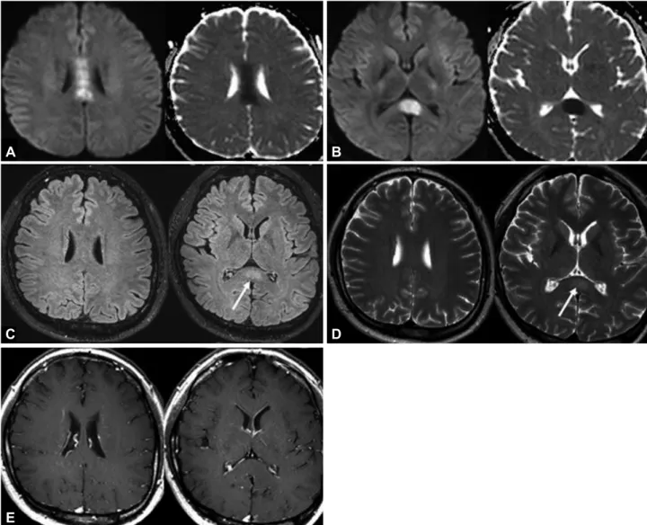
On admission, the patient was in a mildly confused state with definite dysarthria. His blood pressure was 148/89 mm Hg with a heart rate of 141 bpm and body temperature of 40.3°C. Laboratory tests revealed leukocytosis with elevated procalcitonin (PCT) (4.21 ng/mL) and C-reactive protein (CRP) (246.35 mg/L). The findings of other laboratory examinations, including liver function and electrolyte levels, were normal. Diffusion-weighted imaging (DWI) performed at admission showed a focal, well-defined lesion with restricted diffusion in the corpus callosum body and splenium (Fig. 1). The cerebrospinal fluid was normal. The patient was treated with empirical antibiotics due to an impression of sepsis caused by in- fection in the inguinal area. All of his symptoms improved dramatically within 4 days after admission. Magnetic resonance imaging performed 8 days after the initial DWI showed a markedly reduced lesion size without any regional enhancement. Evaluations of the inguinal lesion including a blood culture study all produced normal findings, and the patient was discharged without any symptoms.
The patient had no causative factors associated with a reversible corpus callosum lesion other than adalimumab treatment. Adalimumab is a recombinant human IgG1k anti-tumor necrosis factor (TNF)-α monoclonal antibody that is frequently used in the treatment of autoimmune-mediated inflammatory disorders. For many years it has been considered to be an effective treatment option for UC with fewer adverse effects. However, there have been some reports of CNS complications related to TNF-α inhibitors.2 In vitro studies have sug- gested that TNF-α inhibitors themselves can increase the permeability of the blood-brain barrier.3,4 TNF-α inhibitors share a common mechanism of action, but certain differences be- tween specific drugs such as in their molecular structures and various pharmacologic char- acteristics may cause adverse manifestations.5 One of the possible pathomechanisms is in- flammation by TNF-inhibitor-induced disruption of the balance between effector and Jin-Woo Park
Hung-Yuol Seok Yoohwan Kim Byung-Jo Kim
Department of Neurology, Korea University College of Medicine, Korea University Anam Hospital, Seoul, Korea
pISSN 1738-6586 / eISSN 2005-5013 / J Clin Neurol 2017;13(2):209-211 / https://doi.org/10.3988/jcn.2017.13.2.209
Received December 9, 2016 Revised February 10, 2017 Accepted February 13, 2017 Correspondence Byung-Jo Kim, MD, PhD Department of Neurology,
Korea University College of Medicine, 73 Inchon-ro, Seongbuk-gu, Seoul 02841, Korea Tel +82-2-920-6619 Fax +82-2-925-2472 E-mail [email protected]
cc This is an Open Access article distributed under the terms of the Creative Commons Attribution Non-Com- mercial License (http://creativecommons.org/licenses/by-nc/4.0) which permits unrestricted non-commercial use, distribution, and reproduction in any medium, provided the original work is properly cited.
LETTER TO THE EDITOR
210 J Clin Neurol 2017;13(2):209-211
Reversible Corpus Callosal Lesions Associated with Adalimumab
JCN
regulatory T cells.6
The patient’s inguinal cellulitis was one indication that in- flammation had caused the corpus callosum lesion. The pres- ence of a high fever with elevated PCT and CRP suggested infection due to the decreased immunity associated with the use of adalimumab. However, his symptoms and signs were dramatically improved after receiving antibiotics for only a few days. Blood cultures did not reveal any causative patho- gen. Although PCT can be useful in differentiating bacterial infections, it can also be elevated in autoimmune-related dis- orders. A direct inflammatory reaction induced by the in- creased disease activity of UC was also considered, because treatment was not continued after adalimumab was stopped.
However, there were no symptoms and signs suggesting flare- up of UC.
The time interval of 6 weeks before developing symptoms
after terminating adalimumab treatment may be related to its long half-life of 2–3 weeks, which is the longest half-life of the TNF-α inhibitors. Delayed hypersensitivity may occur as an adverse drug event up to 3 weeks after exposure, but a lon- ger interval is possible since it takes longer than 2 months to eliminate more than 95% of the drug from the body. The rapid improvement of neurologic symptoms in the present patient might have been related to a low drug concentration result- ing from its short-term use. There are also previous reports of a very good prognosis for neurologic complications induced by a TNF-α inhibitor.2,6
The present case indicates that TNF-α inhibitors including adalimumab may cause delayed adverse reactions such as a reversible corpus callosum lesion.
Fig. 1. Diffusion-weighted imaging revealed an ovoid-shaped lesion with restricted diffusion in the corpus callosum and splenium on the day of admission (A and B). Follow-up FLAIR and T2-weighted images (C-E) showed only a subtle remnant of the lesion in the splenium (white arrows).
There was no enhancement of the lesion on gadolinium-enhanced T1-weighted images. FLAIR: fluid attenuated inversion recovery.
A
C
E
B
D
www.thejcn.com 211
Park JW et al.
JCN
Conflicts of Interest
The authors have no financial conflicts of interest.
REFERENCES
1. Liu WM, Lin CH. A reversible stroke-like splenial lesion in viral en- cephalopathy. Acta Neurol Taiwan 2013;22:117-121.
2. Andreadou E, Kemanetzoglou E, Brokalaki Ch, Evangelopoulos ME, Kilidireas C, Rombos A, et al. Demyelinating disease following anti- TNFa treatment: a causal or coincidental association? Report of four cases and review of the literature. Case Rep Neurol Med 2013;2013:
671935.
3. Wong D, Dorovini-Zis K, Vincent SR. Cytokines, nitric oxide, and
cGMP modulate the permeability of an in vitro model of the human blood-brain barrier. Exp Neurol 2004;190:446-455.
4. Bellesi M, Logullo F, Di Bella P, Provinciali L. CNS demyelination during anti-tumor necrosis factor alpha therapy. J Neurol 2006;253:
668-669.
5. Ternant D, Bejan-Angoulvant T, Passot C, Mulleman D, Paintaud G.
Clinical pharmacokinetics and pharmacodynamics of monoclonal antibodies approved to treat rheumatoid arthritis. Clin Pharmacokinet 2015;54:1107-1123.
6. Ali F, Laughlin RS. Asymptomatic CNS demyelination related to TNF-α inhibitor therapy. Neurol Neuroimmunol Neuroinflamm 2016;
4:e298.