JCN
Open AccessCopyright © 2017 Korean Neurological Association 109
Real-Time Detection of Cerebral Artery Rebleeding by Transcranial Doppler Ultrasound: Hemodynamic Changes and Response to Treatment
Dear Editor,
Aneurysmal rupture and subsequent subarachnoid hemorrhage (SAH) lead to multiple complications including vasospasm, delayed cerebral ischemia, cerebral edema, and cerebral herniation.1-3 Transcranial Doppler ultrasound (TCD) is a noninvasive method for evaluat- ing the blood flow velocities in intracranial vessels and the presence of vasospasm.3 An ele- vated intracranial pressure (ICP) leads to decreased mean and diastolic blood flow veloci- ties and increased pulsatility index (PI), which can also be monitored in patients with SAH.3 We present a case report of a patient with SAH and a clipped aneurysm who rebled while undergoing a TCD study, and provide unique images, vitals graphs, and a video of the TCD study before and during the bleeding as well as after applying emergency treatment.
A 68-year-old Caucasian man presented for the evaluation of sudden-onset headache and neck pain. His blood pressure (BP) was initially elevated (240/117 mm Hg) and he had ex- perienced a tonic-clonic seizure for which he received fosphenytoin and was subsequently intubated and started on a propofol drip. Brain computed tomography (CT) revealed SAH.
Brain CT angiography showed an irregular anterior communicating artery aneurysm (2.5×
2×1.5 mm) and increased hemorrhage within the ventricles, septum pellucidum, and corpus callosum (Supplementary Fig. 1 in the online-only Data Supplement).
An attempt to coil the aneurysm was unsuccessful, and so the patient underwent right frontotemporal craniotomy, clipping of the aneurysm, and placement of a ventriculostomy catheter (Supplementary Fig. 2 in the online-only Data Supplement). After surgery he was admitted to the neurointensive care unit (NICU), where an interval decrease in the sub- arachnoid blood was noticed in follow-up brain CT. Postoperatively he remained intubated, but was awake and followed commands briskly, and the results of neurological examination were normal except or a slight drift in the right upper extremity.
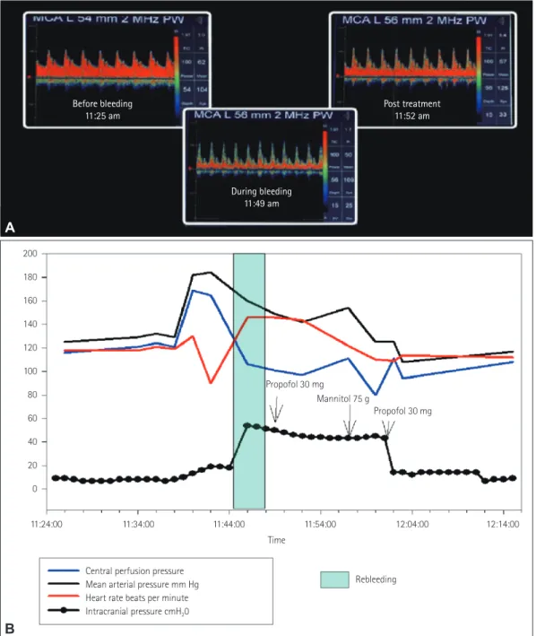
The patient exhibited rebleeding at 8 days after the index event, secondary to clip disloca- tion (Supplementary Fig. 3 in the online-only Data Supplement). TCD waveforms were cap- tured before and during the bleeding as well as after emergency treatment (Fig. 1) (Supple- mentary video 1 in the online-only Data Supplement) in parallel with NICU multimodal monitoring that captured minute-by-minute changes in the vital signs (Fig. 1). PI increased markedly during aneurysmal rebleeding (PI=1.7) compared to before the rebleeding (PI=
1.0) and following rebleeding treatment (PI=1.4) with propofol (two doses of 30 mg) and mannitol (75 g). The patient remained comatose (score on the Glasgow Coma Scale of 4 out of 15) throughout the following few weeks, and the family decided to withdraw life-support treatment at 40 days after admission.
To the best of our knowledge, this is the first case in which the rebleeding of a cerebral an- eurysm was detected in real time using TCD monitoring. This report highlights the poten- Eric J. Marrottea
Panayiotis Mitsiasa Leonard Melvina Asim Mahmoodb Georgios Tsivgoulisc Panayiotis Varelasa,b
a Departments of Neurology and
b Neurosurgery, Henry Ford Hospital, Detroit, MI, USA
c Second Department of Neurology, School of Medicine, University of Athens, Athens, Greece
pISSN 1738-6586 / eISSN 2005-5013 / J Clin Neurol 2017;13(1):109-111 / https://doi.org/10.3988/jcn.2017.13.1.109
Received April 28, 2016 Revised June 13, 2016 Accepted June 16, 2016 Correspondence Georgios Tsivgoulis, MD Second Department of Neurology, School of Medicine, University of Athens, Iras 39, Gerakas Attikis, Athens 15344, Greece
Tel +30 6937178635 Fax +30 2105832471
E-mail tsivgoulisgiorg@yahoo.gr
cc This is an Open Access article distributed under the terms of the Creative Commons Attribution Non-Com- mercial License (http://creativecommons.org/licenses/by-nc/3.0) which permits unrestricted non-commercial use, distribution, and reproduction in any medium, provided the original work is properly cited.
LETTER TO THE EDITOR
110 J Clin Neurol 2017;13(1):109-111
Aneurysm Rebleeding on TCD
JCN
tial of TCD [coupled with multimodal monitoring of the ce- rebral perfusion pressure (CPP), ICP, and vital signs] in documenting hemodynamic changes occurring during clip failure and rebleeding in realtime. It is noteworthy that in ad- dition to the elevation of the mean arterial pressure and CPP, a PI increase preceded the ICP surge, suggesting that hyper- tension caused the dislodgment of the clip and rebleeding.
These observations are consistent with the results of animal studies suggesting that BP elevation triggers rather than fol- lows aneurysm rebleeding.4 The association between high BP and aneurysm rupture is unclear. Although there are reports of patients who rebleed having higher mean BPs,5 a decrease in the rebleeding rate by tight BP control has not been demon- strated definitively. Moreover, several authors have suggested
Fig. 1. A: TCD waveforms before, during the bleed and post treatment (A and B left middle cerebral artery). Prior to bleed: ICP was 8 mm Hg, left MCA flow velocity was 61 cm/sec and PI was 1.0. During bleed: ICP increased to 54 mm Hg and PI to 1.7, with the waveform showing a narrow peak and decreased diastolic and mean velocity. 75 g of mannitol was given and the ventriculostomy was opened to drain. Within 5 minutes the ICP de- creased to 14, PI improved to 1.4, the waveform widened and the velocities returned to previous levels. Please also see supplemental video. B: Teleme- try data, mean arterial pressure increased and cerebral perfusion pressure increased before the rebleed. During rebleed there was a trend for ICP eleva- tion (up to 54 mm Hg, but with poor waveform). After therapy with propofol and mannitol all normalized. ICP: intracranial pressure, MCA: middle cerebral artery, PI: pulsatility index, TCD: Transcranial Doppler ultrasound.
A
B
200 180 160 140 120 100 80 60 40 20 0
Propofol 30 mg
Mannitol 75 g
Propofol 30 mg
11:24:00 11:34:00 11:44:00 11:54:00 12:04:00
Rebleeding During bleeding
11:49 am Before bleeding
11:25 am Post treatment
11:52 am
Central perfusion pressure Mean arterial pressure mm Hg Heart rate beats per minute Intracranial pressure cmH20
12:14:00 Time
www.thejcn.com 111
Marrotte EJ et al.
JCN
that rebleeding could be induced by variations or changes in BP rather than the absolute BP level.6 A very recent case—
control study found that systolic BP variability was associated with increased rates of acute aneurysmal rebleeding, emerg- ing thus as a novel and independent risk factor for rebleeding after SAH.7
The present unique case underscores the diagnostic utility of TCD in offering real-time assessment of the hemodynam- ics and ICP changes during SAH. TCD combined with other imaging modalities such as multimodal magnetic resonance imaging may further enhance our understanding of the patho- physiology of SAH.8 We have also provided evidence that BP elevation leading to an increased PI is the cause of aneurysmal rebleeding, and argue against the hypothesis that a BP increase documented in patients with recurrent SAH is secondary to aneurysmal rerupture. Τhe association between BP control and the risk of aneurysm rebleeding should be evaluated in future randomized clinical trials.
Supplemental Video Legend
Transcranial Doppler ultrasound recordings in left M1 mid- dle cerebral artery before, during aneurysmal rerupture and post elevated intracranial pressure (ICP) treatment. Note the high resistance flow during aneurysmal rerupture in com- parison to normal, low-resistance before the rebleeding of the aneurysm. Also, note the decrease in PI and the increase in diastolic flow following the control of elevated ICP levels complicating the rebleeding of the aneurysm.
Supplementary Materials
The online-only Data Supplement is available with this arti-
cle at https://doi.org/10.3988/jcn.2017.13.1.109.
Conflicts of Interest
The authors have no financial conflicts of interest.
REFERENCES
1. Fugate JE, Rabinstein AA. Intensive care unit management of aneu- rysmal subarachnoid hemorrhage. Curr Neurol Neurosci Rep 2012;
12:1-9.
2. Rowland MJ, Hadjipavlou G, Kelly M, Westbrook J, Pattinson KT.
Delayed cerebral ischaemia after subarachnoid haemorrhage: look- ing beyond vasospasm. Br J Anaesth 2012;109:315-329.
3. Alexandrov AV, Sloan MA, Tegeler CH, Newell DN, Lumsden A, Ga- rami Z, et al. Practice standards for transcranial Doppler (TCD) ul- trasound. Part II. Clinical indications and expected outcomes. J Neu- roimaging 2012;22:215-224.
4. Plesnila N. Pathophysiological role of global cerebral ischemia fol- lowing subarachnoid hemorrhage: the current experimental evi- dence. Stroke Res Treat 2013;2013:651958.
5. Ohkuma H, Tsurutani H, Suzuki S. Incidence and significance of early aneurysmal rebleeding before neurosurgical or neurological manage- ment. Stroke 2001;32:1176-1180.
6. Bederson JB, Connolly ES Jr, Batjer HH, Dacey RG, Dion JE, Diringer MN, et al. Guidelines for the management of aneurysmal subarach- noid hemorrhage: a statement for healthcare professionals from a special writing group of the Stroke Council, American Heart Associ- ation. Stroke 2009;40:994-1025.
7. Lin QS, Ping-Chen, Lin YX, Lin ZY, Yu LH, Dai LS, et al. Systolic blood pressure variability is a novel risk factor for rebleeding in acute subarachnoid hemorrhage: a case-control study. Medicine (Baltimore) 2016;95:e3028.
8. Sun Y, Shen Q, Watts LT, Muir ER, Huang S, Yang GY, et al. Multi- modal MRI characterization of experimental subarachnoid hemor- rhage. Neuroscience 2016;316:53-62.