Original Article
원고 접수일 2013년 2월 27일, 원고 수정일 2013년 4월 5일, 게재 확정일 2013년 7월 25일
책임저자 이정근
(443-380) 수원시 영통구 월드컵로 164, 아주대학교 의과대학 치과학교실 Tel: 031-219-5328, Fax: 031-219-5329, E-mail: [email protected]
RECEIVED February 27, 2013, REVISED April 5, 2013, ACCEPTED July 25, 2013
Correspondence to Jeong Keun Lee
Department of Dentistry, Ajou University School of Medicine 164 WorldCup-ro, Yeongtong-gu, Suwon 443-380, Korea
Tel: 82-31-219-5328, Fax: 82-31-219-5329, E-mail: [email protected]
CC This is an open access article distributed under the terms of the Creative Commons Attribution Non-Commercial License (http://creativecommons.org/licenses/
by-nc/3.0) which permits unrestricted non-commercial use, distribution, and reproduction in any medium, provided the original work is properly cited.
자가치아 뼈 이식재의 유용성에 관한 임상적 연구
한민우ㆍ이정근
아주대학교 의과대학 치과학교실 구강악안면외과
Abstract
Clinical Study on the Efficacy of the Autogenous Tooth Bone Graft Material (AutoBT)
Min Woo Han, Jeong Keun Lee
Division of Oral and Maxillofacial Surgery, Department of Dentistry, Ajou University School of Medicine
Purpose: This study is to evaluate the efficacy of the autogenous tooth bone graft material, clinically and radiologically, as related to implant installation.
Methods: In oral and maxillofacial surgery department of Ajou University Hospital, guided bone regeneration (GBR), implant placement combined with GBR, sinus graft, implant placement combined with sinus graft, and defect filling were performed in 46 patients, using autogenous tooth bone. Among these, 66 implants were inserted with autogenous tooth bone. Implant stability quotient (ISQ) was measured by Osstell Mentor (Integration Diagnostics, Goteborg, Sweden) on 39 implants on the operation date and 4 months later, and on 21 implants 9months on the average at the final setting of restoration. Twenty-eight implants with GBR and sinus graft (GBR group: n=14, sinus graft group: n=14) were evaluated radiologically to measure the resorption of grafted autogenous tooth bone after loading.
Results: The average initial stabilization of the installed implants was 67 ISQ, and the average secondary stabilization at 4 months later was 76. The average bone loss of GBR group as measured 8.0 months after application of prosthesis loading was 0.29 mm and the average bone loss of the sinus graft group as measured 7.6 months after application of prosthesis loading was 0.66 mm, respectively. In the histological assessment, formation of the new bone and continuous trabecular bone pattern was identified around autogenous tooth bone.
Conclusion: Based on these results, we concluded that autogenous tooth bone is an excellent bone graft material that can substitute the autogenous bone.
Key words: Autogenous tooth bone graft material, Efficacy, Dental implant
Table 1. Type of surgery using autogenous tooth bone graft
materialType of surgery Cases
GBR only
GBR with implantation Sinus graft only
Sinus graft with implantation Implantation after bone graft Defect filling
8 34 3 19 13 2 GBR, guided bone regeneration.
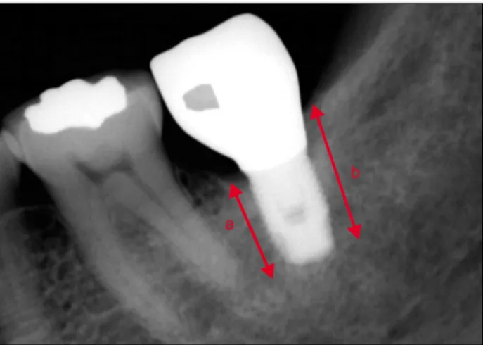
Fig. 1. Radiologic evaluation of GBR group. Bone height was esti-
mated right after implant installation and at the final diagnosis after occlusal loading. The estimated bone height was determined from the mean value of the bone height on both medial (a) and distal (b) surface. GBR, guided bone regeneration.서 론
최근 치조골결손부의 골이식에 자가골, 동종골, 이종골 및 합성 골 등의 골이식재가 사용되고 있다. 이 중 자가골 이식재는 골 결손부의 재건에 있어 가장 이상적인 재료로 알려져 있다. 이는 골형성, 골전도, 골유도 능력을 지녀 빠른 치유를 가능하게 하며, 다른 면역반응을 유발하지 않기 때문이다. 하지만 자가골은 충분 한 양을 채득하기가 어려우며, 공여부의 합병증 가능성이 높고 이식 후 흡수가 많이 발생하는 문제점이 있다. 이러한 문제점을 극복하기 위해 동종골, 이종골, 그리고 합성골 이식재와 같은 다른 골 대체물들이 개발되었으며, 임상에서 다양하게 사용되고 있다. 그러나 동종골, 이종골 이식재는 감염전파의 위험성 및 비싼 가격 등이 문제가 될 수 있고, 합성골은 가격이 저렴하면서 감염전파의 위험성이 전혀 없지만 골형성, 골유도 능력이 결여되 어 생활력 있는 골조직 형성 시 한계가 있다.
이상적인 골이식재는 면역반응이 없고 골생성을 유도할 수 있는 인자들이 많이 포함되어 빠른 골생성 및 재혈관화가 가능해 야 되며, 신생골 침투가 이루어질 수 있도록 공간을 유지할 수 있어야 한다. 또한 골이식재와 임플란트의 골유착이 이루어져야 하며 임플란트 상부 보철물 부하 후에도 골소실 없이 안정적으로 유지되어야 한다. 그 외에도 채취가 용이하여 임상적으로 쉽게 이용 가능하여야 하고 경제적인 이점이 있어야 한다. 이러한 골대 체 재료로서 최근 자기치아 뼈 이식재가 개발되어 기대를 모으고 있다.
자가치아 뼈 이식재는 유기질과 무기질을 모두 함유하고 있고, 다른 골이식재에 비해 치밀골과 가장 유사한 구조 및 물리화학적 특징을 지니고 있어 높은 골재생능력을 보인다. 또 환자 본인의 조직을 이용하여 제작하므로 감염의 전파 위험성이 없으며, 수여 부와 공여부가 동일한 유전자 코드로 이루어져 면역 반응이 일어 나지 않는다. 통상의 발치 술식으로 치아를 채득할 수 있어 임상의 가 쉽게 적용할 수 있으며, 공여부의 합병증 이환을 줄일 수 있는 장점도 있다.
본 연구는 아주대학교병원에서 자가치아 뼈 이식재를 이용한 결손부 이식, 임플란트 주위 골유도재생술 및 상악동 거상술 등의 증례를 보고하고, 임상적, 방사선학적, 조직학적 평가를 통하여
자가치아 뼈 이식재의 임상 적용 시 유용성에 대해 보고하고자 시행하였다.
연구방법
1. 연구 대상
본 연구는 2010년 4월부터 2013년 2월 사이에 아주대학교병원 치과에서 발치 후 자가치아 뼈 이식재를 이용하여 골이식을 시행 한 46명(남자 32명, 여자 14명)의 환자를 대상으로 시행하였다.
환자들의 나이는 19세부터 66세까지 평균 45세였다. 46명의 환자 에서 자가치아 뼈 이식재를 이용하여 골유도재생술만 시행한 증례 8예, 골유도재생술과 동시에 임플란트를 식립한 증례 34예, 상악 동골이식술만 시행한 증례 3예, 상악동골이식술과 동시에 임플란 트를 식립한 증례 19예, 골이식술 후 임플란트를 식립한 증례 13예, 결손부 충전 이식술 2예 등 다양하게 임상에서 적용하였다 (Table 1). 사용한 자가치아 뼈 이식재의 형태는 골유도재생술에 쓰인 1예에서만 블록형이 사용되었으며 나머지 증례에서는 분말 형(powder)이 사용되었다.
2. 연구방법
자가치아 뼈 이식재를 이용하여 골유도재생술, 상악동골이식
술을 시행한 부위의 식립된 임플란트를 대상으로 술 중 및 술
후 합병증, 임플란트의 성공 유무, 임플란트의 안정성, 보철물
부하 후의 임플란트 주위 골 흡수량, 이식된 부위의 조직학적
소견 등을 평가하였다.
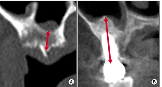
Fig. 2. Radiologic evaluation of sinus
graft group. (A) Pre-operative The estimated bone height was de- termined by the distance between the top of the alveolar crest and the sinus floor before implant installation. (B) Seven months after loading. The esti- mated bone height was determined by the distance between the top of the implant and the bottom of the grafted autogenous tooth bone at the final diagnosis after occlusal loading.Fig. 3. ISQ evaluation. ISQ, implant stability quotient; POD,
post-operative day; GBR, guided bone regeneration.1) 임상적 평가
식립된 임플란트의 술 후의 감염, 혈종, 임플란트 골유착 실패 등의 합병증을 평가하였다. 또한 식립된 임플란트의 수술 직후, 수술 4개월 후 및 보철물 장착 시 Osstell Mentor (Integration Diagnostics, Goteborg, Sweden)를 이용하여 측정한 implant stability quotient (ISQ) 수치를 비교함으로써 임플란트의 고정 정도를 평가하였다.
2) 방사선학적 평가
(1) 골유도재생술 군: 수술 직후 및 보철물 부하 후 최종 경과관 찰 시점에 촬영된 치근단 방사선사진을 이용하여 임플란트 상단부 치조골에서 임플란트 근단부까지의 거리를 측정하여 비교하였다.
치조골 흡수량은 임플란트 근, 원심을 모두 측정하여 평균값으로 결정하였으며, 식립된 임플란트의 길이를 기준으로 방사선사진의 확대율을 계산하였다(Fig. 1).
(2) 상악동골이식 군: 술 전, 수술 직후 및 보철물 부하 후 최종 경과관찰 시점에 촬영된 컴퓨터단층촬영 사진 또는 파노라마 사진을 이용하여 치조골 상단에서 상악동 기저부까지의 거리 및 임플란트 상단에서 상악동 내 이식된 자가치아 뼈 이식재 최상방 부위까지의 거리를 비교하여 상악동골이식 후 증대량과 보철물 부하 후 흡수량을 평가하였다. 식립된 임플란트의 길이를 기준으 로 방사선사진의 확대율을 계산하였다(Fig. 2).
3) 조직학적 평가
치조골 골결손부위의 골유도재생술을 먼저 시행한 2 증례에서 4개월 후 2 mm 트레핀 버를 이용하여 형성된 골을 채득하였다.
채취한 시편은 즉시 10% 포르말린 용액에 24시간 고정한 후 포름산을 이용하여 탈회시켰다. 시편을 에탄올로 탈수시키고 파라 핀에 포매하여 표본을 제작하고, 헤마톡실린-에오신(hematoxylin- eosin)으로 염색 후, 광학현미경 Olympus BX-51 (Olympus
Co., Tokyo, Japan)을 이용하여 100배의 배율로 확대하여 관찰하 였다.
결 과
1. 임상적 평가
평가된 46명의 환자에서 자가치아 뼈 이식재와 함께 식립된
임플란트는 총 66개였으며 이 중 39개의 임플란트에서 수술 직후
및 수술 4개월 후의 ISQ 수치를 비교하였다. 수술 직후의 초기고
정은 평균 67 ISQ였으며, 수술 4개월 후 이차고정은 평균 76
ISQ 값으로 증가하는 양상을 보였다. 그 중 21개의 임플란트에서
수술 평균 9개월 후 보철물 장착 시 ISQ 값을 측정하였으며 평균
78 ISQ 값을 보여 안정적으로 골유착이 되었음을 확인할 수 있었
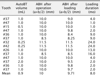
Table 2. Radiologic evaluation of GBR group
ToothAutoBT volume
(mL)
ABH after operation (a+b/2) (mm)
ABH after loading (a+b/2) (mm)
Loading duration
(mo)
#37
#47
#15
#47
#36
#37
#32
#42
#26
#37
#47
#37
#36
#37 Mean
1.0 1.0 0.5 1.0 1.0 1.0 0.25 0.25 1.0 1.0 1.0 2.0 1.0 1.0 0.9
10.0 10.0 10.0 10.0 10.0 8.5 11.5 11.5 10.0 8.5 10.0 10.0 10.0 10.0 10
9.0 10.0 10.0 9.8 8.4 8.25 11.5 11.5 10.0 8.5 10.0 9.5 9.8 9.7 9.71
4.0 1.0 1.0 2.0 9.0 9.0 24.0 24.0 13.0 16.0 4.0 2.0 2.0 2.0 8.0 GBR, guided bone regeneration; ABH, alveolar bone height.
Fig. 4. Radiologic evaluation of GBR group. GBR, guided bone
regeneration; OP, operative.Fig. 5. Radiologic evaluation of sinus graft group. Pre-op, pre-
operative; Post-op, post-operative.Table 3. Radiologic evaluation of sinus graft group
ToothABH before operation
(mm)
ABH after operation
(mm)
ABH after loading
(mm)
Loading duration
(mo)
#25
#26
#27
#17
#26
#27
#16
#15
#16
#26
#27
#15
#16
#17 Mean
14.2 7.5 8.5 6.4 4.0 7.0 5.6 9.0 8.0 8.0 5.8 6.0 4.5 5.6 7.15
16.5 14.5 15.5 15.0 17.5 12.5 12.8 16.0 20.0 21.0 17.9 14.6 18.6 17.4 16.41
16.2 12.0 14.2 14.0 16.5 12.3 12.7 16.0 19.0 20.6 17.5 14.0 18.4 17.2 15.75
12.0 12.0 12.0 1.0 7.0 7.0 1.0 13.0 13.0 5.0 6.0 6.0 6.0 6.0 7.6 ABH, alveolar bone height.
다(Fig. 3). 술 후 1 증례에서 골유착의 실패를 보여 한 단계 큰 크기의 임플란트로 재식립을 시행하였고 현재 안정적으로 유지 되고 있다. 그 외의 감염, 혈종, 창상열개 등 특이할 만한 합병증은 발생하지 않았다.
2. 방사선학적 평가 1) 골유도재생술군
14개의 임플란트에서 평균 0.9 mL의 자가치아 뼈 이식재를 사용하였고, 수술 직후 및 보철물 부하 후의 치근단 방사선사진을 평가한 결과, 평균 8.0개월 보철물 부하 후 평균 0.29 mm의 치조정 골흡수량을 보였다(Table 2, Fig. 4).
2) 상악동골이식술 군
14개의 임플란트에서 술 전 및 수술 직후의 상악동골 증대량을 컴퓨터 단층촬영사진에서 측정한 결과 평균 7.15 mm에서 16.41 mm로 평균 9.26 mm의 증대량을 보였고, 평균 7.6개월 보철물
부하 후 평균 0.66 mm의 골흡수량을 보였다(Table 3, Fig. 5).
3. 조직학적 평가
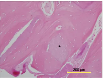
채취한 2개의 시편에서 자가치아 뼈 이식재 주변으로 연속적인 골소주 양상과 신생골의 형성이 관찰되었으며, 100배 확대된 고 배율 사진에서 이식재 주변으로 조골세포의 분포가 확인되었다 (Fig. 6).
고 찰
골이식재료는 골형성, 골유도 및 골전도 능력이 요구되며 자가
골은 3가지 능력을 모두 지니고 있으므로 가장 이상적이라고 할
수 있다. 그러나 공여부에 또 다른 수술이 필요하며 충분한 양을
채득하는 데 한계가 있다. 이러한 단점을 극복하기 위해 동종골,
이종골, 합성골이 소개되었으나 동종골, 이종골은 자가골에 비해
골형성능력이 낮고 교차감염 및 이물반응 등의 우려가 있어 술자
Fig. 6. Optical microscopic view (H&E stain, ×100). Newly formed
bone is identified around autogenous tooth bone graft material.*AutoBT.
의 선호도가 그리 높지 않다. 합성골은 채득량의 한계가 없고 가격이 저렴하며 질병 전염의 위험성이 없으나, 골전도의 기능만 을 가지고 있어 골형성능력이 현저히 떨어지게 된다. 따라서 자가 골의 장점을 지니면서 자가골의 단점을 극복할 수 있는 새로운 골이식 대체물질이 필요하며, 이에 대한 대안으로 최근 자가치아 뼈 이식재가 개발되어 사용되고 있다.
1993년 이후로 치아회분말(toothash)에 관한 임상연구들이 많이 보고되었고[1-4], 치과 임상에서 다양하게 적용할 수 있는 근거가 확보되었다[5]. Kim 등[6]은 10명의 환자에서 낭종적출술 후 발생한 결손부에 치아회분말과 연석고를 혼합한 골이식재료를 적용하여 임상적으로 성공적인 결과를 얻었다. Ku 등[7]은 치아회 분말과 연석고를 혼합한 골이식재료를 개의 치조골 결손부에 적용 시켰고 대조군과 비교 시 우수한 신생골의 형성을 관찰하였으며, Kim 등[5]은 자가치아를 이용하여 뼈이식재를 제조 시 우수한 골형성능력을 보일 수 있음을 보고하였다. 이를 바탕으로 자가치 아를 이용한 뼈 이식재가 개발되었으며, Kim 등[8]은 자가치아 뼈 이식재를 면역반응 및 이물반응이 없고 우수한 골형성능과 골유도능을 지닌 새로운 골이식재로 보고하였다.
치아의 법랑질, 상아질, 백악질 및 치주인대 등에는 골형성에 관여하는 인자가 다수 포함되어 있다는 연구들이 많다. Kim 등[9]
은 치아의 유기질 성분 중 type I collagen이 대부분을 차지하며 이는 골의 석회화 과정에 중요한 역할을 한다고 하였다. Ike와 Urist[10]는 상아질에 포함된 bone morphogenetic protein (BMP)-2가 골형성에 중요한 골유도 효과를 보임을 증명하였고, Gao 등[11]은 상아질은 골과 유사하게 insulin-like growth fac- tor (IGF)-II, BMP-2, transforming growth factors (TGF)-β 같은 성장인자들을 포함한다고 보고하였다. 백악질, 백악질 모세 포 등은 TGF-β, IGF-I, platelet-derived growth factor
(PDGF) 등을 포함한다고 알려져 있고[12], 치주인대 또한 TGF- β, IGF-I, basic fibroblast growth factor, vascular endothe- lial growth factor, BMP-2, PDGF, type I collagen 등을 포함한 다고 발표된 바 있다[13]. Kim 등[14]은 주사 전자현미경(scan- ning electron microscope), X-선 회절분석, Ca/P 용해도 검사 를 통해 자가치아 뼈 이식재의 표면 구조 및 물리화학적 특징들을 분석한 결과, 다른 골이식재료에 비해 자가골과 가장 유사한 특징 을 지니고 있다고 하였다.
Chung과 Lee[15]는 자가치아 뼈 이식재의 발치와 내 이식 후 골 치유 양상을 관찰한 결과, 아무 처치도 하지 않은 대조군의 발치와와 비교하여 방사선학적으로 유의성 있는 골밀도 증가를 확인하였다. 조직형태계측학적 분석에서 역시 대조군과 비교하여 신생골의 면적이 19% 높게 나타났으며, 신생골 면적 중 층판골의 비율이 실험군에서 31% 높게 나타나는 등 통계적으로 유의한 결과를 얻었다. Park 등[16]은 자가치아 뼈 이식재를 적용한 임플 란트를 대상으로 임상적 평가, 방사선학적 평가 및 조직학적 평가 를 시행하여 양호한 결과를 얻었음을 보고하였고, Jeong 등[17]은 미니피그의 하악 치조능에 결손부를 형성하고 자가치아 뼈 이식재 와 수산화인회석을 이식하여 4주 후 조직계측학적으로 평가한 결과 자가치아 뼈 이식재를 이식한 부위의 신생골 형성이 수산화 인회석에 비해 높게 나타남을 확인하였다.
본 연구에서는 자가치아 뼈 이식재와 함께 식립된 임플란트의 ISQ 수치를 수술 직후, 수술 4개월 후 및 보철물 장착 시 측정하 여 임플란트의 고정 정도를 평가하였고 안정적인 골유착 양상을 확인하였다. 또한 보철물 부하 후 임플란트 주위로 이식된 자가 치아 뼈 이식재의 흡수정도를 평가하기 위하여 수술 직후와 보철 물 부하 후 최종경과 관찰시점의 방사선사진을 비교하였으며, 약간의 골흡수 양상을 확인할 수 있었으나 임상적으로 양호한 결과를 얻었다. 임플란트 식립 방향에 따라 보철물 부하 시 발생 하는 임플란트의 피로도는 달라지게 되며 이는 이식된 자가치아 뼈 이식재의 흡수에 영향을 줄 수 있다. 또 환자의 구강 위생 관리 능력 또한 이식재의 흡수에 영향을 줄 수 있다. 이러한 요소가 있음에도 본 연구에서 나온 자가치아 뼈 이식재의 흡수량 은 매우 적었으며 임상적으로 우수한 결과를 보였다. 또한 조직 병리학적 평가 결과 연속적인 골소주 양상 및 조골세포, 신생골 형성을 확인할 수 있었다. 그러나 표본 수가 2 증례뿐인 한계점이 있어 더 많은 증례의 조직병리학적 분석이 필요하다고 생각한다.
1 증례에서 임플란트의 골유착 실패 소견을 보여 한 단계 굵은
크기로 교체하였으며 현재 안정적으로 치유되어 보철물 장착 후
기능 중에 있다. 골유착 실패 원인으로 환자의 골질 불량, 시술
시 과도한 열발생, 초기 고정 불량 등으로 추정되었으며 자가치
아 뼈 이식재에 의한 감염 및 치유 실패로 여겨지지는 않는다.
결 론
본 연구에서는 자가치아 뼈 이식재의 이식과 함께 식립된 임플 란트의 임상적, 방사선학적, 조직학적 평가를 통하여 자가치아 뼈 이식재의 임상적 적용 시 유용성에 대하여 알아보고자 하였다.
평가된 임플란트는 양호한 골유착 소견을 보였고, 보철물 장착 및 부하 후 자가치아 뼈 이식재의 흡수 정도는 매우 미미하여 임상적으로 양호한 결과를 보였으며, 조직학적 소견상 이식된 자가치아 뼈 주위로 신생골 형성을 관찰할 수 있었다. 이상의 결과로 볼 때 자가치아 뼈 이식재는 자가골에 부합되는 골형성 능력을 가지면서 자가골의 단점인 공여부의 합병증 가능성을 극복 할 수 있는 우수한 골이식재로, 이에 대한 더 많은 연구가 필요할 것이라 생각한다.
References