서 론
선천성 결손과 외상 그리고 종양 수술 등에 의한 국소 골
결손은 기능적, 심미적으로 재건을 요한다. 또한 임플란트 시술의 증가로 치아와 치조골의 수복을 위한 골이식이 일반 화되고 있다.
1)골이식의 목적은 이식 부위의 신생골 성장을 정승곤∙박홍주∙유선열
전남대학교 치의학전문대학원 구강악안면외과학교실, 치의학연구소
흰쥐 두개골 결손부에서 베타-트리칼슘 인산염과 탈단백우골의 골형성 효과
The Effect of β -Tricalcium Phosphate and Deproteinized Bovine Bone on Bone Formation in the Defects of Rat Calvaria
Seunggon Jung, Hong-Ju Park, Sun-Youl Ryu
Department of Oral and Maxillofacial Surgery, School of Dentistry, Dental Science Research Institute, Chonnam National University, Gwangju, Korea
Purpose: This study was conducted to evaluate the effect of beta-tricalcium phosphate (Cerasorb
�, Germany) and deproteinized bovine bone (Bio-Oss
�, Switzerland) grafted to the defect of rat calvaria artifi- cially created and the effect of use of absorbable membrane (BioMesh
�, Korea) on new bone formation.
Materials and Methods: Transosseous circular calvarial defects with diameters of 5 mm were prepared in the both parietal bone of 30 rats. In the control group I, no specific treatment was done on the defects. In the control group II, the defects were covered with absorbable membrane. In the experimental group I, depro- teinized bovine bone was grafted without absorbable membrane; in the experimental group II, depro- teinized bovine bone was grafted with absorbable membrane; in the experimental group III, beta-tricalcium phosphate was grafted without absorbable membrane; in the experimental group IV, beta-tricalcium phos- phate was grafted with absorbable membrane. The animals were sacrificed after 3 weeks and 6 weeks respectively, and histologic and histomorphometric evaluations were performed.
Results: Compare to the control groups, the experimental groups showed more newly formed bone. Between the experimental groups, beta-tricalcium phosphate showed more resorption than deproteinized bovine bone. Stabilization of grafted material and interception of the soft tissue invasion was observed in the spec- imen treated with membrane. There was no statistical difference between the experimental group I, III and experimental group II, IV classified by graft material, but statistically significant increase in the amount of newly formed bone was observed in the experimental group I, II and II, IV classified by the use of mem- brane (P < 0.05).
Conclusion: Both beta-tricalcium phosphate and deproteinized bovine bone showed similar osteocon- ductibility, but beta-tricalcium phosphate is thought to be closer to ideal synthetic graft material because it showed higher resorption rate in vivo. Increased new bone formation can be expected in bone graft with use of membrane.
Key words: Bone graft, Deproteinized bovine bone, Tricalcium phosphate, Absorbable membrane
Abstract
자극 또는 촉진하는데 있다.
2)골이식재는 수용부의 초기 출 혈과 염증 상태에서 유지될 수 있어야 하고, 수용부 혈관 조 직의 내성장(ingrowth)과 섬유육아조직의 성장을 허용해 야 한다. 이에 따라 적절한 재혈관화, 파골세포와 골아세포 유도, 골유착의 허용이 이루어진다.
1)골이식에는 자가골, 동종골, 이종골 또는 합성골이 사용된다.
3)자가골을 이식하 는 경우 가장 좋은 임상적 효과를 얻을 수 있지만, 구강 내 에서 채취할 수 있는 자가골 양은 한정되어 있고 구강 외 자 가골은 술후 합병증이 있기 때문에 다른 골이식재가 선택된 다.
4)이종골 하이드록시아파타이트(hydroxyapatite)의 가장 일반적인 출처는 우골이다. 소에서 얻은 이종골 이식재는 특징적인 삼차원 표면 구조를 나타내는 무반응성 골 비계 (bone scaffold)만을 남기고 모든 세포와 단백질을 제거하 기 때문에 자연 골 광물이라고도 불리운다.
5)일반적으로 골 전도성 이식재는 초기에 비계로서 작용한 다음 시간 경과에 따라 흡수되고, 흡수된 부위에서 신생골이 형성되어야 한 다. 최근 탈단백우골(deproteinized bovine bone)인 Bio- Oss
�가 임상에서 널리 사용되고 있으나 이식 후 조직 내 흡 수 여부에 대하여 많은 논란이 야기되고 있다.
6-10)합성골 이식재로는 생활성 유리, 글래스 아이오노머, 산화 알루미늄, 황산 칼슘, 인산 칼슘, 알파 및 베타 트리칼슘 인 산염, 그리고 합성 하이드록시아파타이트 등이 시판되고 있 다.
11)이 중 베타-트리칼슘 인산염(beta-tricalcium phos- phate)은 오랫동안 사용되어 왔고 큰 골전도능을 가져 골 과 직접 결합하고 치유를 촉진시킬 뿐만 아니라 하이드록시 아파타이트에 비해 생체 내에서 높은 흡수율을 가지며 골 성장에 따라 흡수된다.
12-15)한편 골이식 시 골이식재의 불안정성과 결체조직 또는 상 피조직의 침투 때문에 골형성이 원활히 이루어지지 않는 경 우가 있다. 이런 문제를 해결하기 위하여 결체조직이나 상 피세포의 조기 침투는 억제하고 골조직 재생에 필요한 세포 의 유입은 허용하여 양호한 골조직 재생을 도모하는 차폐막 을 이용한 골유도재생술이 사용되고 있다.
16,17)현재 탈단백우골인 Bio-Oss
�가 임상에서 널리 사용되고 있으나 이식 후 생체 내 흡수에 대해 논란이 있다. 그러므로 탈단백우골에 비해 합성골인 베타-트리칼슘 인산염은 어느 정도의 골형성능을 갖고 있는지 알아보고 두 이식재의 생체 내 흡수에 대해서도 비교하여 밝힐 필요가 있다. 그리고 두 이식재를 이식한 다음 흡수성 차폐막을 사용할 경우와 그렇 지 않을 경우의 차이를 비교할 필요가 있다.
본 연구에서는 흰쥐 두개골 전층에 인위적으로 형성한 결 손부에 이식한 베타-트리칼슘 인산염과 탈단백우골이 골형 성에 미치는 영향과 흡수성 차폐막 사용 여부에 따른 차이 를 알아보고자 하였다.
연구 재료 및 방법
1. 연구 재료
생후 8주 경의 체중 250-300 g 내외의 Sprague- Dawley계 암컷 흰쥐 30마리를 선택하여 전남대학교 의과 대학 동물사육사에서 약 2주 동안 사육한 후 실험에 사용하 였다.
골이식재로는 탈단백우골(Bio-Oss
�, Geistlich Co., Wolhusen, Switzerland)과 베타-트리칼슘 인산염 (Cerasorb
�, Curasan AG, Kleinostheim, Germany)을 사용하였다. Polyglycolide와 polylactide의 공중합체 (BioMesh
�, Samyang Co., Seoul, Korea)를 흡수성 차 폐막으로 이용하였다.
2. 연구 방법
1) 동물 실험
Tiletamine과 Zolazepam의 1 : 1 혼합제제(Zoletil
�, Virbac corp, Carros, France) 0.3 mg/kg과 Xylazine (Rompun
�, Bayer Inc., Seoul, Korea) 10 mg/kg을 근 육주사하여 전신마취를 유도하였다.
전기면도기로 흰쥐 두부의 털을 깎고 베타딘을 이용하여 소독한 후 1 : 100,000 에피네프린 함유 2% 리도카인으로 국소마취를 시행하였다. 15번 수술도를 이용하여 흰쥐의 두부 정중선을 따라 2 cm 길이의 전층 절개를 가한 다음, 골막기자로 골막하 박리를 시행하고 피부골막 피판을 양측 으로 견인하여 두개골을 노출시켰다. 뇌경막과 상시상정맥 동 등의 혈관 손상에 주의하면서 치과용 저속 드릴과 직경 5 mm의 trephine bur를 사용하여 생리식염수 주수 하에 양측 흰쥐 두정골에 직경 5 mm의 전층 골결손부를 형성하 였다. 결손부 형성 후 생리식염수로 세척하였다.
모두 30마리의 흰쥐 중 무작위로 10마리를 선택하여 좌
측 두정골 골결손부에 특별한 처치를 하지 않은 것을 대조
군(n = 10)으로 하고, 우측 두정골 골결손부에 차폐막만을
설치한 것을 실험 1군(n = 10)으로 하였다. 다른 10마리
의 흰쥐에서 좌측 두정골 골결손부에 탈단백우골을 이식한
것을 실험 2군(n = 10)으로 하고, 우측 두정골 골결손부에
탈단백우골 이식 후 차폐막을 설치한 것을 실험 3군(n =
10)으로 하였다. 나머지 10마리의 흰쥐에서 좌측 두정골
골결손부에 베타-트리칼슘 인산염을 이식한 것을 실험 4군
(n = 10)으로 하고, 우측 두정골 골결손부에 베타-트리칼
슘 인산염 이식 후 차폐막을 설치한 것을 실험 5군(n =
10)으로 정하였다. 이식재를 결손부 하방의 경막에 압박이
가해지지 않고, 주변 골변연에서 자연스럽게 이행되도록 충
전하였다. 이식재와 차폐막이 움직이지 않도록 주의하며 골
막과 피부를 당겨 4-0 Vicryl
�로 봉합하였다.
골이식 후 감염 예방을 목적으로 aminoglycoside계 항생 제(Fortimycin
�, Yungjin Pharm. Co., Seoul, Korea)를 5일 간 근육주사하였다.
2) 조직표본 제작 및 관찰
각 군당 5마리를 무작위로 선택하여 이식 3주와 6주 경과 후에 ethyl ether로 마취시키고 희생시켰다. 골막을 거상하 여 두개골을 노출시킨 다음, 차폐막을 포함하지 않은 골결 손부와 차폐막을 포함한 골결손부를 인접 건전골을 포함하 여 절제하였다. 절제한 시편을 10% 중성 포르말린에 2일 동안 고정한 다음, 10% formic acid와 10% 중성 포르말 린을 1 : 1로 혼합한 용액에서 7일 간 탈회시켰다. 흰쥐 두 개골 좌우측 골결손부의 중앙부를 중심으로 조직편을 절취 하여 통법에 따라 파라핀에 포매하였다. 파라핀 포매 후 5 μ m 두께의 조직절편을 만들고 hematoxylin-eosin 염색을 시 행하였다. 광학현미경을 이용하여 염증세포 침윤, 결손부 내 섬유조직 성장, 결손부 중앙부 골성장, 결손부 변연 골성 장을 중심으로 관찰하였으며, 양적 평가에 따라 없음 (none), 적음(low), 보통(moderate), 많음(high), 매우 많음(very high)의 다섯 단계로 분류하였다.
3) 조직형태계측학적 분석
제작한 조직 시편에서 골결손부 변연으로부터 중앙부로 형성된 골의 면적을 측정하였다. 조직 시편에서 관찰되는 조직의 면적은 Photoshop CS3 extended (Adobe, San Jose, USA) 프로그램을 이용하여 측정하였으며, 두개골 결손부에 형성된 신생골의 양을 평가하기 위하여 측정된 골 결손부 면적 대 신생골 면적 비율을 백분율로 표시하여 비 교하였다.
4) 통계학적 분석
각 군 간 신생골 형성 면적을 비교하기 위하여 일원배치 분산분석(one-way ANOVA)과 post-hoc Bonferroni’s test를 시행하였고, 이식재에 따른 차이와 차폐막 사용에 따른 차이를 분석하기 위하여 Mann-Whitney U-test을 시행하였다. 통계학적 분석은 SPSS 17.0 (SPSS Inc., USA)을 이용하여 시행하였다.
연구 결과
1. 조직학적 소견
1) 3주 소견
대조군에서는 경도의 염증세포 침윤이 관찰되고, 골결손 부 변연부에서 골아세포와 신생골이 출현하였다. 중앙부는 주로 소성결합조직으로 채워져 있었다(Fig. 2).
실험 1군에서는 결손부 변연에서 미약한 골형성 소견을 볼 수 있었다. 그리고 차폐막이 일부 흡수되어 있었으며 결 손부 방향으로 함몰되어 있었다(Fig. 3).
실험 2군에서는 이식된 탈단백우골 주위로 새롭게 형성된 골이 둘러싸고 있었으나 이식재의 흡수는 관찰되지 않았고, 일부에서는 섬유성 조직에서 연골세포가 관찰되었다(Figs.
4-a, b).
실험 3군에서는 골결손 변연부에서 골아세포에 의한 신생 골 형성과 이식재 주위의 신생골 형성을 볼 수 있었다. 차폐 막은 부분적으로 흡수되어 보였으나 전체적으로 잘 유지되 고 있었다(Figs. 5-a, b).
실험 4군에서는 골결손 변연부에서 신생골 형성 양상과 함께 골 사이에서 신생 혈관이 출현하였으며, 일부에서 소 성결합조직의 침투가 관찰되었다(Figs. 6-a, b).
실험 5군에서도 실험 4군에서와 같이 신생골 형성을 볼 수 있었으며, 차폐막 하방에서 매우 적은 양의 소성결합조 직이 관찰되었으나, 차폐막 상방에서 시작된 연조직 성장은 관찰되지 않았다(Figs. 7-a, b).
2) 6주 소견
대조군에서는 신생골의 성장이 3주에 비하여 증가한 양상 을 보였으나, 골결손 중앙부에서는 섬유성 조직이 형성되어 있었다(Fig. 8).
실험 1군에서는 결손부 변연에 약간의 신생골 형성이 관 찰되나 중앙부는 섬유성 조직으로 채워져 있으며 차폐막의 함몰과 흡수가 관찰되었다(Fig. 9).
실험 2군에서는 이식된 탈단백우골 주위로 신생골 형성이 활발하게 일어났다. 대부분의 이식재는 흡수되지 않고 유지 되고 있었으며 이식재 사이로 연조직 침투를 볼 수 있었다.
이식재의 두께가 비교적 일정하게 유지되고 있었으나 중앙
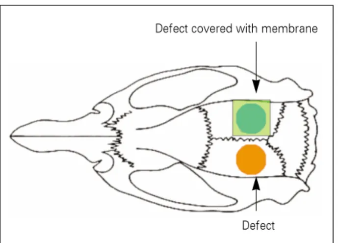
Fig. 1. Diagram of the control group and experimental group.
Defect
Defect covered with membrane
부에서는 변연부에 비해 소량의 신생골이 관찰되었다(Figs.
10-a, b).
실험 3군에서는 일부에서 차폐막의 흡수가 관찰되었으나 연조직 침투는 차폐막에 의해 차단되었으며, 이식된 탈단백 우골의 주위에서 신생골 형성이 보였다(Figs. 11-a, b).
실험 4군에서는 결손부 전체에서 고르게 신생골이 형성되 었으나, 중앙부 골의 양이 변부보다 적었다. 베타-트리칼슘 인산염이 분해되면서 내성장되는 주위의 신생골로 대치되
어 가는 소견을 관찰할 수 있었다(Figs. 12-a, b).
실험 5군에서도 결손부 전체적으로 일정한 양의 골형성과 차폐막 흡수 소견이 나타났다. 또한 베타-트리칼슘 인산염 의 분해와 주위 신생골 형성과 내성장을 관찰할 수 있었으 며, 차폐막 하방에서 연조직 침투는 보이지 않았다(Figs.
13-a, b).
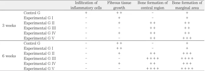
이상 각 군에서 관찰된 조직 소견을 요약 비교하면 Table 1과 같다.
Fig. 4-a. Photomicrograph of the experimental group II at 3 weeks after the graft procedure showing newly formed bone around the graft materials and chondrocyte in the fibrous tissue (Hematoxylin and Eosin, original magnifica- tion x 40).
Fig. 4-b. Photomicrograph of the experimental group II at 3 weeks after the graft procedure showing newly formed bone (N) and chondroid tissue containing chondrocyte (Hematoxylin and Eosin, original magnification x 100).
N
Fig. 2. Photomicrograph of the control group at 3 weeks after the graft procedure showing loose connective tissue filled the space and little bone formation from the defect edges (C) (Hematoxylin and Eosin, original magnification x 40).
Fig. 3. Photomicrograph of the experimental group I after at 3 weeks after the graft procedure showing remained mem- brane (M) and small amount of bone formation with con- nective tissue over the defect (Hematoxylin and Eosin, original magnification x 40).
C
C
C C
M
Fig. 5-a. Photomicrograph of the experimental group III at 3 weeks after the graft procedure showing evenly formed bone beneath the membrane (Hematoxylin and Eosin, original magnification x 40).
Fig. 5-b. Photomicrograph of the experimental group III at 3 weeks after the graft procedure showing slighlty resorbed membrane (M) and newly formed bone (N) (Hematoxylin and Eosin, original magnification x 100).
Fig. 6-a. Photomicrograph of the experimental group IV at 3 weeks after the graft procedure showing newly formed bone from the defect edge with soft tissue invasion (Hematoxylin and Eosin, original magnification x 40).
Fig. 6-b. Photomicrograph of the experimental group IV at 3 weeks after the graft procedure showing newly formed bone (N) and interrupted bony continuity (D) (Hematoxylin and Eosin, original magnification x 100).
Fig. 7-a. Photomicrograph of the experimental group V at 3 weeks after the graft procedure showing slighlty resorbed membrane and newly formed bone containing graft mate- rials (Hematoxylin and Eosin, original magnification x 40).
Fig. 7-a. Photomicrograph of the experimental group V at 3 weeks after the graft procedure showing loose connective tissue (L) and newly formed bone (N) beneath the membrane (M) (Hematoxylin and Eosin, original magnification x 100).
N
D
M L
N N
M
N
Fig. 8. Photomicrograph of the control group at 6 weeks after the graft procedure showing connective tissue covered the defect and limited bone formation from the defect edges (Hematoxylin and Eosin, original magnification x 40).
Fig. 9. Photomicrograph of the experimental group I at 6 weeks after the graft procedure showing resorbed mem- brane (M) and little bone formation (N) near the defect mar- gin (Hematoxylin and Eosin, original magnification x 40).
Fig. 10-a. Photomicrograph of the experimental group II at 6 weeks after the graft procedure showing newly formed bone with irregularity in volume (Hematoxylin and Eosin, original magnification x 40).
Fig. 10-b. Photomicrograph of the experimental group II at 6 weeks after the graft procedure showing newly formed bone (N) and graft materials (B) that most of them show no resorp- tion (Hematoxylin and Eosin, original magnification x 100).
Fig. 11-a. Photomicrograph of the experimental group III at 6 weeks after the graft procedure showing resorbing mem- brane preventing soft tissue invasion and newly formed bone with remained graft material (Hematoxylin and Eosin, original magnification x 40).
Fig. 11-b. Photomicrograph of the experimental group III at 6 weeks after the graft procedure showing membrane (M), newly formed bone (N), and slighlty resorbed graft materi- al (B) (Hematoxylin and Eosin, original magnification x 100).
N M
N
B
N B
M
B N
2. 조직형태계측학적 분석
조직형태계측 결과 대조군과 실험 1군에서는 골결손부에 대한 신생골의 면적비가 낮았다. 실험군 사이에 서로 비교 하였을 때, 이식 3주 후에는 실험 5군의 신생골 면적비가 23.96 ± 4.17%로 가장 높았고, 다음으로 실험 3군 19.88
± 2.66%, 실험 4군 16.58 ± 3.26%, 실험 2군 15.88 ± 2.79%의 순서로 나타났다. 이식 6주 후에도 실험 5군이 34.82 ± 3.08%로 가장 높았고, 실험 3군 33.65 ± 3.43%, 실험 4군 25.97 ± 2.52%, 실험 2군 25.03 ±
1.58%의 순서로 나타났다(Table 2).
계측 결과에 대한 ANOVA 분석 시 각 실험군은 서로 통 계학적으로 유의한 차이를 보였다( P < 0.005)(Table 2).
Bonferroni 방법으로 사후검정을 실시한 결과 대조군∙실 험 1군과 실험 2, 3, 4, 5군, 3주의 실험 2군과 5군, 3주의 실험 4군과 5군, 6주의 실험 2군과 3군, 6주의 실험 2군과 5군 사이에서 유의한 차이가 나타났고, 다른 군 사이에서는 통계적으로 유의한 차이가 발견되지 않았다(P < 0.05).
골 이식시 사용한 재료에 따른 차이를 분석하기 위한 Mann-Whitney 검정에서 차폐막 사용에 따라 분류한 실험
Fig. 12-a. Photomicrograph of the experimental group IV at 6 weeks after the graft procedure showing complete bone bridging along the dura side and newly formed bone from the defect edge (Hematoxylin and Eosin, original magnifi- cation x 40).
Fig. 12-b. Photomicrograph of the experimental group IV at 6 weeks after the graft procedure showing newly formed bone (N) and resorbed graft materials (T) with ingrowth of new bone (Hematoxylin and Eosin, original magnification x 100).
Fig. 13-a. Photomicrograph of the experimental group V at 6 weeks after the graft procedure showing more resorbed membrane and newly formed bone. Most of graft materials were resorbed (Hematoxylin and Eosin, original magnifica- tion x 40).
Fig. 13-b. Photomicrograph of the experimental group V at 6 weeks after the graft procedure showing newly formed bone (N) resorbed graft materials (T) with ingrowth of new bone and resorbed membrane (M) (Hematoxylin and Eosin, original magnification x 100).
T
N T
N
T
M
2, 4군과 실험 3, 5군 간에는 통계적으로 유의한 차이가 나 타났지만, 이식재에 따라 분류한 실험 2, 3군과 실험 4, 5 군 간에는 통계적으로 유의한 차이가 나타나지 않았다(P <
0.05).
고 찰
이종골은 수혜자와 다른 종으로부터 기인한 골대체제이 며, 가장 널리 쓰이고 있는 탈단백우골은 소뼈를 채취한 후 ethylene diamine 등으로 화학 처리하여 단백질 등의 유기 질을 제거하고 멸균처리한 이식재이다. 인간 해면골의 수산 화인회석과 유사한 화학적 조성을 보이며, 형태적∙구조적 특징 또한 인간의 해면골과 유사한 100 μm 직경의 칼슘 결 정체를 갖는다.
5,18,19)합성골은 이상적인 골이식재의 특성 중 골전도와 골유착의 특성을 보인다. 이상적인 합성골 이식재 는 생체에 적합하여야 하고 섬유성 변화를 최소로 일으켜야 한다. 베타-트리칼슘 인산염은 약 90%가 서로 연결된 빈 공간으로 다공성(1-1,000 μm)을 지니는데 이는 내성장을
허용하여, 큰 골전도능을 보인다.
12,20)또한 베타-트리칼슘 인산염은 골에 비해 조금 다른 칼슘/인 비율을 보이지만, 그 특성은 골의 60-70%를 구성하는 무기성분과 유사하다.
21,22)본 연구에서는 인위적으로 형성한 흰쥐 두개골 결손부에 서 베타-트리칼슘 인산염과 탈단백우골 이식이 골형성에 어 떠한 영향을 미치는지 알아보고자 하였다. 최근 임플란트 시술이 보편화됨에 따라 골이식술이 많이 시행되고 있다.
자가골 이식은 골을 채취하기 위한 부가적인 수술이 필요하 므로 술자나 환자 모두 기피하는 경향이 있으며, 따라서 이 종골 또는 합성골 이식이 선호되고 있다. 현재 탈단백우골 인 Bio-Oss
�가 임상에서 널리 사용되고 있으나 이종골에서 추출한 것이므로 화학 처리와 멸균 처리가 필수적이고 생체 내에서 흡수 여부가 문제시 되고 있다. 그러므로 더 좋은 골 이식재를 찾는 과정에서 탈단백우골과 합성골인 베타-트리 칼슘 인산염과 비교하고자 하였다.
흰쥐에서 골결손부의 임계 크기는 8 mm로 보고된 바 있
다.
17,23,24)Furlaneto 등
25)은 흰쥐의 두개골에 5 mm 지름의
결손부를 형성하여 관찰한 연구에서 자발적인 골 재생이 일 Table 1. Comparison of the histologic characteristics of the specimens
Infiltration of Fibrous tissue Bone formation of Bone formation of inflammatory cells growth central region marginal area
Control G + ++ - +
Experimental G I - + - +
3 weeks Experimental G II - + ++ ++
Experimental G III - - ++ ++
Experimental G IV - + ++ ++
Experimental G V - - ++ +++
Control G - ++ - +
Experimental G I - ++ - +
6 weeks Experimental G II - + ++ +++
Experimental G III - - ++++ ++++
Experimental G IV - + ++ +++
Experimental G V - - ++++ ++++
- none; + low; ++ moderate; +++ high; ++++ very high
Table 2. The ratio of new bone to bone defect in each group
3 weeks 6 weeks
mean (%) SD mean (%) SD
Control group 3.98 0.76 5.66 1.42
Experimental group I 7.03 2 8.95 2.08
Experimental group II 15.88 2.79 25.03 1.58
Experimental group III 19.88 2.66 33.65 3.43
Experimental group IV 16.58 3.26 25.97 2.52
Probability * *
* P < 0.005
어나지 않아 임계 크기로서의 기준을 만족시킬 수 있다고 하였다. 본 연구에서는 흰쥐 두개골 결손부의 크기를 Furlaneto 등
25)이 제시한 대로 5 mm로 설정하였으며, 동 일 개체 내에 두 개의 결손부를 형성하여 다른 실험군 간에 비교가 가능하도록 하였다. 본 연구의 대조 1, 2군에서 3주 와 6주 모두 골결손 변연부에서 신생골 형성이 관찰되었지 만 골결손부는 전반적으로 결체조직으로 채워져 있었고, 다 른 실험군과 비교할 때 미약한 수준이었다.
본 연구 결과 대조군에 비해 베타-트리칼슘 인산염과 탈 단백우골을 이식한 실험군에서 골형성 양이 더 많았다. 또 탈단백우골보다 베타-트리칼슘 인산염을 이식한 경우에 더 많은 골형성 양을 나타냈으나 통계적으로 유의하지는 않았 다. 다만 조직학적 소견에서 이식재의 흡수 양상은 이식 3 주 후에는 큰 차이 없었으나, 6주 후에는 베타-트리칼슘 인 산염은 흡수가 많이 진행된 반면 탈단백우골은 흡수가 미미 하였다. 베타-트리칼슘 인산염은 다공성 구조를 가지므로 골의 내성장이 촉진되는데, 다공성 뿐만 아니라 무기성분 조성과 소결 정도 또한 흡수성과 관련이 있다. 베타-트리칼 슘 인산염은 점진적으로 흡수되고 최종적으로 재형성된 골 로 대치된다. 베타-트리칼슘 인산염의 치유는 여러 단계로 나타난다. 첫 단계는 이식재에 신생골이 축적됨에 따라 일 어나는 수용부의 골 흡수이다. 다음 단계는 재형성이 동반 되는 이식재의 흡수이다.
20)베타-트리칼슘 인산염의 경우 초기 분해단계와 분해단계에 동반되는 탐식작용을 하는 세 포 때문에 임플란트의 골유착이 방해받을 수 있으므로, 베 타-트리칼슘 인산염을 이용한 상악동 거상술을 시행할 경우 분해기가 진행된 후에 임플란트를 식립할 것을 권장하기도 한다.
26)동물 모형에 형성된 골결손부에서는 베타-트리칼슘 인산 염이 완전히 흡수되기까지 12주 정도가 소요되며, 사람에 서는 6-8개월이 소요된다.
27,28)그러나 Horch 등
26)에 의하면 방사선상에서 약 12개월 후에 베타-트리칼슘 인산염이 자 가골로 대치되었으며, 조직학적으로는 자가골과 혼합한 경 우 85%가 흡수되었고 단독으로 사용한 경우 65%가 흡수 된다고 하였다.
그에 비해 탈단백우골은 파골세포 작용을 통한 생체 흡수 를 보이기까지 이식 후 3-6년이 소요되기 때문에 비흡수성 재료로 간주되기도 한다.
29)일부 동물 실험을 통한 연구에서 는 탈단백우골의 흡수 양상이 관찰된다고도 하였으나,
7,8)Taylor 등
29)은 탈단백우골의 파골세포가 원래 소뼈의 파골 세포에 비해 크기와 숫자가 감소되었으며 흡수 소와도 적게 생성된다고 하였다. Skoglund 등
9)은 6명의 환자에서 탈단 백우골 이식 후 각기 9개월에서 44개월 사이의 서로 다른 기간에 조직을 채취하여 검사하였음에도 탈단백우골이 관 찰되었음을 보고하였고, Schlegel 등
6)은 71명의 환자에서 탈단백우골 이식 후 골수 및 연조직과 인접한 부위 뿐만 아
니라, 골과 직접적으로 접촉한 부위에서도 탈단백우골의 흡 수소견이 관찰되지 않았다고 하였다. Scarano 등
10)은 자가 골은 이식 후 6개월 경과 후 18%가 이식 부위에 잔존한데 비해 탈단백우골은 31%가 잔존했다고 하였다. 하지만, Zitzmann 등
30)은 6명의 환자에서 치조골 증대술을 위해 탈 단백우골을 흡수성 차폐막과 함께 사용하였으며, 조직학적 으로 탈단백우골이 흡수의 징후를 보였다고 하였다. 본 연 구에서는 탈단백우골을 이식한 실험 1, 2군에서 3주 후에는 흡수 소견을 관찰할 수 없었으며, 6주 후에는 약간의 흡수 소견을 보였으나 대부분 흡수 소견을 관찰할 수 없었다.
신생골 내에서 흡수되지 않은 입자의 존재는 신생골 형성 을 방해하고, 조직 특성을 좋지 않게 하여 치과용 임플란트 골유착능에 영향을 미친다.
31-33)Stavropoulos 등
31)은 생체 흡수가 늦은 재료는 골형성을 촉진하는 대신 방해한다고 주 장했지만, Tamimi 등
34)은 실험적 생체물질 이식군에 비해 이식을 하지 않은 대조군이 항상 더 적은 골 증대를 보여 생 체 내 흡수가 늦더라도 골형성을 방해하지는 않는다고 하였 다. 본 연구에서도 골이식을 시행하지 않은 대조군에 비해 탈단백우골 이식군에서 대부분 흡수 소견을 관찰할 수 없었 으나 3주와 6주 모두 유의하게 많은 골형성 양을 관찰할 수 있었다.
본 연구에서 베타-트리칼슘 인산염 이식군과 탈단백우골 이식군 간에 골형성 양을 비교하였을 때 통계적으로 유의한 차이는 없었다. Jensen 등
2)은 미니돼지의 하악골 우각부에 골결손부를 형성하고 자가골, 베타-트리칼슘 인산염, 탈단 백우골을 이식한 실험에서 탈단백우골이 자가골과 베타-트 리칼슘 인산염에 비하여 골형성 양이 유의하게 적음을 보고 하였다. 또한 베타-트리칼슘 인산염이 신생골로의 완전한 치환과 결손부 내로의 조기 연조직 침투를 방지할 수 있는 오목한 형태의 골결손부에 유용할 수 있다고 하였다. 이상 적인 골대체제는 치유기간 동안 생물학적 지지를 유지하고, 새로 형성되는 골에 의해 점진적으로 대체되어야 한다. 탈 단백 우골의 낮은 흡수율이 득이 될 수 있는데, 이식재가 증 대된 부피를 유지하고, 상부 치은이나 상악동 점막에 의한 압력에 대하여 장기적으로 저항할 수 있다.
2)본 연구에서 Jensen 등
2)의 연구 결과와 달리 베타-트리칼슘 인산염과 탈단백 우골의 골형성 양이 비슷한 것이 골결손부 형태와 두개골막 등의 압력과 관련이 있을 것으로 생각된다.
골이식을 시행하는 경우 연조직의 이식골 내 함입에 의해
신생골 형성이 저해될 수 있는데, 그에 대하여 Nyman 등
35)과 Karring 등
36)은 치주조직 결손부에 물리적 차폐막을 위
치시켜 결체조직의 함입을 방지하고 치주조직의 재생에 필
요한 치주인대 세포나 골아세포의 이동을 도모하는 조직유
도재생술을 소개하였다. 이 때 사용되는 차폐막에는 비흡수
성 차폐막과 흡수성 차폐막이 있다. 본 연구에 사용된 흡수
성 차폐막은 polylactide와 polyglycolide 공중합체 막으로
3주 소견에서 일부 흡수 소견을 관찰할 수 있었으며, 6주 소견에서는 막의 흡수가 더 진행된 소견을 관찰할 수 있었 으나, 전반적인 형태가 비교적 잘 유지되고 있었으며 이식 재 이탈 방지와 함께 연조직 침투를 차단하고 있음을 확인 할 수 있었다. 또한 3주와 6주 모두 차폐막을 이용한 실험 2, 4군에서 차폐막을 이용하지 않은 실험 1, 3군보다 골형 성 양이 많았다. 이는 polylactide와 polyglycolide의 1 : 1 공중합체막으로 만든 흡수성 차폐막의 사용이 효과적이며, 조직소견에서 차폐막이 염증이 적고 생체친화적이고, 실험 4-6주부터 흡수가 시작되지만 5개월까지 차폐막이 유지되 어 골결손 재생에 충분히 사용할 수 있다는 이전의 보고
37,38)를 지지하는 결과라고 생각된다.
이상을 요약하면 탈단백우골과 베타-트리칼슘 인산염은 모두 골전도능을 지니지만 베타-트리칼슘 인산염이 골형성 과정 중 더 많은 흡수를 보이며, 차폐막을 사용할 때 골형성 양이 더 많아짐을 관찰할 수 있었다. 그러나 골형성 양이 결 손부 크기에 비하여 만족스럽지 못하였으므로, 자가골과의 혼합 또는 골형성단백질의 이용 등 골형성 양을 더욱 증대 시킬 수 있는 방법에 대한 연구와 여러 가지 형태의 골결손 부에 대한 연구가 필요할 것으로 생각된다.
결 론
이번 연구에서 골형성양을 비교했을 때 베타-트리칼슘과 차폐막을 사용한 실험군에서 가장 많은 양이, 탈단백우골만 을 사용한 실험군에서 가장 적은 양이 관찰되었다. 그러나 탈단백우골과 베타-트리칼슘 인산염 사이의 유의한 차이는 관찰되지 않았으며, 차폐막 사용에 따라 유의한 차이가 관 찰되었다. 탈단백우골과 베타-트리칼슘 인산염은 골형성양 의 차이가 크지 않았으나, 베타-트리칼슘 인산염 이식군에 서 이식 후 생체 내 흡수 양상이 더 많이 관찰되었다. 그리 고 차폐막을 사용한 경우에 그렇지 않은 경우에 비하여, 조 직학적으로 이식재 안정, 섬유조직 차단을 관찰할 수 있었 다.
이상의 결과로써 탈단백우골과 베타-트리칼슘 인산염 모 두 우수한 골전도능을 보임을 알 수 있었다. 다만, 골형성에 따른 생체 내 흡수의 측면에서 베타-트리칼슘 인산염이 더 이상적인 골 이식재로 생각된다. 그리고 차폐막을 사용할 경우 골형성 양이 더 증가됨을 알 수 있었다.
References