A Comparison of Lateral Abdominal Muscle Activation during Maximum Expiration in Chronic Low Back Pain Patients and Healthy Asymptomatic Subjects
Share "A Comparison of Lateral Abdominal Muscle Activation during Maximum Expiration in Chronic Low Back Pain Patients and Healthy Asymptomatic Subjects"
Share "A Comparison of Lateral Abdominal Muscle Activation during Maximum Expiration in Chronic Low Back Pain Patients and Healthy Asymptomatic Subjects"
A Comparison of Lateral Abdominal Muscle Activation during Maximum Expiration in Chronic Low Back Pain Patients and Healthy Asymptomatic Subjects
Bong-Oh Goo⋅Kang-Hoon Kim 1†
Department of Physical Therapy, Catholic University of Pusan
1 Department of Physical Therapy, General Grauate School, Catholic University of Pusan
Received: October 15, 2013 / Revised: November 10, 2013 / Accepted: January 4, 2014
ⓒ 2014 Journal of Korea Proprioceptive Neuromuscular Facilitation Association
This is an Open Access article distributed under the terms of the Creative Commons Attribution Non-Commercial License (http://creativecommons.org/licenses/by-nc/3.0) which permits unrestricted non-commercial use, distribution, and reproduction in any medium, provided the original work is properly cited.
| Abstract |
Purpose: This study was to examine lateral abdominal muscle activation during maximum expiration exercise between healthy and chronic low back pain(CLBP) patients.
Methods: The subjects were 16 CLBP patients and 16 healthy people between the ages of 22 and 53. The thickness of the abdominal muscles was measured using ultrasonography(LOGIQ Book XP, GE, USA). We instructed the subjects how to perform the exercises and measured changes in thickness of the transversus abdominis(TrA) and internal oblique(IO) muscles during the maximum expiration. The main outcome variables were the ratios of the TrA and IO thickness during the exercise versus in the relaxed position(TrA and IO activation ratios).
Results: There were significant differences between CLBP patients and healthy subjects for TrA in the relaxed position. However there was no difference in the ratio of change in the muscle activity(TrA, IO).
Conclusion: These findings, CLBP patients exhibited atrophy of the TrA muscle, but voluntary TrA muscle activation was similar to that of the normal subjects. Therefore, this exercise could be used during core strengthening in CLBP patients.
Key Words: Chronic low back pain, Maximum expiration, Abdominal muscle
Ⅰ. 서 론
★
요통은 일상생활을 영위하는데 지장을 주는 질환으로 성인 의 50∼80% 정도가 적어도 한 두 번 정도의 통증을 경험한다 (Andersson, 1999; Woolf와 Pfleger, 2003). 요통을 경감시키는 중재방법에는 여러 가지가 있으며 특히 운동치료는 만성 허리 통증에 효과적이다(Bronfort 등, 2011; Van Tulder 등, 2000).
그 중 코어강화(core strengthening)는 요통 관리를 위해서 중요 한 경향으로 계속 발전되어온 운동방법이다.
Fig. 1. Ultrasound imaging of the lateral abdominal muscle in healthy subjects (A) Relaxed, (B) Maximum expiration
Fig. 2. Ultrasound imaging of the lateral abdominal muscle in CLBP patients (A) Relaxed, (B) Maximum expiration 2009; Hides 등, 1996; Hodges와 Richardson, 1996). 이 두 근육은
안정화 기전으로 작용을 할 때 동시적인 수축을 일으키며 특히 다열근에 대한 신경근과 근육에 대한 평가에서는 요통환 자가 정상인보다 지연된 근동원 순서와 감소한 근횡단면적을 보인다고 하였다(Hides 등, 1996; Hodges와 Richardson, 1996).
또 따른 심부 근육인 복횡근도 느린 신경근 반응과 근 두께의 위축을 보인다고 하였다(Hodges와 Richardson, 1996; Ota와 Kaneoka, 2011). 이러한 특징들은 특별한 운동을 시행하지 않 으면 자연적으로 회복되지 않는다(Hides 등, 2001).
요통환자의 지연된 심부근 동원 순서를 개선하는 운동방법 으로 배꼽 넣기 방법(draw-in maneuver)이 제시되고 있다 (Hides 등, 2001). 배꼽 넣기 방법은 가능한 대근육의 수축 없이 심부근육 수축을 유도하는 방법이다 . 배꼽 넣기를 실시 했을 때 신경근 반응과 통증이 감소하였다는 연구들이 있다 (Tsao와 Hodges, 2007). 그렇지만, 배꼽 넣기만을 이용한 중재 는 심부근육의 근횡단면적, 근 두께의 증가를 이루기 어려우 며 이를 위해서는 배꼽 넣기 시행과 함께 체간 강화운동이 필요하다고 하였다(Hides 등, 2001; Danneels 등, 2001).
체간 신전운동과 같은 체간 강화운동은 반복적이고 과도한 허리 신전을 동반하며 이러한 움직임은 척추에 많은 부하를 야기하게 된다(Callaghan 등, 1998; Pollock 등, 1989). 그래서 중립적인 자세를 유지한 상태에서 코어운동을 시행할 필요가 있다 . 중립자세를 유지하는 코어운동에는 배꼽넣기 방법, 복 부근 브레이싱 기법(abdominal bracing), 후방 골반경사 방법 (posterior pelvic tilt)이 있지만 정확한 동작과 선택적인 심부근 육 활성화에는 어려움이 있다(McGill 등, 2003; Vezina와 Hubley-Kozey, 2000).
최근에는 배꼽 넣기 방법보다 수행하기 쉽고 대근육인 내복사근과 외복사근보다 심부근 활동을 증가시켜 주는 최 대호기 방법이 소개되고 있다(Ishida와 Watanabe, 2013). 이 방법은 강력한 호기근인 복횡근을 이용하여 중립 자세에서 체간 안정성을 증대시켜주는 방법이다(Ishida 등, 2012). 하지 만 아직은 요통 환자를 대상으로 최대호기를 적용한 연구는 없다.
그래서 본 연구는 복횡근의 활동을 증가시키는 최대 호기 시 요통환자와 정상인의 복부근 활동 능력을 비교함으로 요통환자에게 적절한 중재방법이 될 수 있는지 알아보고자 한다.
Ⅱ. 연구 방법
1. 연구 대상 및 연구 기간
본 연구는 부산시에 소재한 J한방병원에 입원한 환자를 16명과 정상인 16명으로 하였다.
선정기준은 3개월 이상 요통 증상이 지속된 환자로 하였다.
정상인의 선정기준은 지난 3개월 동안 요통을 호소하지 않은 대상자로 선정하였다. 제외기준은 요추부에 수술 경험이 있 는 자, 암과 같은 계통적 질환으로 인한 요통을 호소하는 자, 심각한 근골격계 질환이 있는 자, 뇌졸중 같은 중추신경계 질환이 있는 자로 하였으며 실험을 진행하기 전에 연구의 목적과 방법에 대하여 대상자들로부터 연구 참여에 자발적인 동의를 얻어 진행하였다.
최대 호기 시에 외측 복부근 수축율 변화를 관찰하기 위해 서 테이블에 무릎을 굽히고 바로 누운 자세를 취하게 하였다.
복부 두께는 초음파를 이용 장골능과 갈비뼈 사이에 중앙부위 에서 2.5cm 전방으로 하여 오른쪽 외측복부를 측정하였다.
휴식 시 측정은 호흡으로 인한 근육두께 변화를 배제하기 위해서 자연스러운 호기 마지막 부분에서 측정을 하였다. 최 대 호기 시 측정은 Ishida 등(2012)이 고안한 방법으로 “복부가 외측으로 부풀러(brace pattern) 오르지 않게 한 상태에서 최대 한 숨을 뱉고 숨을 유지 하세요 .” 구두명령을 주어 5초간 유지 하게 하였다. 복부 브레이싱은 체간 안정성을 증가시키기 위 해서 모든 복부근육을 동시에 수축시켜 허리 외측으로 둘레가 커지게 하는 방법이다(Grenier와 McGill, 2007).
3. 자료 분석
본 연구의 통계는 SPSS 20.0 프로그램을 이용하여 처리하 였으며 독립 변수는 대상자 상태(정상인, 요통환자)로 나누고 종속변수는 근두께와 근활동 비율로 하였다. 근비율 변화=(근 수축 두께-휴식시 두께)/휴식시 두께*100으로 계산하였다 (Teyhen 등, 2009). 그룹간 휴식시 복횡근 두께, 수축율, 근비율 변화(%)는 독립 t-tests를 이용하여 비교하였다. 통계학적 유의 성을 검증하기 위해 유의수준은 α=0.05로 설정하였다.
Ⅲ. 결 과
1. 연구 대상자의 일반적인 특성
본 연구에 참여한 대상자들의 일반적인 특성은 다음과 같으 며 두 군간 통계학적인 차이는 없었다 (Table 1).
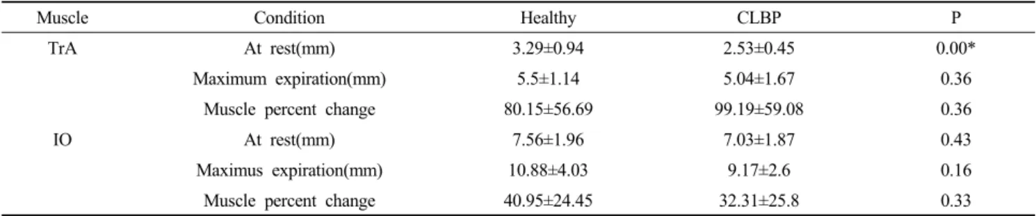
2. 최대 호기시 외측 복부근의 두께변화
정상인과 요통그룹에서 복횡근은 휴식시 두께는 통계적으 로 유의한 차이를 나타났으며(p<0.05), 근비율 변화는 유의한
차이를 나타내지 않았다(p>0.05). 내복사근은 휴식시 두께와 근비율 변화가 통계적으로 유의한 차이가 나타나지 않았다 (p>.05)(Table 2).
Ⅳ. 고 찰
본 연구는 복횡근 활동을 증가시키는 최대 호기 시 요통환 자와 정상인의 외측 복부근 활동 능력을 비교하고자 하였다.
복부 근육활동을 측정함에 초음파는 신뢰성 있는 좋은 장비이 며, 초음파상에서 내복사근과 복횡근의 근 두께 변화는 근전 도상의 근 활동과 선형적인 상관성을 가진다고 하였다 (Hodges 등, 2003; McMeeken 등, 2004). 본 연구에서 외복사근 의 두께 변화는 근전도상의 활동과 초음파상의 두께와 상관성 이 없으므로 측정에서 제외했다(John과 Beith, 2007). 중재방법 에 있어 정상인의 복횡근 비율변화는 80.15%로 나타났으며 이는 선행 연구에서 86∼89% 사이로 나타난 결과(Ishida 등, 2012; Kaneko 등, 2005)와 유사하여 선행 연구와 유사한 최대 호기를 실시했다고 여겨진다.
본 연구 결과에서 요통환자는 일반인보다 안정 시에 근육
두께는 내복사근에서 유의한 차이를 보이지 않았으나 복횡근
에서는 유의하게 감소하였다. Ota와 Kaneoka(2011)는 휴식
시 요통환자의 복횡근 두께는 다른 복부근육과는 다르게 정상
인 비해 얇은 두께를 보이며 이런 결과는 복횡근의 위축을
(4)
의미한다고 하였다. 본 연구에도 요통 환자의 복횡근에 위축 이 있었지만, 복횡근과 내복사근의 비율변화는 두 군간의 차 이를 나타내지 않았다. 또한, 통계적으로 유의하지는 않으나 요통 환자군이 정상인보다 복횡근 비율변화 평균 19%로 높게 나타났으며, 내복사근 비율변화는 정상인보다 평균 8%로 낮 게 나타났다 . 이는 Vasseljen와 Fladmark (2010)가 8주간 시행 한 요부 운동에서 복횡근 비율변화 증가와 내복사근 비율변화 감소는 요통 경감에 긍정적인 영향을 미친다고 하였다. 그러 므로 만성 요통환자에게 장기간 최대 호기 운동을 시행 시 통증의 감소뿐 아니라 복횡근 비율변화 증가로 인해 복횡근의 위축을 방지하는 데 도움이 될 것으로 생각된다. 이런 위축 방지 효과는 복횡근의 계속된 자발적인 활동이 신경근 활동을 증가시켜 줌으로써 이루어질 수 있을 것으로 생각된다.
본 연구는 몇 가지 제한점이 있다. 첫째, 연구 대상자의 근 비율변화에 큰 편차를 보인다. 이는 Ishida 등(2012)의 연구 에서 밝힌 바와 같이 대상자 간의 근력이 다르므로 최대호기 를 수행하기에 개인적인 차이가 난다고 생각된다. 둘째, 본 연구는 단면연구로 16명의 만성요통환자를 대상으로 실험했 으며 이 결과가 모든 만성요통 환자에게 보편화하여 적용하기 는 어렵다 . 섯째, 요통환자와 정상인 간에 외측복부에 일시적 인 근 비율 변화의 차이만을 알아보았다. 코어 운동에 대한 중재방법으로 장기간 중재효과에 대한 연구가 추가로 필요하 리라 여겨진다.
Ⅴ. 결 론
본 연구는 건강한 대상자와 요통 환자를 대상으로 최대 호기 시에 초음파를 이용해 최대 호기 시에 외측 복부근육의 활동능력을 확인하고자 실시하였다. 실험 결과 요통 환자와 정상인의 근변화 비율은 차이가 나지 않았다. 이러한 결과를 통해 최대 호기를 이용한 장기적 안정화 운동이 요통환자의 근위축을 감소시킬 수 있는 효과적인 중재방법이 되리라 생각 된다.
참고문헌
Andersson GB. Epidemiological features of chronic low-back pain.
Lancet. 354(9178): 581-585, 1999.
Akuthota V, Nadler SF. Core strengthening. Arch Phys Med Rehabil.
85(3 Suppl 1):S86-92, 2004.
Bronfort G, Maiers MJ, Evans RL et al. Supervised exercise, spinal manipulation, and home exercise for chronic low back pain:
a randomized clinical trial. Spine J. 11(7):589-598, 2011.
Callaghan JP, Dunning JL, McGill SM. The relationship between lumbar spine load and muscle activity during extensor exercises. Phys Ther. 78(1):8-18, 1998.
Danneels L, Vanderstraeten G, Cambier D et al. Effects of three different training modalities on the cross sectional area of the lumbar multifidus muscle in patients with chronic low back pain.
Br J Sports Med. 35(3):186-191, 2001.
Grenier SG, McGill SM. Quantification of lumbar stability by using 2 different abdominal activation strategies. Arch Phys Med Rehabil. 88(1):54-62, 2007.
Hides JA, Richardson CA, Jull GA. Multifidus muscle recovery is not automatic after resolution of acute, first-episode low back pain. Spine. 21(23):2763-2769, 1996.
Hides JA, Jull GA, Richardson CA. Long-term effects of specific stabilizing exercises for first-episode low back pain. Spine.
26(11):E243-248, 2001.
Hodges PW, Richardson CA. Inefficient muscular stabilization of the lumbar spine associated with low back pain. A motor control evaluation of transversus abdominis. Spine. 15(22):2640-2650, 1996.
Hodges PW Pengel LH, Herbert RD et al. Measuremetn of muscle contraction with ultrasound imaging. Muscle & Nerve.
27(6):682-692, 2003.
Ishida H, Hirose R, Watanabe S. Comparison of changes in the contraction of the lateral abdominal muscles between the abdominal drawing-in maneuver and breathe held at the maximum expiratory level. Man Ther. 17(5):427-431, 2012.
Ishida H, Watanabe S. Changes in lateral abdominal muscles’ thickness immediately after the abdominal drawing-in maneuver and maximum expiration. J Bodyw Mov Ther. 17(2):254-258, 2013.
John EK, Beith ID. Can activity within the external abdominal oblique be measrued using real-time ultrasound imaging? Clin Biomech (Bristol, Avon). 22(9):972-979, 2007.
Kaneko H, Satou H, Maruyama H. Reliability of lateral abdominal muscles thickenss measurement using ultrasonography.
Rigakuryoho Kagaku. 20(3):197-201, 2005.
McGill SM, Grenier S, Kavcic N et al. Coordination of muscle activity to assure stability of the lumbar spine. J Electromyogr Kinesiol.
13(4):353-359, 2003.
McMeeken JM, Beith ID, Newham DJ et al. The relationship between
EMG and change in thickness of transversus abdominis. Clinical
Biomechanics. 19(4):337-342, 2004.
(5)
Norasteh A, Ebarahimi E, Salavati M et al. Reliability of B-mode ultrasonography for abdominal muscles in asymptomatic and patients with acute low back pain. J Bdoyw Mo Ther.
11(1):17-20, 2007.
Ota M, Kaneoka K. Differences in abdominal muscle thicknesses between chronic low back pain patients and healthy subjects. J Phys Ther Sci. 23(6):855-858, 2011.
Otani Y, Itotani K, Maeda N et al. Reliability of the deep abdominal muscle thickness measurements using ultrasonography in normal subjects. J Phys Ther Sci. 23(3):357-359, 2011.
Panjabi MM. A hypothesis of chronic back pain: ligament subfailure injuries lead to muscle control dysfunction. Eur Spine J.
15(5):668-676, 2006.
Pollock ML, Leqqett SH, Graves JE et al. Effect of resistance training on lumbar extension strength. Am J Sports Med. 17(5):624-629, 1989.
Silfies SP, Mehta R, Smith SS et al. Differences in feedforward trunk muscle activity in subgroups of patients with mechanical low back pain. Arch Phys Med Rehail. 90(7):1159-1169, 2009.
Teyhen DS, Bluemle LN, Dolbeer JA et al. Changes in lateral abdominal muscle thickness during the abdominal drawing-in maneuver in those with lumbopelvic pain. J Orthop Sports Phys Ther.
39(11):791-798, 2009.
Tsao H, Hodges PW. Immediate changes in feedforward postural adjustments following voluntary motor training. Exp Brain Res. 181(4):537-546, 2007.
Van Tulder M, Malmivaara A, Esmail R et al. Exercise therapy for low back pain: A systematic review within the framework of the cochrane collaboration back review group. Spine.
25(21):2784-2796, 2000.
Vasseljen O, Fladmark AM. Abdominal muscle contraction thickness and function after specific and general exercises: a randomized controlled trial in chronic low back pain patients. Man Ther.
15(5):482-489, 2010.
Vezina MJ, Hubley-Kozey CL. Muscle activation in therapeutic exercises to improve trunk stability. Arch Phys Med Rehabil.
81(10):1370-1379, 2000.
Woolf AD, Pfleger B. Burden of major musculoskeletal conditions.