• 검색 결과가 없습니다.

Expert tibia nail for subtrochanteric femoral fracture to prevent thermal injury

N/A
N/A
Protected

Academic year: 2021

Share "Expert tibia nail for subtrochanteric femoral fracture to prevent thermal injury"

Copied!
4
0
0

로드 중.... (전체 텍스트 보기)

전체 글

(1)

CASE REPORT – OPEN ACCESS

International Journal of Surgery Case Reports 10 (2015) 158–161

Contents lists available at ScienceDirect

International Journal of Surgery Case Reports

j o u r n a l h o m e p a g e : w w w . c a s e r e p o r t s . c o m

Expert tibia nail for subtrochanteric femoral fracture to prevent thermal injury

Kyung-Jae Lee a,b,∗ , Byung-Woo Min a , Jae-Hoon Jung a , Mi-Kyung Kang a , Min-Ji Kim a

a

Department of Orthopaedic Surgery, School of Medicine, Keimyung University, Daegu, Republic of Korea

b

Pain Research Center, School of Medicine, Keimyung University, Daegu, Republic of Korea

a r t i c l e i n f o

Article history:

Received 5 November 2014

Received in revised form 20 March 2015 Accepted 21 March 2015

Available online 28 March 2015

Keywords:

Subtrochanteric femoreal fracture Intramedullary nailing

Bone marrow Expert tibia nail

a b s t r a c t

INTRODUCTION: Subtrochanteric femoral fractures are relatively uncommon, accounting for 7–15% of all hip fractures and treatment of these fractures are considered challenge for orthopaedic surgeons.

Although several treatment options are reported with up to 90% of satisfactory results, the choice of the appropriate implant is still a matter of debate. Some authors reported thermal injury after reaming for intramedullary nail fixation in patients with narrow medullary canal.

PRESENTATION OF CASE: A 21-year-old female patient was admitted to our hospital because of right subtrochanteric femoral fracture. The narrowest diameter of medullary canal of her femur was about 7 mm but she refused open reduction and internal fixation with plate due to large scar formation. We used expert tibia nail instead of femoral intramedullary nail to prevent thermal injury.

DISCUSSION: Subtrochanteric femoral fractures are difficult to treat because of their biomechanical and anatomical characteristics. Although several implants are reported for the surgical treatment of these fractures, intramedullary nails have been advocated due to their biological and biomechanical advantages.

However, under certain circumstances with associated injury or anatomic difference we might consider another treatment options.

CONCLUSION: Expert tibia nail may be considered one of the treatment options for subtrochanteric femoral fracture with narrow medullary canal. We also emphasize the importance of preoperative eval- uation of the medullary canal size for these risky fractures.

© 2015 The Authors. Published by Elsevier Ltd. on behalf of Surgical Associates Ltd. This is an open access article under the CC BY-NC-ND license (http://creativecommons.org/licenses/by-nc-nd/4.0/).

1. Introduction

Subtrochanteric femoral fractures are relatively rare and their prevalence account for 7–15% of all hip fractures [1]. Anatom- ically, subtrochanteric area consists of hard cortical bones with poor vascularity and biomechanically, it is under high compres- sive and tensile stress [2]. These characteristics can cause high rate of nonunion, malunion, and fixation failure [3,4]. Several treat- ment options are reported with intramedullary or extramedullary implants and up to 90% of reported outcomes have been satisfactory [1,2,5–10]. Although plating techniques offer some advantages, intramedullary nail have been supported for the fixation of these fractures, because of their biological and biomechanical advantages[2].

There were concerns about thermal injury after reaming for intramedullary nail fixation [11–13]. Leunig and Hertel reported three cases of osteocutaneous necrosis induced by heat during

∗ Corresponding author at: Department of Orthopaedic Surgery, School of Medicine, Keimyung University, Dongsan Hospital, 56 Dalsung-Ro, Joong-Gu, Daegu, Korea. Tel.: +82 53 250 8161.

E-mail address: [email protected] (K.-J. Lee).

tibia intramedullary reaming [11]. All of three cases had a narrow medullary cavity. Giannoudis et al. [13], also reported that reaming smaller canals (8 mm) to a larger size may induce a significant heat- ing effect. Now we report a patient with subtrochanteric femoral fracture with narrow medullary canal which was treated unevent- fully by using expert tibia nail (ETN; Synthes

®

, Switzerland) instead of femoral intramedullary nail to prevent thermal injury.

2. Presentation of case

A 21-year-old female patient sustained right subtrochanteric femoral fracture after fall from a height injury (Fig. 1A). Her height was 160 cm, weight was 52 kg and body mass index was 20.3 kg/m

2

. There was no further past or medical history except associated ipsilateral humeral fracture. We planned surgical treatment with cephalomedullary femoral nail but scanogram revealed that nar- rowest diameter of medullary canal was around 7 mm (Fig. 1B). If we tried to use cephalomedullary femoral nail, more than 3 mm of reaming was needed and the small size solid femoral nails that do not require reaming were not available at the time. We explained about the problem of using cephalomedullary femoral nail (thermal injury) and discussed about open reduction and inter-

http://dx.doi.org/10.1016/j.ijscr.2015.03.048

2210-2612/© 2015 The Authors. Published by Elsevier Ltd. on behalf of Surgical Associates Ltd. This is an open access article under the CC BY-NC-ND license (http://

creativecommons.org/licenses/by-nc-nd/4.0/).

(2)

CASE REPORT – OPEN ACCESS

K.-J. Lee et al. / International Journal of Surgery Case Reports 10 (2015) 158–161 159

Fig. 1. Anteroposterior view of right femur shows short transverse fracture with butterfly fragment around subtrochanteric area (A) scanogram of left femur shows relatively narrow medullary canal (B).

nal fixation with extramedullary implant or minimal invasive plate osteosynthesis with locking plate. However, she refused all the extramedullary implant because she worried about relatively large scar formation than intramedullary nail. Her job was a dancer.

We finally decided to use ETN for the fixation of her sub- trochanteric femoral fracture with closed reduction. Patient was placed on the fracture table and closed reduction was tried under guidance of image intensifier. Greater trochanter could be used as an entry point because this nail had 10 degree angulation on its

proximal part. We also put a diameter 5.0 mm locking screw into the femoral head because five locking options in four planes are allowed in proximal part of this nail. Finally, diameter 8 mm and length 330 mm expert tibia nail was inserted (Fig. 2A and B). The patient was allowed to mobilize on the first postoperative day and to weight bear as tolerated with crutches until postoperative 12 weeks. The postoperative rehabilitation was uneventful and radio- graphic union was obtained after 8 months of operation (Fig. 2C).

Removal of implant was done two years after surgery (Fig. 3).

3. Discussion

Treatment of subtrochanteric femoral fractures constitutes a considerable challenge for orthopaedic surgeon. Anatomically, this area consists of hard cortical bones with a slower healing rate than metaphyseal bones. Biomechanically, the proximal femoral shaft is under high stress (compressive force on medial side and ten- sile force on lateral side). Furthermore, extensive comminution and fragment devitalisation can compromise bone healing biologically [1]. These characteristics can cause high rate of nonunion, malu- nion, and fixation failure [3,4]. Although traditional open plating achieves anatomical reduction and rigid fixation, extensive surgical exposure increases delayed union, nonunion, and infection. Open reduction of subtrochanteric femoral fracture is associated with up to 23% of nonunion and with 29% of patients needing bone graft- ing [1,2]. Accordingly, in case of subtrochanteric femoral fractures, focus should be shift from the mechanical aspects toward the bio- logical aspects [14]. Several authors reported superior results of mini-incision or biologic plating than open plating [1,2,7]. More recently, minimal invasive plate osteosynthesis with locking plate technique was introduced for the treatment of subtrochanteric femoral fractures. Oh et al. [2], reported twenty cases of sub- trochanteric femoral fractures treated with minimal invasive plate osteosynthesis with locking plate (LCP-DF, Synthes

®

, Switzerland).

They achieved 100% of bony union without bone graft at an aver- age of 20.1 weeks. They also reported that their technique can be used in patients with narrow medullary canal (7 of 20 patients). We also considered minimal invasive plate osteosynthesis with locking plate for the alternative treatment of our patient, but she refused it because of relatively large scar formation than nail. Compared with extramedullary implant, the advantages of intramedullary

Fig. 2. Postoperative radiographs show well-positioned expert tibia nail greater trochanter was used as an entry point and one locking screw was inserted into the femoral

head (A and B). After 8 months of operation, radiographic union was obtained (C).

(3)

CASE REPORT – OPEN ACCESS

160 K.-J. Lee et al. / International Journal of Surgery Case Reports 10 (2015) 158–161

Fig. 3. After 2 years of operation, removal of implant was done.

nailing consist in a small skin incision, lower operating times, preservation of fracture hematoma, allowance of proximal fixation into the femoral head and the possibility of early weight bear- ing [5]. Burnei et al. [5], compared intramedullary osteosynthesis with plate osteosynthesis in 75 subtrochanteric femoral fractures, they founded that the need of surgical intervention after primary surgery was significantly higher in plating group and recom- mend intramedullary nail especially in patient with medial cortical comminution. Forward et al. [6], compared the biomechanical per- formance of a cephalomedullary nail, a proximal femoral locking plate, and a 95

angled blade plate in a comminuted subtrochanteric fracture model. They concluded that cephalomedullary nail con- struct was biomechanically superior to either the locking plate or 95

angled blade plate construct and the locking plate construct was biomechanically equivalent to the blade plate construct. Thermal injury after reaming of intramedullary nail fixation in patients with narrow medullary canal was reported [11–13]. Also, Eriksson and Albrektsson [15], reported that exposure to a temperature of 47

C for one minute causes bone resorption and subsequent replace- ment and also disturb the middle- and long-term anchorage of implant. Giannoudis et al. [13], determined about the temperature rising during reamed tibia nailing in eighteen patient. The tibial medullary canal diameter ranged from 8 to 11 mm and reaming of the medullary cavity ranged from 9 to 12 mm before nail inser- tion. They found that peek temperature recorded were from 36.1 to 51.6

C and a direct correlation was observed between tempera- ture elevation and amount of reaming. Furthermore, with reaming above 10 mm, tibias with a canal diameter of 8 mm showed a statistically higher temperature rise compared with tibias with a canal diameter of 9, 10, or 11 mm. In our patient, the diameter of medullary canal was around 7 mm and more than 3 mm of reaming was needed if we want to use cepahlomedullary femoral nail.

ETN is a new generation of an intramedullary implant with mul- tiple locking options in different planes at the proximal and distal end. This characteristic can extend the indications and increase rotational stability [16]. In our patient, we can put a locking screw

Fig. 4. Photographs show a diagram of using expert tibia nail for the subtrochanteric femoral fracture. Multiple locking options in different planes with aiming arm can be used.

into the femoral head due to this characteristic and use the great trochanter as an entry point due to proximal angulation (Fig. 4).

4. Conclusion

Expert tibia nail may be considered one of the treatment options for subtrochanteric femoral fracture with narrow medullary canal.

It can be inserted at the greater trochanter as an entry point and locking screw directed to the femoral head also can be used with aiming arm. We also emphasize the importance of preoperative evaluation of the medullary canal size for these risky fractures.

Conflicts of interest Nothing to declare.

Sources of funding Nothing to declare.

Ethical approval

We obtained a written and signed consent for the case, prior to submission. The patient’s detail has been kept anonymous in the manuscript.

Consent

We obtained a written and signed consent for the case, prior to submission. The patient’s detail has been kept anonymous in the manuscript.

Author contribution

Kyung-Jae Lee – performing the surgery, study concept, writing the paper.Byung-Woo Min – data interpretation, study concept.Jae- Hoon Jung – data analysis and interpretation.Mi-Kyung Kang – data collection.Min-Ji Kim – data collection.

Guarantor

Kyung-Jae Lee.

(4)

CASE REPORT – OPEN ACCESS

K.-J. Lee et al. / International Journal of Surgery Case Reports 10 (2015) 158–161 161

References

[1] R. Rohilla, R. Singh, N.K. Magu, R.C. Siwach, S.S. Sangwan, Mini-incision dynamic condylar screw fixation for comminuted subtrochanteric hip fractures, J. Orthop. Surg. (Hong Kong) 16 (2008) 150–155.

[2] C.-W. Oh, J.-J. Kim, Y.-S. Byun, J.-K. Oh, J.-W. Kim, S.-Y. Kim, et al., Minimally invasive plate osteosynthesis of subtrochanteric femur fractures with a locking plate: a prospective series of 20 fractures, Arch. Orthop. Trauma Surg.

129 (2009) 1659–1665.

[3] G.J. Haidukewych, D.J. Berry, Nonunion of fractures of the subtrochanteric region of the femur, Clin. Orthop. Relat. Res. 18 (2004) 5–8.

[4] M.J. Parker, B.K. Dutta, C. Sivaji, G.A. Pryor, Subtrochanteric fractures of the femur, Injury 28 (1997) 91–95.

[5] C. Burnei, G. Popescu, D. Barbu, F. Capraru, Intramedullary osteosynthesis versus plate osteosynthesis in subtrochanteric fractures, J. Med. Life 4 (2011) 324–329.

[6] D.P. Forward, C.J. Doro, R.V. O’Toole, H. Kim, J.C. Floyd, M.F. Sciadini, et al., A biomechanical comparison of a locking plate, a nail, and a 95 angled blade plate for fixation of subtrochanteric femoral fractures, J. Orthop. Trauma 26 (2012) 334–340.

[7] P.C. Lee, P.H. Hsieh, S.W. Yu, C.W. Shiao, H.K. Kao, C.C. Wu, Biologic plating versus intramedullary nailing for comminuted subtrochanteric fractures in young adults: a prospective, randomized study of 66 cases, J. Trauma 63 (2007) 1283–1291.

[8] M.J. Parker, H. Handoll, Gamma and other cephalocondylic intramedullary nails versus extramedullary implants for extracapsular hip fractures in adults, Cochrane Database Syst. Rev. 9 (2010) CD000093.

[9] D.M. Rahme, I.A. Harris, Intramedullary nailing versus fixed angle blade plating for subtrochanteric femoral fractures: a prospective randomised controlled trial, J. Orthop. Surg. (Hong Kong) 15 (2007) 278–281.

[10] J. Wang, X.L. Ma, J.X. Ma, D. Xing, Y. Yang, S.W. Zhu, et al., Biomechanical analysis of four types of internal fixation in subtrochanteric fracture models, Orthop. Surg. 6 (2014) 128–136.

[11] M. Leunig, R. Hertel, Thermal necrosis after tibial reaming for intramedullary nail fixation a report of three cases, J. Bone Joint Surg. Br. 78 (1996) 584–587.

[12] P.E. Ochsner, F. Baumgart, G. Kohler, Heat-induced segmental necrosis after reaming of one humeral and two tibial fractures with a narrow medullary canal, Injury 29 (Suppl. 2) (1998) B1–10.

[13] P.V. Giannoudis, S. Snowden, S.J. Matthews, S.W. Smye, R.M. Smith, Temperature rise during reamed tibial nailing, Clin. Orthop. Relat. Res. 25 (2002) 5–61.

[14] S.M. Perren, Evolution of the internal fixation of long bone fractures: the scientific basis of biological internal fixation, choosing a new balance between stability and biology, J. Bone Joint Surg. Br. 84 (2002) 1093–1110.

[15] A.R. Eriksson, T. Albrektsson, Temperature threshold levels for heat-induced bone tissue injury: a vital-microscopic study in the rabbit, J. Prosthet. Dent. 50 (1983) 101–107.

[16] Y. Li, L. Liu, X. Tang, F. Pei, G. Wang, Y. Fang, et al., Comparison of low multidirectional locked nailing and plating in the treatment of distal tibial metadiaphyseal fractures, Int. Orthop. 36 (2012) 1457–1462, Legend.

Open Access

This article is published Open Access at sciencedirect.com. It is distributed under the IJSCR Supplemental terms and conditions, which

permits unrestricted non commercial use, distribution, and reproduction in any medium, provided the original authors and source are

credited.

수치

Fig. 1. Anteroposterior view of right femur shows short transverse fracture with butterfly fragment around subtrochanteric area (A) scanogram of left femur shows relatively narrow medullary canal (B).
Fig. 3. After 2 years of operation, removal of implant was done.

참조

관련 문서

consumer's behavior behavior behavior behavior of of of of using using using using nail nail nail nail shop shop shop for shop for for women for women

Purpose: Calcaneal fracture is a rare fracture, which accounts for about 2% of all fractures, but is one of the most common fractures in the ankle bone.. There is

The clinical outcome and complication for treating proximal femoral shaft fracture were compared and analyzed through the group treated with closed

82 year-old female patient with traffic accident, (A,B,C) Initial x-rays show open fracture of left distal femur(AO 33-C3, Gustilo-Anderson II) with patella

Nail clipper CD player Vending machine Nail clipper, CD player, Vending machine Mechanical engineering is a field of making a g g g. useful mechanism or machine by using

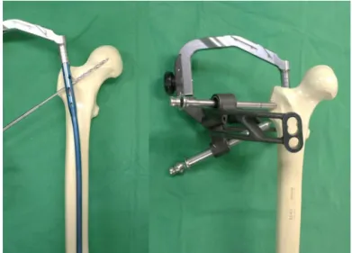
Key Words: Femur, Subtrochanteric fracture, limited open reduction, Proximal femoral nail anti-rotation (PFNA)... 수술 방법 및

No.. ramus insufficiency fracture, B) Thorax and abdomen CT showing multiple spine compression fracture.. ramus insufficiency fracture, B) After 4 months of

 Although van der Waals equation is still less accurate at high