Article http://dx.doi.org/10.14478/ace.2015.1002
쿼세틴과 루틴을 담지한 양이온 리포좀의 특성조사 및 UVA에 대한 세포 보호 효과
구현아⋅김문진⋅김해수⋅하지훈⋅유은령⋅박수남†
서울과학기술대학교 정밀화학과 나노바이오화장품연구실, 화장품종합기술연구소 (2015년 1월 2일 접수, 2015년 2월 2일 심사, 2015년 3월 3일 채택)
Characteristics and Cellular Protective Effects against UVA of Cationic Liposome Loaded with Quercetin and Rutin
Hyun A Gu, Moon Jin Kim, Hae Soo Kim, Ji Hoon Ha, Eun Ryung Yu, and Soo Nam Park
†A Department of Fine Chemistry, Cosemetic R&D Center, Seoul National University of Science and Technology, 232 Gongreung-ro, Nowon-gu, Seoul 139-743, Korea
(Received January 2, 2015; Revised February 2, 2015; Accepted March 3, 2015)
초 록
쿼세틴과 쿼세틴의 배당체인 루틴은 천연 항산화제로 잘 알려진 플라보노이드이다. 본 연구에서는 플라보노이드(쿼세 틴과 루틴)를 담지한 양이온 리포좀을 제조하여 세포 및 피부 투과성과 자외선(UVA)에 대한 HaCaT 세포 보호 효과를 평가하였다. 빈 양이온 리포좀의 입자 크기는 100∼130 nm이며, 입자 표면 전위는 + 33.05 mV를 나타내었다. 포집효 율은 루틴을 담지한 리포좀과 양이온 리포좀이 쿼세틴을 담지한 경우보다 높았다. 세포 내 이입율 비교결과, 양이온 리포좀이 일반 리포좀에 비해 약 5배 정도 높음을 확인했다. In vitro 상에서, 쿼세틴과 루틴이 용해된 PBS (phosphate-buffered saline) 수용액, 동량의 쿼세틴과 루틴을 담지한 리포좀과 양이온 리포좀의 피부투과율을 비교하였 다. 양이온 리포좀에 담지하였을 경우 가장 높은 피부투과율을 보였다. 플라보노이드를 담지한 양이온 리포좀의 자외 선(UVA 25 J/cm
2)에 대한 HaCaT 세포 보호 효과를 측정한 결과, 자외선만 조사한 군에 비해 플라보노이드 담지 양이 온 리포좀을 처리한 군에서 높은 세포 보호 효과를 보였다. 결과적으로, 양이온 리포좀은 플라보노이드를 피부 속으로 전달하는데 있어서 매우 유용한 피부 전달 시스템임을 확인하였다. 따라서, 세포 보호 및 피부 흡수 증진 효과를 가지 는 양이온 리포좀은 항노화 및 항산화 화장품 제형으로써 활용 가능성이 있음을 시사한다.
Abstract
Quercetin and its glycoside, rutin, are flavonoids, which are well known as natural antioxidants. In this study, cationic lip- osomes loaded with flavonoids (quercetin or rutin) were investigated for their effects on cell and skin permeability, and pro- tective effects against UVA. The particle size of the empty cationic liposomes was in the range of 100∼130 nm, and the zeta potential was + 33.05 mV. The entrapment efficiency of 0.5R/CL was higher than that of 0.5 Q/CL. The cellular uptake of the cationic liposomes was five-fold higher than that of liposomes. The skin permeability of quercetin and rutin was inves- tigated using Franz diffusion cells. Compared to the initial loading dose, the amount of quercetin or rutin delivered to the skin by cationic liposomes was higher than that delivered by conventional liposomes or phosphate-buffered saline. From the protective effect of cationic liposomes against UVA (25 J/cm
2), we found that the cell viability in cationic liposomes contain- ing flavonoids was higher than that of using UVA irradiation only. These results indicate that cationic liposomes provide enhanced delivery of flavonoids (quercetin and rutin) into the skin and may be used for antiaging and antioxidant cosmetics.
Keywords: cationic liposome, quercetin, rutin, antioxidant, skin permeation
1. Introduction 1)
Skin aging occurs via two processes : intrinsic aging, in which the
† Corresponding Author: Seoul National University of Science and Technology, A Department of Fine Chemistry, Cosemetic R&D Center, 232 Gongreung-ro, Nowon-gu, Seoul 139-743, Korea
Tel: +82-2-970-6451 e-mail: [email protected]
pISSN: 1225-0112 eISSN: 2288-4505 @ 2014 The Korean Society of Industrial and Engineering Chemistry. All rights reserved.
skin ages in the same way as other internal organs in the body, and ex- trinsic aging, which is caused by various environmental stressors. A ser- ies of deleterious biochemical reactions occur within the skin if it is ex- posed to excess UV radiation : this process is referred to as photoaging.
UV is divided into three classes as determined by wavelength : UVA (320∼400 nm), UVB (280∼320 nm), and UVC (200∼280 nm).
Although UVC comprises the most energetic wavelengths of UV light,
it is mostly absorbed by the ozone layer. Therefore, the classes of UV
light that are able to influence the skin are UVB and UVA.
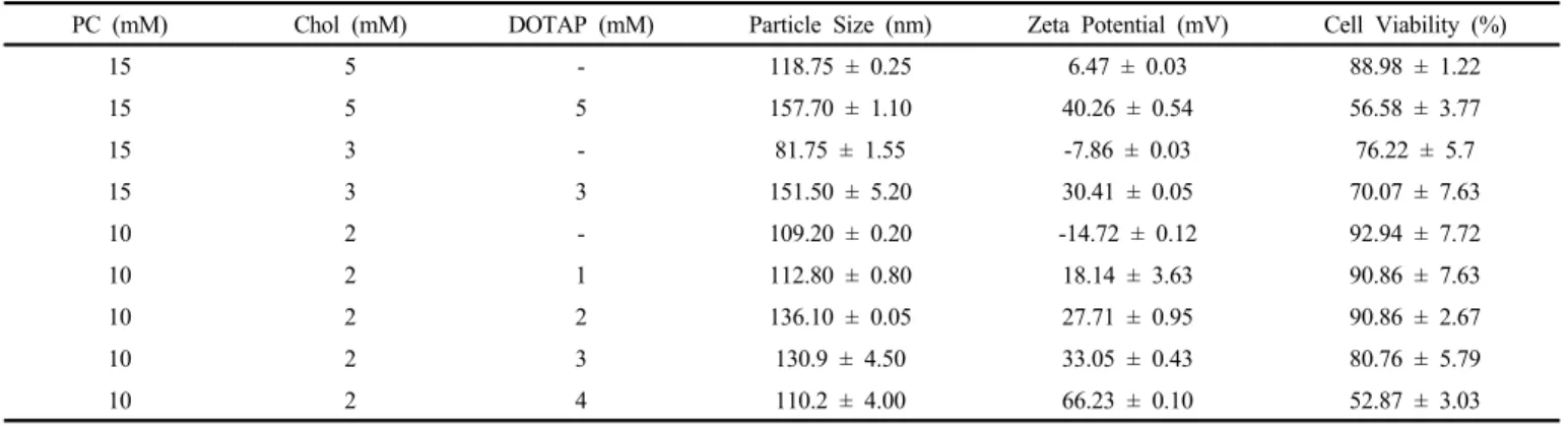
PC (mM) Chol (mM) DOTAP (mM) Particle Size (nm) Zeta Potential (mV) Cell Viability (%)
15 5 - 118.75 ± 0.25 6.47 ± 0.03 88.98 ± 1.22
15 5 5 157.70 ± 1.10 40.26 ± 0.54 56.58 ± 3.77
15 3 - 81.75 ± 1.55 -7.86 ± 0.03 76.22 ± 5.7
15 3 3 151.50 ± 5.20 30.41 ± 0.05 70.07 ± 7.63
10 2 - 109.20 ± 0.20 -14.72 ± 0.12 92.94 ± 7.72
10 2 1 112.80 ± 0.80 18.14 ± 3.63 90.86 ± 7.63
10 2 2 136.10 ± 0.05 27.71 ± 0.95 90.86 ± 2.67
10 2 3 130.9 ± 4.50 33.05 ± 0.43 80.76 ± 5.79
10 2 4 110.2 ± 4.00 66.23 ± 0.10 52.87 ± 3.03
PC : L-α-Phosphatidylcholine from egg yolk (egg PC, ~ 60%), Chol : Cholesterol, DOTAP : 1,2-Dioleoyl-3-trimethylammonium-propane
Table 1. Particle Size, Zeta Potential and Cell Viability of Liposome according to Mole Ratio of Lipids (PC, Chol, DOTAP)
Skin is composed of epidermis, dermis, and subcutaneous tissue.
UVB affects the epidermis, where it destroys DNA in the stratum corneum. In contrast, UVA can penetrate into the dermal layer of the skin, where it generates reactive oxygen species (ROS)[1-2]. ROS cause oxidative damage to nucleic acids, cellular proteins, and lipids and induce the synthesis of a series of matrix metalloproteinases that cause collagen degradation, which results in the formation of wrin- kles[3-4]. To protect the skin against ROS, antioxidant barrier systems consist of enzymatic antioxidants (i.e., catalase and glutathione) and nonenzymatic antioxidants (i.e., vitamin E, vitamin C, and flavonoids) in the skin[5-7]. However, when there is an excessive production of ROS, these natural antioxidant defenses are degraded. Therefore, the homeostasis of antioxidant defenses must be maintained by supplying antioxidants to the skin.
In the epidermis, the stratum corneum plays important roles in pro- tecting the body from harmful elements in the environment and main- taining hydration, but it can also prevent the absorption of active com- ponents[8-9]. Thus, to overcome this barrier and effectively deliver ac- tive components beyond it, various drug delivery systems have been studied[10-13].
Cationic liposomes made of positively charged lipids are increas- ingly being researched for use in gene therapy, due to their favorable interactions with negatively charged nucleic acids, plasmids, mRNA, and proteins via electrostatic attraction[14]. 1,2-Dioleoyl-3 trimethy- lammonium-propane (DOTAP) is the most widely used cationic lipid for in vitro and in vivo applications. Thus, we used DOTAP together with phosphatidyl choline (PC) and cholesterol (Chol) at various molar ratios to prepare the cationic liposomes used in this study[15].
Quercetin and its glycoside, rutin are typical flavonoids that are re- ported to act as strong antioxidants. These flavonoids, have been wide- ly used as anti-oxidants in cosmetics, but their use is limited because it makes its formulation difficult and have poorly water solubility.
In this study, the cationic liposomes containing quercetin or rutin were prepared and the physical characteristics compared with conven- tional liposomes. And the cationic liposomes containing flavonoids were investigated for protective ingredient against UVA, in HaCaT cells and skin permeability to investigate whether cationic liposomes
containing flavonoids could be an efficient drug delivery system.
2. Materials and Methods
2.1. Materials
DOTAP was purchased from Avanti Polar Lipids Inc. (Alabaster, USA). L-α-Phosphatidylcholine from egg yolk (egg PC : ∼60%), Chol (≥ 99.0%), quercetin, rutin, paraformaldehyde, phosphotungstic acid (PTA), fluorescein isothiocyanate (FITC), rhodamine B, 4ʹ,6-dia- midino-2-phenylindole (DAPI), and polyvinyl alcohol mounting were purchased from Sigma (USA). Dulbecco’s modified Eagle’s medium (DMEM), fetal bovine serum (FBS), and penicillin-streptomycin were purchased from PAA Co. (Pasching, Austria). Solvents used such as Na
2H
2PO
4⋅2H
2O, Na
2HPO
4⋅12H
2O, 1,3-butyleneglycol, ethanol, chloroform, and dimethyl sulfoxide (DMSO) were extra pure grade.
2.2. Cationic liposomes
2.2.1. Preparation of cationic liposomes
Cationic liposomes were prepared by the thin-film hydration method.
Egg PC, DOTAP, Chol, quercetin and rutin were used in various molar ratios (Table 1). All components were dissolved in 10 mL chloroform.
The solvent in the tube was subsequently removed using a rotary evap- orator (Buchi, Switzerland). The film was hydrated with 10 mL phos- phate-buffered saline (PBS; pH 7.4). Then, the vesicle suspension was homogenized using a digital probe sonicator (Branson, USA) for 15 min to produce a homogenous mixture and pass through a 1.20 µm fil- ter (Minisart CA 26 mm).
2.2.2. Cell viability
Cell viability was assessed using the MTT assay. HaCaT cells were cultured using DMEM medium supplemented with glutamine (2 mM), penicillin (400 U/mL), streptomycin (50 mg/mL), and 10% FBS for 24 h in 96-well plates (37 ℃, 5% CO
2). Liposomes were diluted 10-fold with serum-free medium and then incubated with HaCaT cells for 1 h.
MTT solution (2 µg/mL) was added to the cells, which were then in-
cubated for 3 h (37 ℃, 5% CO
2). The formazan crystals produced
were dissolved in DMSO and quantified by measuring their optical
density at 570 nm using an ELISA reader. Non-treated cells were used as the negative control, and the cell viability was calculated using the following equation :
Cell viablility A
con tr olA
ex per i m en t× (1)
2.2.3. Particle size and zeta potential
Particle size and distribution in the liposome solution was assessed 70 times with 3 repeated measurements using a particle size analyzer (Otsuka ELS-Z2, Otsuka Electronics, Japan) at 25 ℃ with a scattering angle of 165° using an argon laser. The average particle size was de- termined by cumulative analysis, and the distribution was resolved us- ing the Contin method.
Zeta potential was measured at 25 ℃ with a scattering angle of 165°
using an argon laser. The measurement was repeated three times and was resolved using the Smoluchowski method.
2.2.4. Transmission electron microscopy
Each liposome was negatively stained using 4% PTA (pH 7.4). The ratio of liposome and PTA was 1 : 1. 20 µL of the sample was applied to a grid (200 mesh). Liposomes that passed through the grid were re- moved using filter paper. The prepared sample was observed by using transmission electron microscopy (TEM, JEM1010, Japan).
2.2.5. Flow cytometry
HaCaT cells were cultured in 6-well plates for 24 h (37 ℃, 5%
CO
2). 0.03% FITC was loaded in each liposome and dissolved in PBS.
Prepared samples were diluted 10-fold with serum-free medium and then incubated with the cells for 1 h. After that, cells were washed with PBS twice and then treated with 0.25% trypsin to harvest the cells. The resulting cell suspension was centrifuged at 3000 rpm. The cell pellet was resuspended in 0.1% BSA and then fixed with paraf- ormaldehyde (4%, PBS) at 4 ℃. Fluorescence analysis to identify cel- lular uptake was performed with a FACS AriaIII (BD Biosciences, USA). FITC uptake was determined using excitation at 488 nm using an argon laser (emission wavelength = 520 nm).
2.2.6. Cell imaging system
HaCaT cells were cultured on cover glass in 6-well plates for 24 h (37 ℃, 5% CO
2). 0.03% FITC was loaded in each liposome and dis- solved in PBS. Prepared samples were diluted 10-fold with serum-free medium and then incubated with the cells for 1 h. After that, the cells were washed with PBS twice and then treated with 10 µg/mL DAPI for 20 min at room temperature to stain the nuclei. The cells were then treated with mounting solution (BioMeda) and fixed on the slide glass.
Fluorescence analysis to identify cellular uptake was performed with a DeltaVision system (Applied Precision). FITC uptake in cells was measured via excitation at 488 nm using an argon laser (emission wavelength = 520 nm). DAPI was excited at 408 nm using an argon laser (emission wavelength = 460 nm).
2.2.7. Confocal laser scanning microscopy
Confocal laser scanning microscopy (CLSM) was used to investigate the delivery to the skin by cationic liposome. Each sample was pre- pared using 0.03% FITC. Full-thickness skin was removed from the dorsal side of an ICR hairy albino mouse (eight weeks, female). The bristles and subcutaneous fat were carefully removed from the skin.
Diffusion studies were carried out as described below (12 h). The skin surface was washed with PBS and the diffusion area was punched out.
The diffusion area was then incorporated into an OCT compound and frozen at -70 ℃. The frozen skin was then sectioned with a cryostat into 10 µm slices. These tissues were fixed onto slide glasses and in- cubated with 10 µg/mL DAPI for 20 min at room temperature to stain the nuclei. To improve the adhesion between the tissues and cover glass, a polyvinyl alcohol-mounting medium with DABCO (Sigma) was applied. The degree of penetration by FITC was assessed by CLSM. FITC was excited with the 520 nm line from an argon laser.
To distinguish dermis and epidermis, DAPI-stained cell nuclei were ex- cited at 405 nm using an argon laser (emission wavelength = 460 nm).
2.3. Cationic liposomes loaded with quercetin or rutin.
2.3.1. Entrapment efficiency of quercetin or rutin in cationic liposomes
Unloaded quercetin or rutin was removed from 1 mL of a cationic liposome solution using a 1.20 µm filter. The filtered liposome solution was then degraded with 15 mL ethanol. The ethanol was evaporated in a rotary evaporator, and then the quercetin or rutin was redissolved in 1 mL ethanol. The concentration of quercetin or rutin was de- termined using HPLC (Shimazu, Japan) at 370 nm and 355 nm, re- spectively, and equation (2) was used to calculate the entrapment effi- ciency :
Entrapment efficiency C
iC
e× (2)
C
e: the concentration of encapsulated drug (encapsulated concen- tration, µM),
C
i: the initial concentration of the drug (initial concentration, µM).
2.3.2. Determination of the stability of cationic liposomes
The stability of cationic liposomes loaded with quercetin or rutin was evaluated by measuring the change of average particle size and ze- ta potential for three weeks. All prepared cationic liposomes were ob- served by naked eye on precipitation.
2.3.3. In vitro skin permeation study
The in vitro skin permeation study for cationic liposomes loaded with quercetin or rutin was carried out using 9-mm Franz diffusion cells (receptor volume 5 mL). Full-thickness skin was removed from the dorsal side of an ICR albino mouse (eight weeks, female). The bristles and subcutaneous fat were carefully removed from the skin.
The skin was fixed between the donor and the receptor phase with the
stratum corneum side facing the donor compartment. The receptor
phase was prepared using a ratio of HCO-60 : ethanol : PBS = 2 :
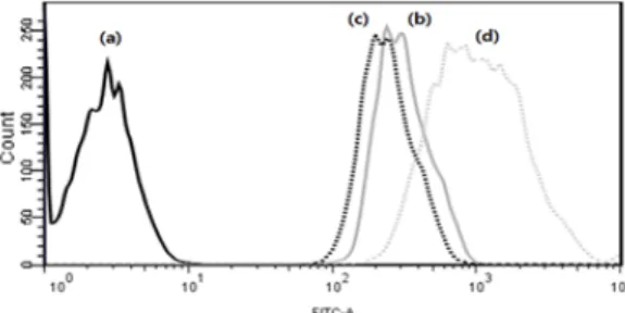
Figure 1. Flow cytometric analysis; intracellular uptake of FITC-loaded in catinic liposomes; (a) negative cell, (b) PB solution, (c) liposome, (d) cationic liposome (DOTAP 3 mM).
20 : 78 (w/w/w %). After adding 5 mL of the receptor phase into the receptor chamber, the receptor phase was continuously stirred using a V6A Stirrer (Permegear, USA) at 150 rpm for 24 h. The temperature was maintained at 37 ℃. Samples (0.2 mL) were applied to the skin in the donor compartment. Receptor solution samples were withdrawn through the sampling port of the receptor compartment at 4, 8, 12, and 24 h. The receptor phase was immediately replenished with an equal volume of fresh receptor phase. The withdrawn sample was analyzed by HPLC.
The amount of quercetin or rutin retained in skin was determined at the end of the in vitro permeation experiment (24 h). The skin surface was washed three times with PBS to remove residual donor sample.
The stratum corneum was removed by the stripping method using Scotch tape (3M, Korea). The quercetin or rutin present in each skin sample was extracted in 10 mL ethanol using a sonicator. The concen- tration of quercetin or rutin was determined by HPLC[16-17].
2.3.4. Protective effect against UVA
Each liposome was diluted 10-fold with serum-free medium and then added to HaCaT cells in 96 well plates for 1 h. After that, cells were washed with PBS twice and then covered with PBS prior to UVA exposure. For irradiation, a CL-1000UVA Crosslinker (USA) was used, and cells were irradiated at 25 J/cm
2. The cells were then kept in the dark in an incubator (37 ℃, 5% CO
2) for 24 h, and the cell viability was calculated using the MTT assay and the following formula :
Cell viablility A
con tr olA
ex per i m en t× (3)
2.4. Statistical analysis
Experiments were carried out in triplicate and Paired t-test was used to assess the differences.
3. Results and Discussion
3.1. Cationic liposome 3.1.1. Cell viability
We prepared cationic liposomes by varying the molar ratio of PC, Chol and DOTAP to find the condition which not showed cytotoxicity (Table 1). The standard of cytotoxicity was above 80% of cell viability. As a result, when the content of total lipid or DOTAP in- crease the cell viability decreased. When the content of DOTAP in- crease, the zeta potential of cationic liposomes increased and influ- enced in cell viability. A high zeta potential caused cytotoxicity in var- ious cell types was reported[18]. Thus, we reduced the total content of lipids in general liposomes and then prepared cationic liposomes by in- creasing the content of DOTAP. Therefore, we investigated that opti- mal ratio of PC : Chol was 10 mM : 2 mM and the content of DOTAP not showed cytotoxicity until 3 mM.
3.1.2. Particle size and zeta potential
When the ratio of PC : Chol : DOTAP are various, the particle size
and zeta potential were compared (Table 1). When the content of DOTAP increased, the particle size and zeta potential of cationic lip- osomes increased. The average particle size of prepared liposomes was 110.89 nm and shown the monodisperse. Moreover, each zeta potential of cationic liposome (PC : Chol : DOTAP = 10 : 2 : 3) and conven- tional liposome (PC : Chol = 10 : 2 ) were 33.05 ± 0.43 mV and -14.72 ± 0.12 mV, respectively. The maximam difference between them was 47 mV.
3.1.3. Flow cytometry (FACS)
We used flow cytometry to compare the intracellular uptake of cati- onic liposomes prepared with different amounts of cationic lipids. The experiment was processed by using the same amount of poorly wa- ter-soluble fluorescent material. As a result, when the content of DOTAP increased, the amount of intracellular uptake increased. These results indicate that the optimal formulation of cationic liposomes was PC : Chol : DOTAP = 10 : 2 : 3 (data not shown). In addition, the amount of cellular uptake of cationic liposomes was 5.03-fold higher than that of conventional liposomes or PBS (Figure 1). These result has been widely considered that as the cell membrane is negatively charged, the electrostatic attraction between cationic liposomes and the cell membrane facilitates uptake.
3.1.4. Cell imaging system
We used a cell imaging system to confirm the location of fluo- rescent material in HaCaT cells (Figure 2). We found that when cati- onic liposome was treated on cell, the intensity of fluorescence around the cells was high. In addition, the fluorescence was localized around the nuclei. These results indicated the intracellular uptake of the fluo- rescent material and accorded with previous results from flow cytometry.
3.1.5. Transmission electron microscopy (TEM)
We used TEM to compare the morphology of liposome and cationic liposome (Figure 3). The particle size of each liposome showed 100 nm according to results of particle size. The morphology of liposome was a mono-layer and it of cationic liposome was multi-layer.
3.1.6. Confocal Laser Scanning Microscope (CLSM)
To compare the efficiencies of delivery to the skin by cationic lip
Liposome Formulation (mM) Particle Size (nm)
Zeta Potential (mV)
Entrapment Efficiency
PC Chol DOTAP Q R (%)
L 10 2 - 109.20 ± 0.20
*-14.72 ± 0.12 -
CL 10 2 3 130.90 ± 4.50
*33.05 ± 0.43 -
Q/L 10 2 - 1 155.50 ± 0.50 -16.74 ± 0.69 38.07 ± 0.01
Q/CL 10 2 3 1 140.80 ± 1.40 32.54 ± 0.18 63.58 ± 0.01
R/L 10 2 - 1 262.10 ± 1.10 -16.80 ± 0.04 64.87 ± 0.02
R/CL 10 2 3 1 202.05 ± 0.05 35.21 ± 0.40 67.51 ± 0.01
0.5Q/L 10 2 - 0.5 119.50 ± 2.20
*-16.74 ± 0.45 57.69 ± 0.02
0.5Q/CL 10 2 3 0.5 122.70 ± 9.45
*39.15 ± 0.15 59.64 ± 0.02
0.5R/L 10 2 - 0.5 83.05 ± 2.25
*-15.23 ± 0.03 69.61 ± 0.01
0.5R/CL 10 2 3 0.5 121.75 ± 1.15
*35.74 ± 4.70 71.64 ± 0.01
*