Received: June 21, 2021 Revised: August 16, 2021 Accepted: August 20, 2021 Journal of
Trauma and InJury
CASE REPORT
J Trauma Inj 2021;34(3):183-186 https://doi.org/10.20408/jti.2021.0045
Correspondence to Dae Sung Ma, M.D.
Trauma Center, Department of Thoracic and Cardiovascular Surgery, Dankook University Hospital, 201 Manghyang-ro, Dongnam-gu, Cheonan 31116, Korea Tel: +82-41-550-7640
Fax: +82-41-550-7060 E-mail: [email protected] ORCID: https://orcid.org/0000-0001- 7521-3949
http://www.jtraumainj.org eISSN 2287-1683
pISSN 2799-4317
Copyright © 2021 The Korean Society of Traumatology
This is an Open Access article distributed under the terms of the Creative Commons Attribution Non-Commercial License (http://creativecommons.org/licenses/by-nc/4.0/) which permits unrestricted noncommercial use, distribution, and reproduction in any medium, provided the original work is properly cited.
Extra-Pericardial Tamponade due to Internal Thoracic artery rupture after Blunt Trauma: a Case report
Donsub Noh, M.D., Sung Wook Chang, M.D., Ph.D., Dae Sung Ma, M.D.
Trauma Center, Department of Thoracic and Cardiovascular Surgery, Dankook University Hospital, Cheonan, Korea
Cardiac tamponade is an acute life-threatening condition that predominantly involves the intra-pericardial space; however, an expanding mediastinal hematoma can also sometimes cause cardiac tamponade. Here we describe the case of a 45-year-old male driver in whom a traffic accident resulted in rupture of the left internal thoracic ar- tery (ITA), extra-pericardial hematoma, and sternal fracture. After resuscitation, he was scheduled to undergo angio-embolization to repair the ruptured left ITA, but he suddenly developed cardiac tamponade that required a decompressive sternotomy.
Nevertheless, the patient had an uncomplicated recovery, and this case suggests that extra-pericardial cardiac tamponade should be considered as a possible consequence of retro-sternal hematoma due to traumatic ITA rupture.
Keywords: Blunt injury; Internal thoracic arteries; Cardiac tamponade
INTRODUCTION
Traumatic internal thoracic artery (ITA) injury is exceedingly rare; it occurs due to
blunt trauma in most cases and is accompanied by injuries such as rib fracture (17%)
and sternal fracture (17%) [1]. Traumatic ITA injury typically presents as hemotho-
rax, anterior mediastinal hematoma, or pseudoaneurysm, and very rarely presents as
extra-pleural hematoma, extra-pericardial hematoma, or arteriovenous fistula. Nota-
bly, most of these conditions are managed by angio-embolization [1]. Nevertheless, a
growing extra-pericardial hematoma can result in cardiac arrest and requires surgical
treatment [2]. Here, we describe the successful management of extra-pericardial car-
diac tamponade caused by a large retro-sternal hematoma due to traumatic rupture of
the ITA.
184
https://doi.org/10.20408/jti.2021.0045Journal of Trauma and Injury Volume 34, Number 3, September 2021
CASE REPORT
A 45-year-old male driver was transferred to Regional Trauma Center of Dankook University Hospital after he had crashed into a fence. At presentation, he was mildly drowsy and had sustained an approximately 20 cm long deep laceration on his right knee, and his distal femur was
exposed. His vital signs were a blood pressure of 77/56 mmHg, a pulse rate of 103 beats/minute, a respiratory rate of 22 breaths/minute, and a digital oxygen satu- ration of 98%. The right dorsalis pedis pulse was weak and his Glasgow Coma Scale score was 14 (E3V5M6).
An extended focused assessment with sonography did not show the sliding sign, which is a characteristic of left
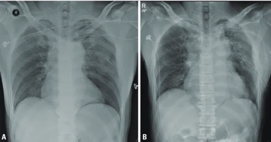
Fig. 1. (A) Left pneumothorax, as seen on the initial chest X-ray. (B) Mediastinal shadow widening was observed on computed tomography.
A b
Fig. 2. Chest computed tomography showed active bleeding from the left internal thoracic artery in the anterior mediastinum, along with a sternal
fracture.
185
http://www.jtraumainj.org
Dongsub Noh, et al. Extra-Pericardial Tamponade after Trauma
hemithorax, and was otherwise unremarkable; however, a chest X-ray examination revealed left pneumothorax (Fig. 1A). After resuscitation with two units of red blood cells and a left-sided closed thoracostomy, his blood pres- sure increased to 90/42 mmHg. Although computed to- mography (CT) of the brain and the abdomen showed no specific injuries related to this trauma, contrast-enhanced chest CT showed active bleeding from the left ITA in the anterior mediastinum along with a multiple-segment ster- nal fracture (Fig. 2). Therefore, angiographic embolization of the left ITA and evaluation of the injured artery around the right knee were planned. However, while waiting for angio-embolization, his vital signs deteriorated to a blood pressure of 67/32 mmHg and a pulse rate of 120 beats/
minute, and a follow-up chest X-ray showed widening of the mediastinal shadow (Fig. 1B). Fluid management and blood transfusion did not restore his blood pressure and pulse rate. Based on his deterioration, we reasoned that the active bleeding from the ruptured left ITA could have led to extra-pericardial cardiac tamponade. Therefore, rather than angio-embolization of the ITA, we performed a median sternotomy, both to immediately relieve the
tamponade by removing the substernal hematoma and to ligate the ruptured ITA. When sternotomy was per- formed, we found two sites of transverse fractures on the sternum with bone bleeding, a large hematoma compress- ing the heart, and active bleeding from two rupture sites of the ITA. Removal of the hematoma (about 2,000 mL) restored the patient’s vital signs and the proximal part of the left ITA was ligated. The pericardium was opened, and a gross examination did not reveal any injuries to the heart or the great vessels. After controlling bleeding, the sternotomy site was fixed with SternaLock
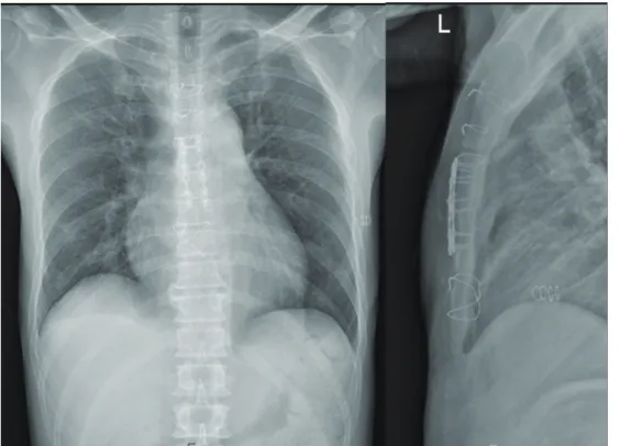
®(Biomet Microfixation Inc., Jacksonville, FL, USA), and the ster- notomy area was fixed with a figure-of-eight suture using steel wires (Fig. 3). The patient’s immediate postoperative course was uneventful, and he underwent knee repair surgery 3 days later. He was discharged after a 3-month hospital stay.
DISCUSSION
Cardiac tamponade is an acute, life-threatening condition
Fig. 3. The fractured sternum was fixed using SternaLock® (Biomet Microfixation Inc., Jacksonville, FL, USA), a steel wire, and figure-of-eight sutures.