경상대학교 의과대학 내과학교실, 진단방사선과학교실
함현석, 이승준, 조유지, 정이영, 전경녀1, 김호철, 이종덕, 황영실
A Case of Lung Cancer Obscured by Endobronchial Aspergilloma
Hyun Seok Ham, M.D., Seung Jun Lee, M.D., Yu Ji Cho, M.D., Kyoung Nyeo Jeon M.D.1, Yi Yeong Jeong M.D., Ho Cheol Kim, M.D., Jong Deok Lee M.D., Young Sil Hwang M.D.
Department of Internal Medicine and Diagnostic Radiology, College of Medicine, Gyeongsang National University, Jinju, Korea
A 70-year-old man was referred to the department of pulmonology due to blood tinged sputum and an abnormal chest X-ray. The chest X-ray and CT scans revealed a lobulated contour mass-like lesion in the left upper lung field. The bronchoscopic examination showed a whitish and polypoid mass occluding the left upper lobe bronchus. A biopsy specimen from the lesion revealed many aspergillus hyphae. Intravenous and oral itraconozole were administered over a 4 weeks period. Several months later, the size of the mass on chest X-ray increased and a percutaneous lung biopsy revealed a sarcomatoid carcinoma. We reported a case of lung cancer that was obscured by an endobronchial aspergilloma with a review of the relevant literature. (Tuberc Respir Dis 2006; 61: 157-161)
Key words: Endobronchial aspergilloma, Lung cancer.
Address for correspondence: Ho Cheol Kim, M.D.
Department of Internal Medicine, College of Medicine, Gyeongsang National University. 92 Chilam Dong, Jinju, 660-751, Korea.
Phone: 055-750-8684 Fax: 055-758-9122 E-mail: [email protected]
Received: Apr. 18. 2006 Accepted: Jun. 23. 2006
서 론
아스페르길루스는 호흡기를 가장 흔히 침범하는 진균으로 환자의 면역상태에 따라 침윤성 아스페르 길루스증, 만성 괴사성 폐 아스페르길루스증, 알레르 기성 기관지폐 아스페르길루스증, 아스페르길루스종 등의 다양한 임상양상으로 나타난다1. 정상적인 면역 기능을 가진 환자에서 폐의 공동성 병변없이 기관지 내에만 국한되어 나타나는 기관지내 아스페르길루스 종(endobronchial aspergilloma)은 기존의 분류와는 구별이 어려운 특이한 형태이며2, 면역 저하 환자에서 침습성 아스페르길루스증과 동반되어 나타나는 괴사 성 기관지내 아스페르길루스증(necrotizing endob- ronchial aspergillosis)과는 다른 질환이다3.
저자들은 각혈과 폐 종괴로 내원한 70세 남자 환자 에서 기관지내시경을 이용한 조직 검사로 기관지내 아스페르길루스종으로 진단하고 치료하였으나 호전
을 보이지 않아 이후 시행한 경피적폐생검을 통해 폐 암으로 진단된 1예를 경험하여 문헌과 함께 보고하는 바이다.
증 례
70세 남자 환자가 간헐적인 각혈과 단순흉부방사 선에서 종괴가 관찰되어 호흡기 내과로 전원되었다.
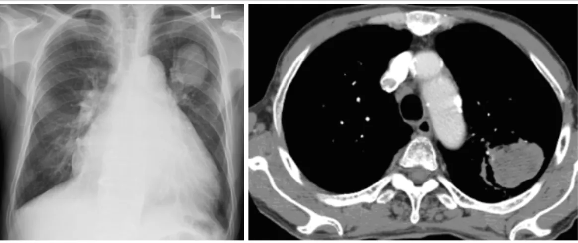
환자는 약 4년 전부터 울혈성심부전과 심방세동으로 본원 심장내과에서 지속적으로 치료를 받았으며 최 근에 갑작스런 호흡곤란의 악화로 심장내과에 입원 하여 치료하던 중 각혈과 흉부방사선의 이상으로 전 원되었다. 평소 울혈성 심부전과 심방 세동으로 Dig- oxin과 warfarin, 칼슘통로 길항제, 이뇨제를 지속적 으로 복용 중이었다. 가족력상 특이 소견은 없었으며 폐결핵이나 당료의 병력은 없었다. 이학적 소견에서 호흡음은 정상이었으나 심장 청진소견에서 수축기 잡음이 관찰되었으며 생체 활력증후는 정상 범위였 다. 단순흉부방사선 사진에서 좌상엽에 약 3.5cm 정 도의 바벨 모양의 종괴가 관찰되었으며 흉부 전산화 단층촬영에서는 좌상엽에 소엽형상의 모서리를 가지 고 조영증강이 되지 않는 저음영의 종괴가 관찰되었 다(Figure 1, 2). 일반혈액 검사상 백혈구 6500/mm3, 혈색소 9.5g/dL, 적혈구용적율 29%, 혈소판 175,000/
Figure 1. Initial chest radiograph showed mild cardiomegaly and barbell shaped mass-like opacity in left upper lung field.
Figure 2. A CT scan showed multi-lobulated contour and non-enhancing soft tissue mass –like lesion in left upper lobe posterior segment.
Figure 3. Bronchoscopic examination showed whitish and polypoid mass occluding left upper lobe apicoposterior segment.
Figure 4. Histologic finding of the mass showed numerous branched, septated aspergillus hyphae with adjacent fibrous exudates in PAS stain(x 100).
mm3 이었고 생화학 검사는 정상이었다. 대기중에서 시행한 동맥혈가스 검사상 pH 7.45, 이산화탄소분압 38mmHg, 산소분압 78mmHg, 중탄산염 24mmol/L, 산소포화도 92%이었다. 객담도말 검사상 결핵균은 발견되지 않았으며 혈액 및 객담의 세균 배양검사에 서도 이상소견은 없었다. 기관지내시경 소견에서 좌 측 기관지의 상부 기시부의 꼭대기뒤구역(apicopos- terior)을 막고 있는 흰색의 폴립모양의 종괴가 관찰 되었으며(Figure 3) 조직검사에서 격막이 있고, 급성
분지를 이루는 진균성 균사가 다량으로 관찰되어 기 관지내 아스페르길루스종으로 진단하였다(Figure 4).
이후 intraconazole 100mg을 하루 2회 정맥으로 약 2 주간 사용한 뒤 경구 제제로 교체한 뒤 2주간 복용하 면서 경과를 관찰하였다. 치료 6주 경과 뒤 기관지내 시경에서 병변의 변화가 없었고 간헐적인 객혈 이외 증상의 변화가 없어서 특별한 치료를 하지 않고 경과 를 관찰하였다. 약 6개월 뒤 확인한 단순흉부방사선
Figure 5. Several months later, the size of mass increased in chest radiograph and CT scan.
Figure 6. Immunohistochemical staining showed that a pleomorphic tumor with spindle cells, positive for vimentin, are invading into normal large round pale pink muscle fibers(x400)
소견에서 좌측 폐야의 종괴가 커지는 양상을 보여 경 피적 폐생검을 시행하였으며 면역화학조직염색에서 vimetin이 양성을 보인 육종양 암종(sarcomatoid carcinoma)으로 진단되었다(Figure 5, 6). 환자는 불 량한 전신상태와 동반된 중증의 울혈성 심부전으로 수술은 시행하지 못하였다.
고 찰
본 증례는 종괴와 폐허탈로 내원한 환자에서 기관 지내 아스페르길루스종이 동반되어 폐암의 진단이
늦어진 예로 국외 문헌에는 본 증례와 비슷한 증례가 보고되었다4,5. 국내에서는 대량 객혈6과 우중엽의 폐 허탈7을 보인 기관지내 아스페르길루스종이 보고되 어 있으며 본 증례와는 반대로 기관지 폐쇄를 유발한 종괴로 폐암이 의심되어 시행한 폐절제술에서 아스 페르길루스종으로 진단된 예가 보고되어 있다8. 본 증 례처럼 기관지내시경을 통한 조직검사로 기관지내 아스페르길루스종으로 오인되었다가 폐암으로 밝혀 진 경우는 처음 보고되는 것이다.
본 증례는 아스페르길루스과 관련된 폐질환 중에 서 Franquet 등9의 분류에 의하면 기도침습형 아스페 르길루스(airway-invasive aspergillosis)의 한 형태 인 폐쇄성 기관지폐 아스페르길루스(obstructing br- onchopulmonary aspergillosis)와 가장 유사한 것으 로 판단된다. 이것은 비침습적인 형태를 보이고 아스 페르길루스가 다량으로 기관지내에서 과성장을 하여 방사선 소견에서 기관지 또는 세기관지의 확장, 점액 질의 매복증(mucoid impaction) 및 폐쇄에 의한 무기 폐를 특징으로 한다. 본 증례에서도 흉부단층촬영에 서 점액질의 매복증에 의한 무기폐 소견으로 생각하 여 기관지내시경을 시행하였다. 기관지내 아스페르길 루스의 한 형태인 괴사성 기관지 아스페르길루스는 임상적, 병리학적으로 침습적 아스페르길루스의 한 형태로 분류하며, 폐실질의 침범없이 기관지 상피와 점막을 침범하여 기관지를 폐쇄하거나 기관지 주위 조직까지 파괴, 괴사시키는 상당히 드문 형태의 아스
페르길루스와 관련된 폐질환이다. 본 증례의 경우는 조직검사를 통해 기관지내 아스페르길루스가 확인이 되었지만 괴사성 기관지내 아스페르길루스라기 보다 는 단순한 집락으로 생각하는 것이 타당한 것으로 판 단된다. 본 증례에서 처음 아스페르길루스가 증명되 어 itraconazole를 약 2주간 정맥으로 투여하고 추가 적으로 2주 이상 경구로 투여하였지만 치료 6주 후에 시행한 기관지내시경 소견에서 병변이 전혀 변화가 없었으며 조직소견에서도 아스페르길루스 균사가 동 일하게 검출되었다. 이후 추가적인 경과 관찰에서도 침습적 아스페르길루스증으로 생각되는 임상양상은 보이지 않았다.
본 증례의 환자는 동반된 심장 질환은 있었으나 면 역 저하 상태이거나 아토피의 과거력이 없었는데 아 스페르길루스가 집락을 형성하는 데는 악성 종양이 기관지를 막고 있어 기관지 내부의 공기 흐름에 정체 를 유발하여 아스페르길루스가 집락을 형성하기 쉬 운 환경이 만들어졌거나, 원래 공동을 형성한 폐종양 이 있는 상태에서 집락을 형성했을 가능성이 있다. 폐 절제술 후 잘린끝(stump)에서 발생한 아스페르길루 스증은 드물지만 임상에서 관찰할 수 있으며 이것은 잘린끝에 매복 또는 돌출되어 있는 봉합사가 아스페 르길루스에 의한 감염의 핵으로 작용하여 발생하는 것으로 생각하고 있다10.
기관지내 아스페르길루스종의 치료는 확립된 것이 없으며 폐에 생기는 아스페르길루스종과 동일하게 항진균제를 경구 복용하거나 흡입, 경피적 또는 기관 지내로 주입하는 치료를 시도하거나 수술적으로 제 거하는 방법이 있다11. 공동을 가진 소세포성 폐암과 기관지내 잘린끝 아스페르길루스에서 고용량의 itra- conazole을 장기간 사용하여 호전된 예는 보고되어
있다12,13. 본 증례와 유사한 국외 보고에서는 3개월 동
안의 경구 itraconazole과 기관지내로 fluconazole의 점적 주입 후 기관지내시경을 통해 숨어있던 편평세 포암을 진단하였다. 본 증례의 환자에서도 이런 치료 를 좀 더 적극적으로 한 후 기관지내시경을 하였다면 조기 진단이 가능하였을 것으로 판단된다.
본 증례와 같이 기관지내 아스페르길루스종은 흔 하지는 않지만 기관지 폐쇄가 있는 경우 집락을 형성
하여 과성장을 유발할 수 있으므로 조직소견에서 아 르페르길루스가 증명이 된 경우에도 기관지 폐쇄를 유발하는 악성 종양이 있는 지를 염두해 두어야 할 것으로 사료된다.
요 약
저자들은 종괴와 폐허탈로 내원한 환자에서 기관 지내시경을 이용한 조직검사에서 기관지내 아스페르 길루스종으로 진단되어 치료하였으나 종괴의 크기가 증가하여 시행한 경피적 폐생검을 통해 악성 폐종양 으로 진단된 예를 경험하여 보고하는 바이다.
참 고 문 헌
1. Soubani AO, Chandrasekar PH. The clinical spectrum of pulmonary aspergillosis. Chest 2002;121:1988-99.
2. Kim JS, Rhee Y, Kang SM, Ko WK, Kim YS, Lee JG, et al. A case of endobronchial aspergilloma. Yonsei Med J 2000;41:422-5.
3. Pervez NK, Kleinerman J, Kattan M, Freed JA, Harris MB, Rosen MJ, et al. Pseudomembranous necrotizing bronchial aspergillosis: a variant of in- vasive aspergillosis in a patient with hemophilia and acquired immune deficiency syndrome. Am Rev Respir Dis 1985;131:961-3.
4. Yoshitomi A, Kuwata H, Suzuki T, Narushima M, Nakajima T, Yogo Y, et al. Lung cancer obscured by aspergillus hyphae. Nihon Kokyuki Gakkai Zasshi 2000;38:321-4.
5. Quoix E, Gasser B, Apprill M, Gourdon C, Pauli G, Roegel E. Endobronchial aspergillosis associated with a carcinoid tumor. Rev Mal Respir 1990;7:609-12.
6. Kim TH, Yong BJ, Kim YK, Lee YM, Kim KU, Uh ST, et al. A case of endobronchial aspergilloma with massive hemoptysis. Tuberc Respir Dis 2004;57:
589-93.
7. Eom WY, Kim NI, Kim SW, Lee BH, Kim SH, Ahn YS, et al. A case of endobronchial aspergilloma in patient with collapse of right middle lobe. Korean J Med 2006;70:221-5.
8. Park BJ, Kim YK, Kim H, Kim YH, Lee HI, Kang HM, et al. A case of endobronchial aspergillosis completely obstructing lobar bronchus. Tuberc Respir Dis 2005;59:311-4.
9. Franquet T, Muller NL, Gimenez A, Guembe P, de la Torre J, Bague S. Spectrum of pulmonary asper-
gillosis: histologic, clinical, and radiologic findings.
Radiographics 2001;21:825-37.
10. e Rochais JP, Icard P, Simon T, Poirier P, Evrard C.
Bronchial stump aspergillosis. Ann Thorac Surg 2000;
70:302-4.
11. Yamada H, Kohno S, Koga H, Maesaki S, Kaku M.
Topical treatment of pulmonary aspergilloma by antifungals: relationship between duration of the disease and efficacy of therapy. Chest 1993;103:
1421-5.
12. Impens N, de Greve J, de Beule K, Meysman M, de Beuckelaere S, Schandevyl W. Oral treatment with itraconazole of aspergilloma in cavitary lung cancer.
Eur Respir J 1990;3:837-9.
13. Noppen M, Claes I, Maillet B, Meysman M, Monsieur I, Vincken W. Three cases of bronchial stump aspergillosis: unusual clinical presentations and bene- ficial effect of oral itraconazole. Eur Respir J 1995;8:
477-80.