Ⅰ. 서 론
암은 정상적인 세포의 기능에 필요하나 기능을 상실하였을 때 종양 유전자로 변하는 종양억제유전자1)(Tumor suppressor gene) 와 전 종양유전자(proto-oncogene)의 유전적 변화에 의해 발생된
DMBA로 유도된 햄스터 협낭암종에서 p53 유전자 변이와 mdm-2 단백의 발현에 관한 연구
박용선∙김경욱∙이재훈∙김창진*
단국대학교 치과대학 구강악안면외과학교실, 순천향대학교 의과대학 병리학교실*
Abstract
박 용 선
330-716, 충남 천안시 신부동 산7-1 단국대학교 치과대학 구강악안면외과학교실 Yong-Sun Park
Dept. of OMFS, College of Dentistry, Dankook Univ.
san7-1, Shinbu-dong, Chon-an, Chung-Nam, 330-716, Korea Tel:82-417-550-1991 Fax:82-417-551-8988
STUDY ON MUTATION OF P53 AND EXPRESSION OF MDM-2 IN DMBA INDUCED CARCINOMA OF HAMSTER BUCCAL POUCH
Yong-Sun Park, Kyung-Wook Kim, Jae-Hoon Lee, Chang-Jin Kim* Department of Oral and Maxillofacial Surgery, College of Dentistry, Dankook University
Department of Pathology, College of Medicine, Soonchunhyang University*
Cellular proliferation is an intricately regulated process mediated by the coordinated interactions of critical growth control genes.
Two of these factors in mammalian cells are the p53 and mdm-2 genes. A protein product of the mem-2 oncogene has been recently shown to associate with the protein encoded by the tumor suppressor gene p53.
The p53 tumor suppressor protein is stabilized in response to DNA damage and other stress signals and causes the cell to undergo growth arrest or apoptosis, thus preventing the establishment of mutations in future cellular generations. Mutation or loss of p53 is a very common event in tumor progression. It occurs in about 50% of all tumors analysed including of colon, lung, breast and liver.
The cellular mdm-2 gene, which has potential transforming activity that can be activated by overexpression, is amplified in a signifi- cant percentage of human sarcoma and in other mammalian tumors. Proteins encoded by the mdm-2 gene are able to bind to the p53 protein and, when overexpressed, can inhibit p53′s transcriptional activation function, thus mdm-2 can act as a negative regulator of p53 function.
Experimental study was performed to observe the relationship between p53 gene mutation and mdm-2 protein expression and apply the results to the clinical activity.
36 golden syrian hamster each weighing 60�80g were used and painted with 0.5% DMBA by 3 times weekly on the right buccal cheek(experimental side) for 6, 8, 10, 12, 14 and 16 weeks. Left buccal cheek(control side) was treated with mineral oil as the same manner to the right side. The hamsters were sacrificed on the 6, 8, 10, 12, 14 & 16 weeks.
Normal and tumor tissues from paraffin block were examined for histology and immunohistochemistry observation, and were com- pletely dissected by microdissection and DNA from both tissue were isolated by proteins K/phenol/chloroform extraction. Segments of the hamster p53 exons 5, 6, 7 and 8 were amplified by PCR using the oligonucleotide primers, and then confirmational change was observed by SSCP respectively.
The results were as follows :
1. Dysplasia at 6 weeks, carcinoma in situ at 8 weeks and invasive carcinoma from 10 weeks could be observed in experimental groups.
2. p53 mutations were detected in 10 of the 36(28%) and the exons 6(6 of the 10 : 60%) was the most hot spot area among the highy conserved region(exons 5, 6, 7 & 8).
3. Immunohistochemical study confirmed 22 of the 36(61%) of p53 expression involving 10 of p53 mutations.
4. mdm-2 expression of was showed in 3 of the 36(8%) involving 1 of the 22 of p53 expression and 2 of the 14 of p53 non-expres- sion.
From the above results, mutation of p53 gene or expression of p53 protein may have the influence of the DMBA induced carcinoma of hamster buccal pouch but the expression of mdm-2 protein may not have relationship with tumorigenesis.
다. 즉, 전 종양유전자의 비정상적인 활성화나 종양억제유전자 의 비 활성화가 일어나 정상세포가 비정상적인 염색체의 구조, 수 및 형태를 나타내고 세포분열을 자주하는 형질전환(transfor- mation)된 암세포로 변하게 된다2-4).
인간에서 발생되는 전체 암종의 약 3%를 차지하며 95% 이상이 편평세포암종을 이루는 구강암에 대한 분자생물학적 연구에 의 한 전 종양유전자나 활성화된 종양 유전자(oncogene)에 대한 보 고들5-8)은 많이 있지만, 인간의 상피세포, 간엽세포, 조혈기관, 림 프조직 및 신경계등에서 발생되는 악성종양의 약 50% 이상9)에서 변이를 보이는 p53 종양억제유전자와 이 단백의 기능을 조절해 주는 다른 조절인자들과의 관계가 암 발생과정(tumorigenesis)에 미치는 영향에 대한 연구 특히 구강암에 대해서는 거의 없는 실 정이다.
p53은 처음에는 쥐의 태생기 섬유모세포에서 변이가 일어나 형질전환을 나타내므로 종앙유전자라고 생각되었으나, 정상 의 p53이 다른 종양유전자에 의해서 나타나는 형질전환을 감 소시킨다는 것을 관찰한 후부터 종양억제 유전자로 알려졌 다10).
정상 p53 유전자는 정상상태에서 손상받은 세포를 세포주기의 G1/S상태에서 세포복구를 유발하며11,12), 비가역적인 세포 손상의 경우 예정된 자연사(apotosis)를 유발하여 종양발생을 억제한 다13,14).
p53은 자외선, 방사선등 DNA 상해요인에 의해 단백질의 양 및 활성이 증가하는 상해유도 유전자(damage-inducible)로 DNA의 절단부위나 상해부위에 결합하여 성장저해유전자들을 활성화 시켜 세포주기를 조절하는데 mdm-2가 대표적 성장저해유전자 로서 p53 유전자의 음성조절자로서 역할을 한다15).
mdm-2는 분자량 90KD(p90)의 단백질로 쥐 3T3 cell line의 10번 염색체에서 형질전환되어(3T3-DM) 유전자 증폭이 일어난 유전 자라고 최초로 보고되었고16)골이나 연조직 육종17-22), 교모세포종 (glioblastoma)이나, 선상세포종(astrocytoma)17,23), 여러 종류의 혈액
암24,25)구강암과 유방암26,27)및 악성흑색종28)등 인간의 암종에서 유
전자증폭17,18), 전사 및 번역의 증가24,25,29,30)등에 의해 발현된 양상을
나타내고 있다17).
실험동물에서 암유발물질을 이용해서 악성종양을 유발시킬수 있는 방법이 소개된 이후로 점막에서 암을 유발하고자 하는 시 도가 많이 이루어져 Salley는 점막성 암종의 유발을 위해서는 햄 스터 협낭(Hamster Buccal Pouch ; HBP)이 가장 이상적인 동물 모 델이라고 소개하였고32,33), Pyrene 화합물로 조직단백과 강한 결합 을 하여 강력한 발암효과를 유발시키는 9, 12-dimethyl benzan- thracene(DMBA)을 미네랄 오일을 용매로 하여 사용한 후에 효과 적인 암 발생이 나타나 대표적 암유발물질로 이용하고 있다34).
본 연구에서는 햄스터 협낭에 DMBA를 정기적으로 도포하여 암을 유발시킨 후 p53의 변이가 많이 일어나는 유전적으로 잘 보 존된 지역(Highly conserved region)35,36)에서 p53 유전자 변이와 p53의 음성조절자인 mdm-2 단백의 발현을 관찰하여 구강암의 임상적 응용에 도움이 되고자 하였다.
Ⅱ. 연구재료 및 방법 1. 실험동물 및 수술 방법
체중 60�80g의 웅성 햄스터를 실험동물로 사용하였다. 실험 군으로 햄스터의 우측 협낭에 미네랄 오일에 녹인 0.5% DMBA를 0.2ml씩 주 3회 도포하였고, 대조군으로 좌측협낭에는 미네랄 오 일만을 같은 방법으로 도포하면서 도포 6주, 8주, 10주, 12주, 14 주 및 16주째에 6마리씩 희생하였다.
2. 연구방법
1) 조직학적 관찰
도살 후 좌우 협낭 점막을 절제하여 차가운 4% paraformalde- hyde에 8시간 고정 후 통상적인 방법에 의해 파라핀 포매를 하였 다. 이 포매에서 4
μ
m의 절편을 만들어 통상적인 방법으로 hema- toxylin-eosin염색을 하여 종양발생 유무를 관찰하였다(Fig. 2).2) 면역조직화학적 관찰
p53와 mdm2의 면역 조직화학적 염색을 위한 일차 항체는 각 각 Zymed(California, USA)사의 단일클론항체를 사용하였다.
염색은 paraffin절편을 xylene에 탈 파라핀 한 후 graded alcohol 로 함수과정을 거쳐 10mM citrate buffer(pH 6.0)에 넣고 microwave oven에 700W에서 20분간 끓여 항원을 노출시킨 후 내 인성 peroxidase를 차단하기 위하여 methanol 용액에 3% H2O2를 첨가시켜 실온에서 20분간 처리하였다. 비특이성 결합을 방지하 기 위하여 10% 정상 goat혈청과 30분간 실온에서 반응시켰다. 일 차 항체를 10% BSA(bovine serum albumin)의 PBS(phosphate buffered saline, pH 7.4)에 희석하여 실온에서 1시간 반응시키고 PBS로 3회 수세하고 biotin이 부착된 antimouse goat 면역글로부 린을 떨구고 실온에서 30분간 반응시켰다. PBS로 3회 수세 후 peroxidase가 부착된 streptavidin에 30분간 실온에서 반응시킨 후 PBS로 수세 후 0.003% 3′3-diaminobenzidine(DAB)로 발색 후 흐르 는 물로 수세하고 hematoxylin으로 대조염색하였다. 음성대조군 은 일차항원대신 정상 mouse serum을 사용하여 같은 과정을 거 쳤다.
면역조직화학 염색상 양성반응은 음성 대조군과 비교하여 암 세포의 10% 이상에서 핵내의 갈색반응을 보인 것을 양성으로 하 였다.
3) DNA추출
암세포와 정상세포에서 각각 DNA를 추출하기 위하여 microdissection 방법으로 암 조직과 정상조직을 구분하였다. 이 를 위하여 paraffin block에서 5개의 7
μ
m절편을 만들어 통상적인 방법으로 탈파라핀하고 degraded alcohol로 함수시킨 후 조직을 건조시켰다. 건조된 조직을 10× 현미경하에 28guage 바늘로 암 조직과 정상조직을 긁어(Fig. 3, 4), 300μ
l의 digestion buffer (50mM Tris HCl, pH 8.5, 1mM EDTA, 0.5% Tween 20, 400μ
gproteinase K)가 들은 1.5ml Eppendorf tube에 넣어 55℃에서 24시간 부란하였 다. 부란 후 proteinase K의 활성을 억제하기 위하여 97℃에서 5분 간 처리하고 phenol chloroform으로 DNA를 추출하였다. 추출과 정은 65℃의 0.5% sodium sarcosin의 buffer-saturated phenol을 300
μ
l씩 시료에 넣어 vortex시켜 homogenous emulsion을 만든 후 3,500g로 원심분리 하고 상층액을 깨끗한 Eppendorf tube에 옮긴 후 이에 300μ
l의 chloroform isoamylalcohol을 첨가하여 vortex로 잘 섞은 후 역시 3500g로 원심분리 하였고, 이 과정을 3회 반복하 였다. 그 후 상층액을 새 tube에 옮긴 후 1/10 volume의 3M sodi- um acetate와 2× volume의 100% ethanol을 첨가하여 조심스럽게 섞은 후 -70℃에 1시간 방치하여 DNA를 침전시켰다. 그 후 3500g 로 5분간 원심분리 한 후 상층액을 버리고 침전물을 70% cold ethanol로 수세 후 재원심분리하여 DNA를 침전시키고 DNA pel- let을 공기 중에 건조시킨 후 멸균 증류수로 용해시켰다.추출한 DNA의 순도는 photometer로 260nm와 280nm의 파장에 서 수치를 읽어 이 수치의 비 260/280가 1.8 이상인 DNA를 시료 로 사용하였고 순도가 이 수치 이하인 경우에는 phenol/chloro- form추출을 다시 하였다.
이렇게 추출한 DNA는 1% agarose gel에 전기영동하여 dsDNA 를 확인하였다37).
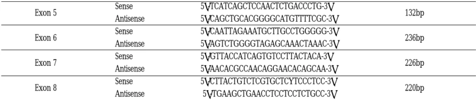
4) Polymerase Chain Reaction(PCR) Amplification PCR 반응을 위한 primer는 다음과 같었다38).
Exon 5 : Upstream 5′-TCATCAGCTCCAACTCTGACCCTG-3′
Down stream 5′-CAGCTGCACGGGGCATGTTTTCGC-3′
Exon 6 : Upstream 5′-CAATTAGAAATGCTTGCCTGGGGG-3′
Down stream 5′-AGTCTGGGGTAGAGCAAACTAAAC-3′
Exon 7 : Upstream 5′-GTTACCATCAGTGTCCTTACTACA-3′
Down stream 5′-AACACGCCAACAGGAACACAGCAA-3′
Exon 8 : Upstream 5′-CTTACTGTCTCGTGCTCYTCCCTCC-3′
Down stream 5′-TGAAGCTGAACCTCCTCCTCTGCC-3′
반응은 20
μ
l의 반응으로 1x PCR buffer에 dNTP 각각 250mM, 각 각의 primer 10pM, DNA/modified DNA 50ng, 1.25mM MgCl2, Taq polymerase 0.5U들을 넣어 반응 시켰고 반응조건은 95℃에서 5분 반응 후 95℃에서 30초, 55℃에서 30초, 72℃에서 30초 반응시키 고 이 과정을 36회 반복하였다. 그 후 72℃에서 5분 연장한 후 반 응을 종결하였다. 이렇게 반응시켜 얻은 PCR 산물은 1% agarose gel에 전기영동하여 각각의 bp를 확인하였다39).(Abbreviation ; T : Thymine, A : Adenine, C : Cytosine, G : Guanine)
5) Single Strand Conformation Polymorphism(SSCP)에 의한 변이 검사.
각각의 PCR증폭 산물 10
μ
l에 0.2M NaOH, 20mM EDTA의 alka- line denaturing solution 1μ
l를 넣은 후 42℃ 수조에 5분간 거치하 여 double strand DNA를 single strand DNA로 변성시킨 후 빨리 어름에 꽂아두어 single strand가 고유의 conformation으로 바뀌게 하였다. 그 후 0.5% bromophenol blue, 0.5% xylene cyanol in for- mamide로 구성된 loading solution 1
μ
l를 섞어 loading 하였다. Gel 은 5%의 acrylamide gel을 사용하여 상온에서 200V에서 15mA정 도로 3시간 전기영동하였다. Ethidium bromide로 염색하여 UV광 하에 관찰하여40), 정상과 종양에서의 PCR 산물을 비교하여 정상 에 비하여 다른 band shift를 보인 종양의 변이를 검사하였다.Ⅲ. 연구결과 1. 조직학적 소견
1) 정상군
정상군의 햄스터 협낭 상피는 4�6층으로 중층배열은 한 편평 상피로 이루어져 있고, 낭을 형성하고 있다(Fig. 1). 이들 정상군 에서는 p53과 mdm-2 단백의 발현은 관찰되지 않았다(Fig. 11, 18).
2) 실험군 가. 6주 소견
전 실험동물에서 상피세포의 증식으로 세포층이 증식되기 시 작하였으며 이들 증식세포들은 핵의 농염, 다형성(pleomor- phism)과 핵과 세포질이 비율이 증가하는 이형성(dysplasia) 소견 을 보였다(Fig. 5). 이러한 이형성 부위의 상피에서는 기저세포층 에서부터 p53 단백의 발현이 관찰되었고(Fig. 12). 한편 mdm-2의 발현은 관찰되지 않았다.
나. 8주 소견
이형성의 정도가 심해지며 국소적으로 상피내암의 소견이 관 찰되었다(Fig. 6). 이러한 상피내암 세포에서는 비균일적으로 p53 단백의 발현이 관찰되었고(Fig. 13), mdm-2 단백의 발현도 관찰 할 수 있었다(Fig. 19).
다. 10주 소견
침윤암으로 진행된 소견을 관찰할 수 있었고(Fig. 7), p53 단백 의 발현을 관찰할 수 있었고(Fig. 14), mdm-2 단백의 발현은 관찰 되지 않았다.
라. 12주, 14주 및 16주 소견
10주 소견과 마찬가지로 침윤성 암으로 발전된 양상(Fig. 8~10) 을 관찰할 수 있었고 p53 단백의 발현을 관찰할 수 있었다(Fig.
15~17). mdm-2 단백의 발현은 14주에서만 관찰할 수 있었다(Fig.
20).
2. Microdissection에 의한 DNA 추출
현미경 하에 종양조직과 정상조직을 구분하여 microdissection 으로 얻은 파라핀 포매절편에서 추출한 DNA는 1% agarose gel 전 기영동상에 smear pattern을 보였는데 이는 조직이 paraformalde-
hyde에 고정된 것이고 또한 paraffin block 제작 시 여러 단계의 유기용매에 조직이 접촉되었기 때문에 DNA가 분절(fragmenta- tion)된 결과인 것이었다. 그러나 이러한 분절된 DNA들도 300bp 이하의 DNA를 PCR로 증폭하는 데는 어려움이 없었다.
3. p53 유전자의 Polymerase Chain Reaction(PCR) Amplification
PCR 증폭 산물들을 1% agarose gel에 전기 영동하여 각각의 bp 를 확인하여 exons 5에서는 132 bp, exons 6은 236 bp, exons 7은 226 bp 그리고 exons 8은 220 bp로 각각 확인할 수 있었다(Table 1 , Fig. 21).
4. Single Strand Comformation Polymorphism (SSCP)
실험대상 36예 중 10주 실험군 exons 5에서 1예, 실험군 6, 8 ,12, 14 및 16주 exons 6에서 6예, 실험군 8주, 14주 exons 7에서 2예, 실 험군 12주 exons 8에서 1예로 모두 10예서 p53 유전자의 변이를 관찰할 수 있었다(Table 2, Fig. 22).
5. p53 단백과 mdm-2 단백의 발현
p53의 발현은 실험군 6주에서 3예, 8주에서 4예, 10주에서 4예, 12주에서 2예, 14주에서 5예 그리고 16주에서 4예 모두 22예서 관 찰되었고 이 22예에는 p53 유전자 변이를 보인 10예가 모두 포함 되었다.
mdm-2 단백의 발현은 p53 발현이 관찰된 22예 중 1예, 발현되 지 않은 14예중 2예 모두 3예에서 관찰되었다(Table 2).
Table 1.Primer Sequence of Oligonucleotide
Exon 5 Sense 5′-TCATCAGCTCCAACTCTGACCCTG-3′
132bp Antisense 5′-CAGCTGCACGGGGCATGTTTTCGC-3′
Exon 6 Sense 5′-CAATTAGAAATGCTTGCCTGGGGG-3′
236bp Antisense 5′-AGTCTGGGGTAGAGCAAACTAAAC-3′
Exon 7 Sense 5′-GTTACCATCAGTGTCCTTACTACA-3′
226bp Antisense 5′-AACACGCCAACAGGAACACAGCAA-3′
Exon 8 Sense 5′-CTTACTGTCTCGTGCTCYTCCCTCC-3′
220bp Antisense 5′-TGAAGCTGAACCTCCTCCTCTGCC-3′
Abbreviation : T : Thymine A : Adenine C : Cytosine G : Guanine
Table 2.Mutations of p53 and Expression of p53 & mdm-2.
6 weeks (n=6) 1 3 0
8 weeks (n=6) 1 1 4 2
10 weeks (n=6) 1 4 0
12 weeks (n=6) 1 1 2 0
14 weeks (n=6) 1 1 5 1
16 weeks (n=6) 2 4 0
합계 10 22 3
Abbreviation
Exp : Experiment Expr : Expression
Exp. Date p53 Gene Mutation Exons
p53 Expr. mdm-2 Expr.
5 6 7 8
Ⅳ. 총괄 및 고찰
암의 발생빈도는 계속 증가하여 사망에서의 순위는 순환기계 질환과 더불어 수위를 차지하고 있다. 두경부 암으로 분류되는 구강암은 전체 암의 발생빈도에서 차지하는 비율은 남자에서 약 4%, 여자에서 약 2% 정도이며 암에 의한 사망 원인 중에서는 남 자에서 약 2%, 여자에서 약 1% 정도이며 대부분이 편평세포암종 으로 아직도 원인과 치료가 뚜렷이 제시하지 않고 있는 대표적 질환 중이 하나이다41).
분자 생물학의 발달로 암의 발생을 유전자 변이로 이해하기 시 작하여42), 암은 전종양유전자의 비정상적인 활성화나 종양억제 유전자의 비활성화가 일어나 비정상적인 염색체의 구조, 수 및 형태를 나타내고 세포분열을 자극하는 형질전환된 암세포로 변 하여 종양이 발생되는 것으로 밝혀지고 있다2-4).
종양유전자는 현재까지 약 30개 이상의 실체가 규명되고 있고43), 종양억제유전자는 세포의 성장과 게놈의 변이를 억제시켜 클론 (clone)의 팽창을 저지하는 기능을 갖고 있으며 p53과 망막모세 포종(retiroblastoma : Rb) 억제 유전자등이 대표적 종양억제 유전 자로 알려지고 있다44).
p53 유전자는 인간의 17번 염색체 짧은 팔에 위치하고 있으며 11개의 exon으로 구성되어 있으며 p53 단백은 분자량 53KD으로 375개의 아미노산을 갖고 있는 인산화 단백이고10), SV 40에 의해 형질전환된 세포에서 T-antigen과 결합된 상태로 1979년 처음으 로 발견되어 종양유전자로 생각되었으나 정상의 p53 유전자가 ras나 adenovirus E1B 같은 종양유전자에 의해 형질전환된 섬유모 세포의 성장을 G1phase에서 멈추게 하고45), 동물이나 인간에서 발생된 여러 암종에서 p53 유전자의 변이의 관찰46)그리고 p53 유전자가 비활성화 혹은 변이된 상태로 SV 40 T-antigen, aden- ovirus E1B 및 papillomavirus E6에서 유래된 종양유발단백질과 결 합된 것을 관찰한47)후 정상의 p53은 종양억제유전자로 알려지 기 시작하였다.
체세포의 세포분열에서 특징적인 양상이 반복하여 나타나는 데 이를 세포주기(cell cycle)라 한다. 세포주기는 Howard와 Pelc 의 방법에 따라 G1, S 및 G2단계를 포함하는 간기(interphase)와 분열기(mitosis)로 구분된다. 간기의 첫 단계인 G1(Gap1)에서는 단 백질과 여러 세포구성물질이 합성되기 시작하고 S기에서는 DNA의 합성이 시작되고 완성되면서 G2(Gap2)에서는 세포분열 에 필요한 단백질 합성이 일어나는 짧은 시기이며 이어 M기 (mitosis)로 들어간다. 이 과정을 통해 세포분열이 완성되면 생성 된 두 개의 세포는 다시 다음 간기의 G1기를 시작한다48-50). 이와 같은 각기의 기능은 두 개의 검문소(check point)에 의해 엄격하 고 정확하게 통제되는데 검문소 1(check point 1)은 G1/S사이에 위치하며 시작(start)시점 혹은 제한점(restriction point)라 하고 검 문소 2 (check point 2)는 G2/M 상에 위치하며 G1/S에 비하여 보조 적 역할을 하고 있다51).
세포주기를 조절하는 유전자군은 크게 3가지로 구분되는데 첫 번째 군은 직접 세포주기를 조절하는 기본 조절유전자군으로서 G1/S기 경계에 관련된 유전자군이고 두번째 군에 속하는 유전자
들은 그 자신이 직접 세포주기에 관여하기 보다는 첫번째에 속 한 유전자의 활성을 변화시키는 유전자이고 마지막으로 세번째 군은 DNA 복제등의 다른 활성에 관여하는 유전자군으로15), p53 유전자는 첫 번째 군에 속하는 유전자로 세포가 변이를 일으킬 수 있는 물질에 폭로되면 cyclin kinase inhibitors인 p21CIP를 유도하 여 이것이 cyclin dependen kinases (CDKS)와 cyclin이 결합된 복합 제의 기능을 억제시켜 세포주기중 G1기의 진행을 방해하여 DNA 회복을 위한 시간을 제공하여 S기로 진입을 억제하여 성장을 정 지키거나, 상태가 너무 심하여 회복의 가치가 없는 세포를 프로 그램에 따라 죽이는 예정사(apoptosis)로 유도하여 종양억제유전 자로서의 역할을 하게 되나52-57), 정상의 p53이 SV 40 large T anti- gen이나 adenovirus E1B, “high risk”virus인 human papillomavirus (HPV) type 16, 18의 E6protein과 같은 viral oncoprotein과 결합하 면 반감기가 증가되어 비활성화되거나 변성이 초래되어 변형된 p53이 마치 viral oncoprotein 같이 작용하여 p53의 작용을 못하게 하거나47), p53 유전자 변이에 의한 기능상실에 의해 종양이 형성 된다58). p53 종양억제 유전자는 이론적으로 두 개의 대립 유전자 중 어느 한쪽만 정상이라도 종양을 억제시키는 능력을 가지므로 유전자의 비활성화는 일종의 이형성 상실(Loss of heterozygosity : LOH)에 기인하나 실제로는 한쪽의 대림유전자에만 변이가 생겨 도 이것에 의해 변이 단백질이 형성되어 정상단백의 기능을 비 활성화시키는 우성음성조절 능력(Dominant negative activigty)을 변이단백질이 가지고 있는 것으로 알려져 있다59-61).
p53은 자외선 및 전리방사선등의 DNA 상해요인에 의해 단백 질의 양 및 활성이 증가하는 상해유도 유전자로 DNA의 절단부 위 또는 DNA 상해에 결합하여 성장저해 유전자들을 활성화시키 는데 활성화되는 성장저해 유전자들로는 세포주기의 억제에 관 여하는 GADD 45, 사이클린 의존성 인산화 효소(cyclin dependent kinase)인 CdK2와 복합체를 형성하여 CdK2의 활성을 억제하여 G1 기에서 S기로 진행을 방해하는 WAF1 그리고 p53의 음성조절자 (negative regulator)인 mdm-2등이 있다15). mdm-2는 murine doulble minute 2의 약어로 쥐의 정상 cell line인 BALB/C 3T3을 형질전환 (3T3-DM) 시키는데 주된 역할을 하고 double minutes라고 불리우 는 중심에서 벗어난 두 개의 염색질체가 1-2 megabase당 3개씩 나타나는데 2번째 유전자가 mdm-2라고 보고되기 시작하였다16).
정상의 p53은 반감기가 짧아 p53이 활성화되면 mdm-2의 전사 가 일어나고 p53의 퇴행이 일어나 mdm-2가 증가되는 자동조절 이 이루어진다. 그러나 DNA 손상이 야기되면 p53의 양이 급격히 증가되어 반감기가 늘어나 정상상태에서 잘 조절되던 자동조절 현상은 무너지고 손상받은 DNA의 보수작용이 일어나는 동안 p53은 mdm-2 유전자를 대량으로 전사시켜 mdm-2 단백을 만들 고 이것은 p53의 양을 조절하여 정상적인 자동조절작용 (autotegulatory feed back loop)이 일어나는데62), 이것을 mdm-2의 p53에 대한 음성조절 작용이라 한다.
1954년 Salley가 처음으로 햄스터를 대상으로 편평세포 암종을 발생시킨 후32)pyrene 화합물로 조직단백과 강한 결합을 하며 강 력한 발암효과를 유발하는 DMBA를 햄스터 협낭에 도포하는 방 법이 구강암 발생의 모델로 이용되고 있으며 DMBA는 대표적 화
학적 암유발 물질로 알려져 있다34).
본 연구에서는 종양유무를 관찰하기 위한 조직학적 표본을 만 들어 종양의 발생과정과 p53 단백과 mdm-2 단백의 발현을 관찰 하기 위한 면역조직 화학적 검사를 시행하였으며 Greer 등37)의 방법으로 파라핀 조직에서 DNA를 추출하여 Saiki 등39)이 소개한 PCR 방법을 이용하여 변이가 많이 일어나는 부위를 유전적으로 변이가 잘 보존된 지역인 exons 5, 6, 7 및 8의 크기를 각각 132bp, 236bp, 226bp 및 220bp로 증폭하여 SSCP 방법으로 유전자 변이를 검사하였다.
본 실험의 조직학적 관찰에서 DMBA 도포 6주째 실험군에서 편평상피세포층의 증식과 이형성이 관찰되면서 시간이 경과함 에 따라 두터워지고 14주에는 침윤소견을 보이는 침윤성 암으로 발전된 양상을 보여 비슷한 실험방법으로 햄스터 협낭점막 유도 종양과정을 보고한 Eisenberg63)와 김 등64)의 결과와 유사하였다.
실험군을 대상으로 한 p53 유전자의 exons 5, 6, 7 및 8에서의 변이가 각 1예, 6예, 2예 그리고 1예 모두 10예로 전체 실험대상 36예에 대해 약 28% 비율로 나타나 실험동물을 대상으로 한 Chang 등의 보고38)(8예중 2예로 25%)와 비슷한 수치를 보였지만 인간의 암종을 대상으로 한 여러 보고들1-4)에 비해 낮은 수치를 보였고, exons 6에서 p53 유전자 변이를 보인 10예중 6예로 60%
를 차지하여 exons 6가 변이가 잘 일어나는 hot spot area 임을 시 사하였다.
p53 단백의 발현검사는 상피의 암세포중 10% 이상에서 핵내에 서 갈색 반응을 보인 것을 양성으로 판단하여 p53 유전자 변이를 보인 10예를 포함한 22예서 p53의 발현이 나타나 전체 36예에 대 해 약 61%로 나타나 인간에서 발생되는 암종 중 식도암의 63%, 식도암의 77%, 직장암의 67% 간암의 30% 및 방광암의 75%에서 p53 단백의 발현을 보고한 연구들65-69)과 유사하였다. p53 단백의 발현에 나타난 22예에서 p53 유전자가 정상의 w-p53 유전자라고 는 말할 수 없다. 앞에서 기술하였듯이 본 연구는 p53 유전자가 갖고 있는 11개의 exon 모든 부분을 검사하지 않고 대부분의 변 이가 일어나는 exons 5, 6, 7 및 8에서 조사하였고, RNA work을 하 지 않았기 때문이다.
mdm-2 단백의 발현은 p53 단백의 발현이 관찰된 22예중 1예, 발현되지 않은 14예중 2예 모두 3예로 전체 36예중 8%의 발현율 을 보여 Chang 등의 보고38)와 유사하였고, mdm-2의 발현은 세포 를 형질시킬 수 있는 종양유도 단백질로 작용되어 인간의 육종 (sarcoma)에서 대부분 증폭되어 나타난다는 보고들17-22)의 신뢰성 을 간접적으로 입증하고 있다.
이상의 결과를 종합하면 p53 유전자의 변이나 p53 단백의 발현 이 DMBA 유도 햄스터 협낭 암종의 발생에 관계가 있으나 mdm- 2 단백은 큰 관계가 없으리라 생각되었고 p53 유전자의 RNA work, mdm-2 유전자의 DNA 및 RNA work 그리고 상피조직이 아 닌 결체조직의 구강조직을 대상으로한 더 많은 연구가 뒤 따라 야 구강암에 대한 포괄적이고 체계적인 진단 및 치료에 이용될 수 있으리라 사료된다.
Ⅴ. 결 론
DMBA로 유도된 햄스터 협낭암종에서 p53 유전자 변이와 mdm-2 발현을 조사하여 다음의 결과를 얻었다.
1. 실험군 6주부터 상피세포의 이형성증식이 관찰되었고, 8주에는 상피 내암이 그리고 실험 10주째부터 침윤성 암이 관찰되었다.
2. 실험군의 p53 유전자 변이는 36예의 실험동물 중 10예(28%)에 서 관찰되었고, exons 5, 6, 7 및 8 중 exons 6의 변이가 6예 (60%)로 가장 빈번한 변이를 보였다.
3. 실험군에서 p53의 발현은 36예의 실험동물 중 p53의 유전자 변이를 보인 10예를 포함한 22예(61%)에서 관찰되었다.
4. mdm-2의 발현은 36예중 p53의 발현이 관찰된 22예 중 1예 (4.5%), 발현되지 않은 14예 중 2예(14%)를 포함한 3예(8%)에 서 관찰되었다.
이상의 결과를 종합하면 p53 유전자 변이나 발현은 DMBA로 유도된 협낭암종의 발생에 밀접한 관계가 있으나 mdm-2 단백은 큰 관계가 없는 것으로 사료되었다.
참고문헌
1. Bishop JM : The molecular genetics of cancer. Science 235 : 305, 1987.
2. Cohen SM. Ellwein LB : Genetic errors, cell proliferation, and carcinogenesis. Cancer Res 51 : 6493, 1991.
3. Bos JL. ras oncogenes in human cancer : A review. Cancer Res 49 : 4682, 1989.
4. Harris CC : Chemical and physical carcinogenesis : Advances and perspectives for the 1990S. Cancer Res 51(suppl) : 5023, 1991.
5. Guerrero I, Pellicer A : Mutational activation of oncogenes in animal model systems of carcinogenesis. Mutation Res 185 : 293, 1987.
6. Ellis RW, Defeo D, Shih TY, et al : The p21 src genes of Harvey and Kirsten sarcoma viruses originate from divergent members of a fami- ly of normal vertebrate genes. Nature 292 : 506, 1981.
7. McGrath JP, Capon DH, Smith EY, et al : Structure and organization of the human Ki-ras proto- oncogene and a related processed.
Nature (London) 304 : 501, 1983.
8. Shimizu KM, Goldfarb Y, Guard M, et al : Three human transform- ing genes arc related to the viral ras oncogenes. Proc Natl Acad Sci USA 80 : 2112, 1983.
9. Vogelstein B : Cancer A deadly inheritance. Nature 348 : 681, 1990.
10. Fuju Chang, S. Syrjanen, K. Kurvinen, et al : The p53 tumor sup- pressor gene as a common cellular target in human carcinogenesis.
American J. Gasteroenterogy 88 : 174, 1993.
11. Hinds PW, Finlay CA, Levine AJ : Mutation is required to activate the p53 gene for cooperation with the ras oncogene and transformation. J Virol 63 : 739, 1989.
12. Nigro JM : Mutations in the p53 gene occur in diverse human tumour types. Nature 342 : 705, 1989.
12. Levine AJ : Tumor Suppressor Genes, the Cell Cycle and Cancer, Cold Spring Harbor Laboratory Press, Cold Spring Harbor, 1992.
13. Levine AJ, Chang A, Dittmer D, et al : The p53 tumor suppressor gene. J. Lab Clin Med 123 : 817, 1994.
14. Ryu JW, Koo BH, Chae YS : A study of the p53 gene mutation in human breast cancer. JKSS 48 : 160, 1995.
15. Sang-dae P, Dong-Sun K, Sung-Ho Hur, et al : Chemical reaction and regulator. Molecular biology 357, 1998.
16. Cahilly-Snyder L, Yang-Feng T, Francke U, et al : Molecuar analysis and chromosomal mapping of amplified genes isolated from a transformed mouse 3T3 cell line. Som Cell Mo Gen 13 : 235, 1987.
17. Oliner JD, Kinzler KW, Meltzer PS, et al : Amplification of a gene encoding a p53 associated protein in human sarcomas. Nature 358 : 80, 1992.
18. Meddeb M, Valent A, Danglot G, et al : mdm-2 amplification in a primary alveolar rhabdomyosarcoma displaying a t(2:13)(q35:q14).
Cytogenet Cell Genet 73 : 325, 1996.
19. Leach FS, Tokino T, Meltzer P, et al : p53 mutation and mdm-2 amplification in human soft tissue sarcomas. Cancer Res 53 : 2231, 1993.
20. Cordon-Cardo C, Latres E, Drobnjak M, et al : Molecular abnormalities of mdm-2 and p53 genes in adult soft tissue sarcomas. Cancer Res 54 : 794, 1994.
21. Ladanyi M, Cha C, Lewis R, et al : mdm-2 gene amplification in metastatic osteosarcoma. Cancer Res 53 : 16, 1993.
22. Lonardo F, Ueda T, Huvos AG, et al : p53 and mdm-2 alterations in osteosarcomas : correlation with clinicopathologic features and proliferative rate. Cancer 79 : 1541, 1997.
23. Reienverger G, Ichimura K, Reifenberger J, et al : Refined mapping of 12q13-q15 amplicons in human malignant gliomas suggests CDK4 SAS and mdm-2 as independent amplification targets. Cancer Res 56 : 5141, 1996.
24. Bueso-Ramos CE, Yang Y, deLeon E, et al : The mdm-2 oncogene overespression in chronic lvmphocytic leukemia and low-grade lymphoma of B-cell origin. Blood 84 : 3158, 1994.
25. Watanabe T, Hotta T, Ichikawa A, et al : The mdm-2 oncogene overespression in chronic lymphocytic leukemia and low-grade lymphoma of B-cell origin. Blood 84 : 3158, 1994.
26. Matsumura T, Yoshihama Y, Kimura T, et al : p53 and mdm-2 expression in oral squamous cell carcinoma. Oncology 53 : 308, 1996.
27. Bueso-Ramos CE, Manshouri T, Haidar MA, et al : Abnormal expression of mdm-2 in breast carcinomas. Breast Cancer Res Treat 37 : 179, 1996.
28. Poremba C, Yandell DW, Metze D, et al : Immunohistochemical detection of p53 in melanomas with rare p53 gene mutations is associated with mdm-2 overespression. Oncol Res 7 : 331, 1995 29. Landers JE, Haines DS, Strauss JF, et al : Enhanced translation : a
novel mechanism of mdm-2 oncogene overespression identified in human tumour cells. Oncogene 9 : 2745, 1994.
30. Landers JE, Cassel SL, George DL : Translational enhancement of mdm-2 oncogene expression in human tumour cells containing a stabilized wild-type p53 protein. Cancer Res 57 : 3562, 1997.
31. Momand J, Zambetti GP, Olson, DC, et al : The mdm-2 oncogene product forms a complex with the p53 protein and inhibits p53-mediated transactivation. Cell 69 : 1237, 1992.
32. Salley JJ : Histologic changes in hamster cheek pouch during early hydrocarbon carcinogenesis, JD Res 36 : 48, 1957.
33. Salley JJ : Experimental careinogencesis in the cheek pouch of the syrian hamster, JD Res 33 : 253, 1954.
34. Steidler NE, Reade PC : Initiantion and promotion of experimental oral mucosal carcinogenesis in mice. J Oral Pathol 15 : 43, 1986.
35. Hollingsworth RE, Lee WH : Tumor suppressor genes : New prospects for cancer research. J Natl Cancer Inst 83 : 91, 1991.
36. Puisieux A, Lim S, Groopman J, et al : Selective targeting of p53 gene mutational hotspots in human cancers by etiologically defined carcinogens. Cancer Res 51 : 6185, 1991.
37. Greer CE, Peterson SL, Kiviat NB : PCR amplification from paraffin embedded tissues : Effect of fixative and fixation time. Am. J. Clin Pathol 95 : 117, 1991.
38. Chang KW, Lin SC, Koos S, et al : p53 and H-ras mutations in chemically induced hamster buccal pouch carcinoma.
Carcinogenesis 17 : 595, 1996.
39. Saiki RK, Gelfnad OH, Stoftel S, et al : Primer-directed enzymatic amplification on DNA with a thermostable DNA polymerase.
Science 239 : 487, 1988.
40. Orita M, Iwahana H, Kanazawa H, et al : Detection of polymor- phisms of human DNA by gel electrophoresis as single strand con- formation polymorpisms. Proc Natl Acad Sci USA 86 : 2766, 1989.
41. Hong-Geun Woo : Combination of radiation of & chemo therapy in oral cancer. Abstr Oral Cancer Workshop KAOMS : 81, 1999.
42. Nowell PC, Hungerford DA : A minute chromosome in human chronic granulocytic leukemia. Science 132 : 1497, 1960.
43. Bishop JM : Viral oncogenes. Cell 42 : 23, 1985.
44. Myers EN, Suen JY : Cancer of the head and neck. WB Saumders : 782, 1996.
45. Eliyahu D, Michalovitz D, Eliyahu S, et al : Wild-type p53 can inhibit oncogene-mediated focus formation. Proc Natl Acad Sci USA 86 : 8769, 1989.
46. Hinds P, Finlay C, Levine AJ : Mutation is required to activate the p53 gene for cooperation with the ras oncogene and transformation.
J Virol 63 : 739, 1989.
47. Levine AJ : The p53 protein and its interactions with the oncogene products of the small DNA tumor viruses. Virology 177 : 419, 1990.
48. Dynlacht BD : Regulation of transcription by proteins that control the cell cycle. Natrue 389 : 149, 1997.
49. Ford HL, AB Pardee : The S phase : beginning, middle, and end : a perspective. J Cell Biochem Suppl 30 : 1, 1998.
50. Planas-Silva MD, RA Weinberg : The restriction point and control of cell proliferation. Curr Opin Cell Biol 9 : 768, 1997.
51. Elledge SJ : Cell cycle checkpoints : prevention an identity crisis.
Science 274 : 1664, 1996.
52. Bargonetti J, Friedman PN, Kern SE, et al : Wild-type but not mutant p53 immunopurified proteins bind to sepuences adjacent to the SV40 origin of replication. Cell 65 : 1083, 1991.
53. EL-Deiry Ws, Kern SE, Pietenpol JA, et al : Definition of a consensus binding site for p53. Nature Genet l : 44, 1992.
54. Farmer GE, Bargonetti J, Zhu H, et al : Wild-type p53 activates transcription in vitro. Natrue 358 : 83, 1992.
55. Funk WD, Pak DJ, Karas RH, et al : A transcriptionally active DNA-binding site for human p53 protein complexes. Mol Cell Biol 12 : 2866, 1992.
56. Kern SE, Kinzler KW Bruskin A, et al : Oncogenic forms of p53 ingibit p53-regulated gene expression. Science 252 : 178, 1991.
57. Zambetti GP, Bargonetti J, Walker K, et al : Wild-type p53 mediates positive regulation of gene expression through a specific DNA sequence element. Genes Dev 6 : 1143, 1992.
58. Levine AJ, Momand J, Finlay CA : The p53 tumour suppressor gene.
Nature 351 : 453, 1991.
59. Chen PL, Chen Y, Bookstein R, et al : Genetic mechanisma of tumor suppression by the human p53 gene. Sciencie 250 : 1576, 1990.
60. Davidoff AM, Humphrey PA, Lglehart D, et al : Genetic basis for p53 overexpression in human breast cancer. Proc Natl Acad Sci USA 88 : 5006, 1991.
61. Nigro JM, Baker SJ, Preisionoger AC, et al : Mutations in the p53 gene occur in diverse human tumor types. Nature 342 : 705, 1989.
62. Freedman DA, Wu L, Levine AJ : Functions of the MDM2 oncoprotein. Cell Mol Life Sci 55 : 96, 1999.
63. Eisenberg E : Neoplasia following cessation of DMB applicotiion to hamster buccal pouch. J Deat Res 56 : 1430, 1997.
64. Kim JW, Kim KW, Shim JJ : Study on tenascin Expression in Experimentally induced carcinoma of hamster buccal pouch Abstr.
KAOMS 37th : 115, 1996.
65. Hollstein M, Sidransky D, Vogelstein B, et al : p53 mutations in human cancers. Science 253 : 49, 1991.
66. Bartek J, Bartkova J, Vojtesek B, et al : Aberrant expression of the p53 oncoprotein is a common feature of a wide spectrum of human malignancies. Oncogene 6 : 1699, 1991.
67. Porter PL, Gown AM, Kramp SG, et al : Widespread p53 overexpression in human malignant tumors : An immunohistochem- ical study using methacam-fixed, embedded tissue. Am J Pathol 140 : 145, 1992.
68. Gusterson BA, Anbazhagan R, Warren W, et al : Expression of p53 in premalignant and malignant squamous epithelium. Oncogene 6 : 1785, 1991.
69. Cossman J, Schlegel R : p53 in the diagnosis of human neoplasia. J Natl Cancer Inst 83 : 980, 1991.
사진부도 설명
Fig. 1. Normal hamster buccal pouch shows stratified squamous epithelium composed of several layers of eqithelial cells (H&E, ×100).
Fig. 2. Squamous cell carcinoma before microdissection shows dysplastic cells in whole thickness of the epithelium (H&E, ×100).
Fig. 3. Normal buccal pouch after microdissection shows completely removed epithelium (H&E, ×100).
Fig. 4. Squamons cell carcinoma after microdissection shows completely removed carcinoma (H&E, ×100).
Fig. 5. Experimental group of 6 weeks treatment shows carcinoma in situ lesion with hyperkeratosis (H&E, ×100).
Fig. 6. Experimental group of 8 weeks treatment shows carcinoma in situ lesion (H&E, ×100).
Fig. 7. Experimental group of 10 weeks treatment shows invasive squamous cell carcinoma (H&E, ×100).
Fig. 8. Experimental group of 12 weeks treatment shows invasive squamous cell carcinoma (H&E, ×100).
Fig. 9. Experimental group of 14 weeks treatment shows invasive squamous cell carcinoma (H&E, ×100).
Fig. 10. Experimental group of 16 weeks treatment shows invasive squamous cell carcinoma (H&E, ×100).
Fig. 11. Normal epithelium show negative reaction for p53 (Immunostain for p53, ×100).
Fig. 12. The nuclei of the squamous cell carcinoma of experimental group (8 weeks) shows brown positive reaction for p53 (Immunostain for p53, ×100)
Fig. 13. The nuclei of the squamous cell carcinoma of experimental group (8 weeks) shows brown positive reaction for p53 (Immunostain for p53, ×100).
Fig. 14. The nuclei of the invasive squamous cell carcinoma of experimental group (10 weeks) shows brown positive reaction for p53 (Immunostain for p53, ×100).
Fig. 15. The nuclei of the invasive squamous cell carcinoma of experimental group (12 weeks) shows brown positive reaction for p53 (Immunostain for p53, ×100).
Fig. 16. The nuclei of the invasive squamous cell carcinoma of experimental group (14 weeks) shows brown positive reaction for p53 (Immunostain for p53, ×100).
Fig. 17. The nuclei of the invasive squamous cell carcinoma of experimental group (16 weeks) shows brown positive reaction for p53 (Immunostain for p53, ×100).
Fig. 18. The normal epithelial cells show negative reaction for mdm-2 (Immunostain for mdm-2, ×100).
Fig. 19. The nuclei of the squamous cell carcinoma of experimental group (6 weeks) show brown positive reaction for mdm-2 (Immunostain for mdm-2, ×100).
Fig. 20. The nuclei of the invasive squamous cell carcinoma of experimental group (14 weeks) show brown positive reaction for mdm-2 (Immunostain for mdm-2, ×100).
Fig. 21. Agarose gel electrophorosis of PCR products of exons 5, 6, 7 and 8 (1% agarose, ethidium bromide stain)
Fig. 22. In SSCP of exons 5, 6, 7 and 8, each exons shows different mobility shift between tumor and normal tissue (5% polyacrylamide gel electrophorosis, silver stain).
Abbreviation : Exp : experiments W : weeks
SCC : squamous cell carcinoma H-E : hematoxylin & eosin Epith : epithelium SM : size marker E5 : exons 5 E6 : exons 6 E7 : exons 7 E8 : exons 8 bp : base pair N : normal tissue T : tumor tissue
사진 부도 1
1 2
3 4
사진 부도 2
5 6
7 8
9 10
사진 부도 3
11 12
13 14
15
17
16
사진 부도 4