선천성 기관지 폐쇄증
한양대학교 의과대학 진단방사선과학교실1, 호흡기내과학교실2
최요원1, 윤호주2, 신동호2, 박성수2
=Abstract=
Congenital Bronchial Atresia
Yo Won Choi, M.D.1, Ho Joo Yoon, M.D.2, Dong Ho Shin, M.D.2, Sung Soo Park, M.D.2
Departments of Diagnostic Radiology1 and Internal Medicine2, Hanyang University College of Medicine, Seoul, Korea.
Congenital bronchial atresia is a rare anomaly, which results from a congenital focal obliteration of a proximal segmental or subsegmental bronchus, with normal development of the distal structures. The short atretic segment leads to the accumulation of mucus within the distal bronchi, forming a bronchocele and air trapping of the alveoli supplied by these bronchi. The diagnostic CT features include the presence of a branching opacity and the bronchocele, which radiate from the hilum and are surrounded by an area of hyperlucency.. (Tuberculosis and Respiratory Diseases 2004, 56:343-347)
Key words : Bronchial disease; Tomography, X-Ray Computed; Tomography, spiral computed.
Address for correspondence:
Yo Won Choi, M.D.
Department of Radiology, Hanyang University Hospital 17 Haengdang-dong, Sungdong-ku, Seoul 133-792, Korea.
Phone : (822) 2290-9161 Fax : (822) 2293-2111 Email : [email protected] 서 론
선천성 기관지 폐쇄증(congenital bronchial atre
sia)은 드문 선천성 기형으로 병리학적으로는 구역 기관지(segmental bronchus)의 기시부위가 선천성 으로 막히고 그 이하 부위의 구조물은 정상인 질 환이다. 이 질환은 단순흉부방사선에 단일성 폐결 절로 보여 악성 종양과 구별을 요하나 흉부 전산
화단층촬영에서는 특징적인 소견을 보이므로 대개 조직검사나 폐절제 수술을 필요치 않는다. 따라서 호흡기학을 전공하는 의사로서는 꼭 알 필요가 있 다고 할 수 있다.
증 례
52세 여자로 3개월전 타병원에서의 건강 검진에서
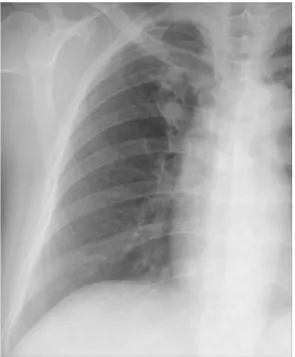
Fig. 1. Magnified chest radiograph of the right lung shows a pulmonary nodule in the right upper lobe. A surrounding radiolucent area is not evident.
우연히 발견된 폐종괴가 폐암 가능성이 있다는 이 야기를 듣고 내원하였다. 3개월 전에 당뇨병 진단 을 받은 과거력이 있었다. 이학적 소견이나 검사 소견에서 특이 소견은 없었다. 흉부방사선에서 우 상폐야에 경계가 불분명한 폐결절이 관찰되었으며 주위에 다른 소견은 관찰되지 않았다(Fig. 1). 이 폐결절은 흉부 전산화단층촬영에서 분지 모양을 하고, 석회화가 없으며, 조영 증강이 안되는, 저음 영의 연조직이었다(Fig. 2). 흡기 상태에서 촬영한
고해상전산화단층촬영에서 우상엽의 폐첨분절
(apical segment)에 분지되는 형태의 연조직 음영 이 관찰되고 주위의 폐실질에는 저음영이 관찰되 었다(Figs. 2b and 2c). 호기 상태에 촬영한 고해 상전산화단층촬영에서는 저음영 부위가 흡기 때에 비해 주위보다 훨씬 더 낮게 보여 저음영 부위가 공기포획(air trapping) 때문임을 시사하였다(Fig.
2d). 관상면 재구성(coronal reformatted) 전산화단 층촬영에서 우상엽의 폐첨분절이의 구역기관지 개 구부가 없었고(Fig. 2e) 가상기관지내시경 영상과 (Fig. 2f) 기관지 내시경에서도 같은 소견을 보였 다(Fig. 3). 선천성 기관지 폐쇄증 진단을 내렸고 다른 증상이 없어서 환자는 퇴원하였다.
고 찰
선천성 기관지 폐쇄증은 구역기관지의 근위 관강 (lumen)이 폐쇄된 기형으로 대개 한 구역기관지에 국한되어 폐쇄 혹은 협착이 있다. 폐쇄된 기관지의 원위부는 개방되어 있어 이 기관지는 점액으로 차 게 되어 기관지 내 점액고착(mucoid impaction)이 생긴다. 이 협착된 기관지에 연결된 폐포는 측부환 기(collateral ventilation)로 인해 공기포획의 형태 가 되어 결국 원위 기관지 주위에 과팽창된 부위 가 생긴다. 가장 흔히 생기는 부위는 좌상엽의 첨 부와 후방 구역(apical and post segments)이며 그 다음으로 우상엽의 구역 기관지, 우중엽, 그리고 하엽의 순서이다1.
선천성 기관지 폐쇄증은 방사선학적 소견이 특징 적이다. 요약하면, 점액이 가득 차서 늘어난 기관지 가 폐문으로부터 뻗어 나가고, 둥근, 분지 모양으로 나타나며 주위에는 저음영이 둘러싼 것이 방사선학 적 검사에 보이는 것이다2,3. 흉부단순촬영에서 폐 저음영이 90%, 폐문 종괴가 80%, 두 소견 모두가 70%에서 보였다는 보고도 있다4. 폐실질의 저음영 은 협착된 기관지 원위부 폐 실질 내의 혈량감소 (oligemia)와 공기 용적의 증가로 생긴 것이다.
흉부 전산화단층촬영에서도 늘어난 기관지 내의 점액고착, 구역 과팽창, 점액고착된 기관지 근위부 기관지의 폐쇄가 보이고, 이 소견들이 있으면 진단
적이다3,5-8. 늘어난 기관지 내부의 점액고착은 기관
지가 있을 위치를 따라 분지하는 모양의 연조직 음영으로 보이는 것이 보통이지만 기수면(air-fluid
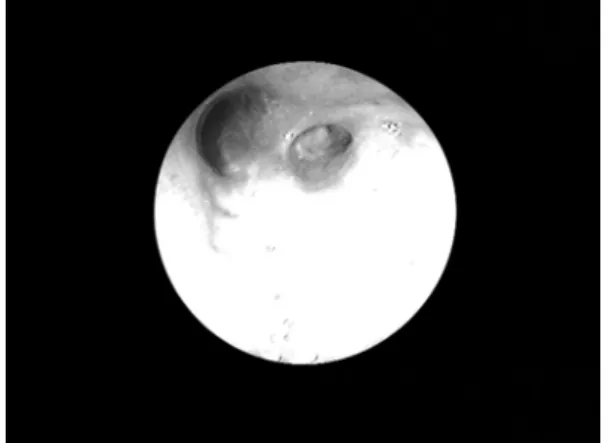
Fig. 2. (a-f) Chest CT scans showing the abnormality on a chest radiograph. The lesion is a poorly enhanced (a and e), branching (b and c), soft tissue opacity along the expected course of the apical segmental bronchi of the right upper lobe. CT scans obtained at end inspiration (b) and expiration (d) demonstrate air trapping in the same segment. A coronal reformatted CT scan (e) shows an ovoid soft tissue shadow emanating from an expected opening site of the apical segmental bronchus of the right upper lobe. Note the smooth upper surface of the right upper lobar bronchus, suggesting obliteration of the apical segmental bronchial opening (e). Virtual bronchoscopic image of the right upper lobar bronchus does not show the opening of the apical segmental bronchus (f).
level)을 보이거나 완전히 공기로 차여 있기도 한 다. 기관지 폐쇄가 좌상엽의 첨부와 후방 구역에 있으면 단순촬영 소견만으로도 상당히 특징적이어 서 진단이 쉽다. 그러나 다른 부위의 경우에는 흉 부단순촬영에 국소적 저음영만 보이는 등 비특이 적일 수 있으며 그럴 경우에도 전산화단층촬영에 서는 기관지 내 점액고착이 대부분 보이기 때문에 더 유용하다7,8.
나선형 전산화단층촬영이 나오기 이전에는 영상 단면이 두껍기 때문에 실제로 기관지 개구 부위가 어떤 병변으로 인해 후천적으로 막혔는지 선천성 기관지 폐쇄처럼 원래 없는지 알 수 없는 경우가
많다. 하지만 본 예에서와 같이 최근의 다중절편 (multislice) 전산화단층촬영에서는 단면 두께가 굉 장히 얇아서 이를 이용한 이차원 혹은 3차원 재구 성 영상에서는 위의 두가지가 어느 정도 구별될 잠재적 가능성이 있다(Fig. 2).
전산화단층촬영이 개발되기 전에 진단은 폐쇄된 구역 기관지를 보여주는 기관지조영술(broncho
graphy)로 하였다. 그러나 현재는 기관지 내시경 혹은 기관지조영술 같은 침습적 검사는 적합치 않 다6. 하지만 종양, 이물질, 혹은 염증성 협착 같은 후천적인 원인에 의한 근위부 기관지의 폐쇄의 가 능성을 배제하기 위해 기관지내시경 검사가 시행
(a) (b) (c)
(d) (e) (f)
Fig 3. Fiberoptic bronchoscopy of the right upper lobar bronchus confirms the obliteration of the apical segmental bronchus, which was seen on Fig 2f.
되기도 한다9. 본 증례에서의 기관지 내시경검사는 선천성 기관지 폐쇄증이 의심되기 전에 시행되었 다.
환자들은 대개 증상이 없어 약 50%에서 우연히 발견된다. 따라서 대부분의 경우에 있어 어떠한 처 치도 하지 않는 것이 보통이다. 그리고 추적 검사 는 단순촬영과 전산화단층촬영으로 할 수 있다10. 약 1/3의 환자에서는 호흡 곤란, 기침 등 비특이적 증상을 호소하며 드물게 병변 부위의 감염을 동반 하기도 하는데11, 감염이 반복되면 수술의 대상이 될 수 있다12.
요 약
선천성 기관지 폐쇄증은 단순촬영에서 폐 결절로 보여 악성 종양으로 오인될 수 있다. 그러나 흉부 단순촬영과 특히 전산화단층촬영에서는 늘어난 기 관지 내의 점액고착, 구역 과팽창 등이 특징적으로 보여 더 이상의 침습적 검사 없이 선천성 기관지 폐쇄증으로 진단할 수 있다.
참 고 문 헌
1. Fraser RG, Pare JAP, Pare PD, Fraser RS, Genereux GP. Congenital bronchial atresia.
In: Diagnosis of diseases of the chest. 3rd ed. Philadelphia: Saunders; 1989;721-2.
2. Meng RL, Jensik RJ, Faber LP, Matthew GR, Kittle CF. Bronchial atresia. Ann Thorac Surg 1978;25:184-92.
3. Matsushima H, Takayanagi N, Satoh M, Kurashima K, Kanauchi T, Hoshi T, et al.
Congenital bronchial atresia: radiologic fin
dings in nine patients. J Comput Assist Tomogr 2002;26:860-4.
4. Jederlinic PJ, Sicilian LS, Baigelman W, Gaensler EA. Congenital bronchial atresia: a report of 4 cases and a review of the lite
rature. Medicine 1986;66:73-83.
5. Kinsella BD, Sissons G, Williams MP. The radiological imagings of bronchial atresia. Br J Radiol 1992;65:681-5.
6. Cohen AM, Solomon EH, Alfidi RJ.
Computed tomography in bronchial atresia.
Am J Roentgenol 1980;135:1097-9.
7. Remy-Jardin M, Remy J, Ribet M, Gosselin B. Bronchial atresia: diagnostic criteria and embryologic considerations. Diagn Intervent Radiol 1989;1:45-51.
8. Mata JM, Caceres J, Lucaya J, Garcia- Conesa JA. CT of congenital malformation of the lung. RadioGraphics 1990;10:651-74.
9. Ward S, Morcos SK. Congenital bronchial atresia-presentation of three cases and a pictorial review. Clin Radiol 1999;54:144-8.
10. Caceres J, Mata J, Palmer J, Zidan A, Donoso L. General case of the day. Radio
Graphics 1991;11:1143-5.
11. Scully RE, Galdabini JJ, McNeely BU. Hyper
inflation of the left upper in a five-year-old
boy. N Engl J Med 1979;301:829-35.
12. Zylak CJ, Eyler WR, Spizarny DL, Stone CH.
Developmental lung anomalies in the adult:
radiologic-pathologic correlation. Radiographics 2002;22:S25-43.