• 검색 결과가 없습니다.

The BMPs expression and histomorphometric study of ${\beta}-TCP$ / rhBMP-2 Grafting on the rabbit cranial bone defects

N/A
N/A
Protected

Academic year: 2021

Share "The BMPs expression and histomorphometric study of ${\beta}-TCP$ / rhBMP-2 Grafting on the rabbit cranial bone defects"

Copied!
10
0
0

로드 중.... (전체 텍스트 보기)

전체 글

(1)

INTRODUCTION

Oral and maxillofacial bone defect can result from many causes such as trauma, tumor, resective surgery, and atrophy with aging. The goal of bone graft is to stimulate or facilitate the growth of new bone into the defect. The gold standard for recovering these defects to use the autogenous bone. Although highly effective, it has some disadvantages such as limitation of quantity, donor site morbidity, fear of patient, need for additional surgery and various complication (such as pain, bleed- ing, infection and paresthesia). Therefore graft of syn-

thetic biomaterials come into common use with unlimit- ed availability and without the risk of transmitting infec- tion.

The ideal bone substitute should maintains biologic supports during the healing period and is then gradually replaced by the newly formed bone. Several alloplastic materials have been examined as a substitute for autoge- nous bone, such as allografts

1)

, ceramics , and various osteoinductive agents. Synthetic ceramics, such as hydroxyapatite, tricalcium phosphate (TCP)

2)

, and bipha- sic calcium phosphate are commonly used as bone sub- stitute

3)

.

However, alloplastic materials have insufficient activi- ty in osteogenic or osteoinductive activity, so it takes long period in bone remodeling, it is needed therefore add the signaling agent of cell growth in order to rein- force the new bone formation.

The bone morphogenic protein (BMP) is known as

* Corresponding author

Kyung-Gyun Hwang, DDS, MSD, PhD.

Dept. of Dentistry/OMFS, College of Medicine, Hanyang Univ.

#17 Haengdang-dong, Seongdong- gu, 133-792, Seoul, Korea Tel: 82-2-2290-8676 Fax: 82-2-2290-8673

E-mail: hkg@hanyang.ac.kr

The BMPs expression and histomorphometric study of β β-TCP / rhBMP-2 Grafting on the rabbit cranial bone defects

Byung-Sup Lim, Jae-Yoon Jeon, Chang-Joo Park, Jae-Jung Im, Kyung-Gyun Hwang, Kwang-Sup Shim

Department of Dentistry/Oral and Maxillofacial Surgery, College of Medicine, Hanyang University, Korea

Objective: The Purpose of the study was to investigate the bone morphogenic protein expression of rhBMP-2(recombinant human bone morphogenic protein-2) as singnaling molecule and β-TCP(Tricalcium phosphate) as a bone substitute and carrier medium of rhBMP-2.

Materials and Methods: 16 rabbits divided into 2 group of each 8 rabbit. Two standardized bone defect, round bilateral defect was made in the cranium of the 8 rabbit of first group, and was grafted with 150~500μm diameter β-TCP 0.25g in one side, which was soaked with rhBMP-2, and autogenous bone was grafted on another side as a positive control. Second group of 8 rabbit, only β-TCP was grafted with same size and same manner. After 2, 4, 8, and 12 weeks, specimen was taken for microscopic immunohiostochemical and histomorphometric analysis.

Result: Grafting β-TCP with rhBMP show the early formation of the bone regenerative factor (BMP-4) and more quantity of new bone formation than only use of β-TCP (8,12 week), even show less new bone formation than autogenous bone.

Conclusion : The experimental study result that β-TCP graft combination with rhBMP-2 as a delivery system is an effective with osteoinductive capacity and biodegradable properties, so that provide clinical availibility of composite use in reconstruction of bony defect.

Key words

Beta-Tricalcium phosphate, bone graft, Bone Morphogenic Protein, bone substitute, histomorphometry, Immunohistochemistry

Abstract

(2)

continued in oral and maxillofacial surgery, and the use of BMP alone or in conjunction with other graft modali- ties shows great experimental result in the reconstruction of defects and deficiencies in recents

6,7)

. According to Wozney

8)

, BMP combination with a graft material effects as biologic catalyst that proliferate the cell and promote chondrogenesis and osteogensis. BMPs are growth factor proteins secreted by cells and serve as signaling agent.

They work as part of a large cellular communication net- work that influences the cell functions of division, matrix synthesis, and tissue differentiation

9)

.

The ideal material of BMP delivery system should have a function as prevent invasion by the surrounding soft tissue until induction of new bone, and are then gradual- ly absorbed with new bone formation, and many carrier have already been examined, such as TCP, Hydro- xyapatite, collagen, Polylactic acid-polyglycolic acid copolymer, and titanium

10-14)

. β-TCP is a synthetic porous ceramic which has shown osteoconduction and very favorable substitution rate in standardized bone defects

15)

, and has good tissue tolerance with no immuno- logical or toxic reactions, good biocompatibility, charac- teristics of absorption, and is effectively replaced by nat- ural bone in an appropriated period of time

16-19)

.

Since there are little study of grafting β-TCP in conjunc- tion with rhBMP-2 with incremental new bone formation by period, we evaluate immunologic and histomorpho- metric changes of bone grafting material and new bone formation in sequence of weeks in this study. In addi- tion, we investigate the osteoinductive activity of β- TCP/rhBMP-2 through immunohistochemical expres- sion of the BMP-2, -4. -7.

MATERIALS AND METHODS

The protocol of animal study was approved by Hanyang University Hospital Animal Laboratory and the study was performed in 16 Newzealand White rabbits.

was shaved and local anesthesia was induced with 2%

lidocaine, 1:100000 epinephrine (1.8ml). Disinfection with povidone iodine solution and drap was done.

Through the central incision, the cranial bone was suffi- ciently exposed and bilateral round and standardized cranial defect was designed with ready-made round tem- plate (15mm diameter) and preperated throught saline irrigation with low speed round bur (106 Komet

, Germeny) to depth of meninges. Lateral aspect of subpe- riosteal incision extend to periorbital area, and symmetr- cial round defect does not invade cranial suture line. The harveseted corticocancellous bone blocks were crushed and ground to a particulate size about 1~1.5mm using a bone mill and filled in left side of cranial bone defect.

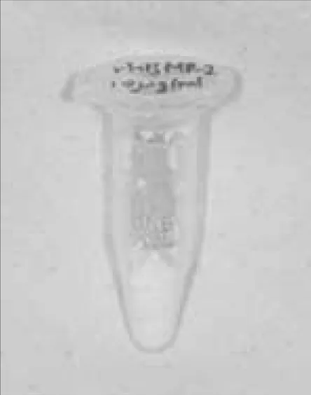
And in first group of 8 rabbits, in Right side, distilled water diluted 0.01% rhBMP-2(Fig. 1) solution was soaked to 0.25g of β-TCP(Fig. 2) and filled in another right side of cranial defect. In second group of 8 rabbits, only β- TCP was grafted in the defect on one side, and autoge- nous bone graft was done in the other side with same method. Periosteal approximation was done with 4-0 vicryl(Ethicon, Norderstedt, Germany) and dermal suture by 3-0 silk(Fig. 3-5). During the first 3 day, antibi- otic (Gentamycin

. Dong Hwa. Korea 30mg/kg body weight) was injected throught intramuscular for preven- tion of infection andanimals were checked daily.

2) Healing period

The 16 animals were divied into 2 groups of 8 animals following periods of grafting operation : 2, 4, 8, 12 weeks

At the end of each periods, the animals were sacrificed and cranial bone defect specimen contained to round grafted defect with 2mm margin were obtained by drill.

3) rhBMP-2 and β-TCP

rhBMP-2 (CHO cell derived, RND system, Minea-

police. USA : 10μg/ml >90% purity) was diluted by 0.1ml

distilled water into 0.01% volume concentration (10μ

g/ml), and soaked to 150~500μm of β-TCP (Cerasorb

,

Curasan AG, Kleinostheim, Germany ; Particle size

(3)

4) Histologic analysis

The specimen was fixed by 4% neutral buffered paraformaldehyde solution for 4 weeks and were washed with 0.1M phosphoric buffer solution(PBS) for 30 minutes, and it was demineralized with EDTA 10%

with 0.1M PBS for 80 days and embedded in methyl methacrylate. Specimen was stained with H&E, analyzed by microscope (U-MOD1083, Olympus Japan ; ×100 magnification)

5) Histomorphometric analysis

20μm sections of specimen stained with H&E were ana- lyzed by OPTIMUS (6.5 system, Nethelands), and remaining bone, new bone, soft tissue area was detected and divided by total defect area and expressed as per- centage.

6) Immunohistochemistry

Streptavidin-biotin method was used for immunohisto- chemical stain and analyzing specimen. Paraffin of slide Fig. 1. rhBMP-2(CHO cell derived,

RND system. mineapolice).

Fig. 2. β -TCP (Cerasorb

, Curasan AG, Kleinostheim, Germany ; Particle size 150~500μm).

Fig. 3. Bilateral round and standard- ized cranial defect (15mm diameter).

Fig. 4. Grafted in rabbit cranial defects(β-TCP(or rhBMP-2 with β-TCP) in Rt. side,Autogenous particulated bone in Lt. side).

Fig. 5. Perosteal approximation. Fig. 6. rhBMP-2 was diluted by 0.1ml

.0 distilled water with 0.01% volume

concentration (10μg/ml), and soaked

to 150~500μm of β-TCP.

(4)

IgG crossaction. In room temperature, BMP-2, -4, -7 pri- mary antibody was applied for 1 hour and washed three times by distilled water. Polyclonal goat anti-BMP-2, -4, - 7 (SantaCruz, U.S.A) was used for primary antibody with 1:50 solution. Later, biotin marked secondary anti- body was incubated for 15 min and washed by PBS.

Peroxidase marked streptavidin was incubated for 15 minutes and washed by PBS. Specimen was dyed by brown DAB(3,3-diaminobenzidine) for 5 minutes and washed by distilled water, and counter-colored by Meyer’s hematoxyline, and permount was examined by light microscopic. Each specimen was examined, and amount of color was divided into 4 steps for dyed area percantage of total, 0-5%(+/-), 6-25%(+), 26-50%(++), more than 50%(+++). Two pathologic specialists were studied and total score was integrated.

RESULT

Sixteen Newzealand rabbits have no vital problems and signs of infection in experimental period, and showed no other complications. For histomorphometry, total size were examined by two examiners twice for each specimens by microscopy (magnification ×20), and mean volume percent were noted with standard devia- tion.

For immunohistochemistry, ×200 magnification was applied, and two pathologic specielists choose the specif- ic area and analyze.

activated fibroblast and proliferation of endothelial ves- sel cell were found and after 8 week were not founded.

At 8 week, reduction of grafted bone was seen.

2) β-TCP graft + rhBMP-2(Fig. 11-14)

The more week went by, the more new bone formation was finding around β-TCP and some of them were linked each other and expanded. At 4 week, slightly resorption of β-TCP granule was found. At 8 week, acti- vated osteoblast and connective tissue with vessel were found around the new bone.

3) β-TCP only(Fig. 15-18)

At 4 week, slightly resorption of β-TCP granule was found and new bone was found around β-TCP, but the connection of new bone was not found. At 8 week, new bone was found around resorbed β-TCP and some of them were linked each other at some area.

2. Histomorphometric Comparison of New Bone Formation(Table 1)

(1) 2 week

New bone formation in autogenous bone was 12.88%

and it was more than other β-TCP graft group(6.7%) and β-TCP graft with rhBMP-2 grafted group(8.24%) with significance (p>0.05). But there was no difference between β-TCP graft group and β-TCP graft with rhBMP- 2 grafted group.

Table 1. Histomorphometric analysis

Autogenous bone graft β-TCP graft + rhBMP-2 β-TCP only

Graft Bone New Bone Soft Tissue Graft Bone New Bone Soft Tissue Graft Bone New Bone Soft Tissue

2 W 67.06±7.95 12.88±3.11 20.06±4.84 46.30±3.97 8.24±4.70 51.20±8.66 84.52±1.95 6.79±0.92 8.69±1.04

4 W 66.91±8.74 20.39±5.37 12.70±3.38 50.20±14.06 15.05±7.73 34.74±20.03 78.91±0.98 10.28±4.34 10.81±3.36

8 W 47.65±11.36 47.83±10.37 4.51±0.99 19.42±3.75 35.50±6.77 44.08±4.40 49.30±3.36 14.00±2.24 36.68±4.52

12 W 29.09±9.19 67.31±8.82 2.78±0.36 14.79±18.03 37.54±10.92 47.67±10.50 51.30±9.68 21.91±9.42 26.77±6.44

(Area Mean Volume Percent ± Standard deviation)

(5)

Fig. 7. Microscopic exam. shows the au- togenous bone graft, 2 weeks (× 20, H&E).

Fig. 8. Microscopic exam. shows the au- togenous bone graft, 4 weeks (× 20, H&E).

Fig. 9. Microscopic exam. shows the au- togenous bone graft, 8 weeks (× 20, H&E).

Fig. 10. Microscopic exam. shows the autogenous bone graft, 12 weeks (× 20, H&E).

Fig. 11. Microscopic exam. shows the β- TCP with rhBMP-2 graft, 2 weeks (×

20, H&E).

Fig. 12. Microscopic exam. shows the β- TCP with rhBMP-2 graft, 4 weeks (×

20, H&E).

Fig. 13. Microscopic exam. shows the β- TCP with rhBMP-2 graft, 8 weeks (×

20, H&E).

Fig. 14. Microscopic exam. shows the β- TCP with rhBMP-2 graft, 12 weeks (×

20, H&E).

Fig. 15. Microscopic exam. shows the β- TCP graft, 2 weeks (× 20, H&E).

Fig. 16. Microscopic exam. shows the β- TCP graft, 4 weeks (× 20, H&E).

Fig. 17. Microscopic exam. shows the β- TCP graft, 8 weeks (× 20, H&E).

Fig. 18. Microscopic exam. shows the β-

TCP graft, 12 weeks (× 20, H&E).

(6)

Fig. 19. Immunohistochemical exam.

shows BMP-2 expression on the auto- genous bone graft, 2 weeks (× 200).

Fig. 20. Immunohistochemical exam.

shows BMP-2 expression on the auto- genous bone graft, 8 weeks (× 200).

Fig. 21. Immunohistochemical exam.

shows BMP-2 expression on the auto- genous bone graft, 12 weeks (× 200).

Fig. 22. Immunohistochemical exam.

shows BMP-4 expression on the auto- genous bone graft, 2 weeks (× 200).

Fig. 23. Immunohistochemical exam.

shows BMP-4 expression on the auto- genous bone graft, 4 weeks (× 200).

Fig. 24. Immunohistochemical exam.

shows BMP-4 expression on the auto- genous bone graft, 8 weeks (× 200).

Fig. 25. Immunohistochemical exam.

shows BMP-4 expression on the auto- genous bone graft, 12 weeks (× 200).

Fig. 26. Immunohistochemical exam.

shows BMP-7 expression on the auto- genous bone graft, 8 weeks (× 200).

Fig. 27. Immunohistochemical exam.

shows BMP-2 expression on the β-TCP with rhBMP-2 graft, 8 weeks (× 200).

Fig. 28. Immunohistochemical exam.

shows BMP-2 expression on the β-TCP with rhBMP-2 graft, 12 weeks (× 200).

Fig. 29. Immunohistochemical exam.

shows BMP-4 expression on the β-TCP with rhBMP-2 graft, 2 weeks (× 200).

Fig. 30. Immunohistochemical exam.

shows BMP-4 expression on the β-TCP

with rhBMP-2 graft, 4 weeks (× 200).

(7)

Fig. 31. Immunohistochemical exam.

shows BMP-4 expression on the β-TCP with rhBMP-2 graft, 8 weeks (× 200).

Fig. 32. Immunohistochemical exam.

shows BMP-4 expression on the β-TCP with rhBMP-2 graft, 12 weeks (× 200).

Fig. 33. Immunohistochemical exam.

shows BMP-7 expression on the β-TCP with rhBMP-2 graft, 8 weeks (× 200).

Fig. 34. Immunohistochemical exam.

shows BMP-2 expression on the β-TCP graft, 4 weeks (× 200).

Fig. 35. Immunohistochemical exam.

shows BMP-4 expression on the β-TCP graft, 4 weeks (× 200).

Fig. 36. Immunohistochemical exam.

shows BMP-4 expression on the β-TCP graft, 12 weeks (× 200).

Table 2. Immunohistochemical study on BMP-2 expression

Autogenous bone graft β-TCP graft + rhBMP-2 β-TCP only

2 W 4 W 8 W 12W 2 W 4 W 8 W 12W 2 W 4 W 6 W 8W

Sample 1 +/- - + + - - - + - - + +/-

Sample 2 +++/- - - ++ - +/- - ++ - - +/- +

Positive was graded on a scale, with - representing no staining, +/- indistinct, + weak stainig, ++ moderate staining, and +++ strong staining.

Table 3. Immunohistochemical study on BMP-4 expression

Autogenous bone graft β-TCP graft + rhBMP-2 β-TCP only

2 W 4 W 8 W 12W 2 W 4 W 8 W 12W 2 W 4 W 6 W 8W

Sample 1 ++ + + + + ++ +++ ++ - + + +

Sample 2 + ++ + ++ +/- ++ +++ + +/- +/- +/- +

Positive was graded on a scale, with - representing no staining, +/- indistinct, + weak stainig, ++ moderate staining, and +++ strong

staining.

(8)

New bone formation of autogenous bone graft (47.83%) and β-TCP graft with rhBMP-2 grafted group (35.50%) were more than other β-TCP graft group (14.00%) with significance (p>0.05) but there is no difference between Autogenous bone graft and β-TCP graft with rhBMP-2 grafted group (p>0.05).

(4) 12 week

New bone formation of autogenous bone was 67.31%

and it was more than other β-TCP graft group (21.91%) and β-TCP graft with rhBMP-2 grafted group (37.54%) but there was no difference of significance (p>0.05).

3. Immunohistochemical study on BMP expression (Table 2, 3)

1) Autogenous bone graft(Fig. 19-26)

BMP-2 (except at 4week), BMP-4 were founded around grated bone but BMP-7 was not founded except at 4week.

2) β-TCP graft + rhBMP-2(Fig. 27-33)

BMP-4 was founded in osteoblast round grafted bone, and no BMP-2 and BMP-7 were not founded at 2,4,8 week. At 12 week, BMP-2 and BMP-4 were founded but BMP-4 was decreased than 8 week.

3) β-TCP only(Fig. 34-36)

Less than +/- of BMP-2, -4 were founded around osteoblast with grafted bone. At 12 week, BMP-2, -4 were slightly dercrease than 8 week.

DISCUSSION

Calcium phosphate is on the major substances compris- ing hard tisseue, tooth and bone. It is non-toxic, osteo- conductive, and biocompatible to bone and soft tissue.

Molecular structure of TCP is similar to bone and TCP has a good boicompitibility, and optimal resorption peri-

porous, resorbable, dense and bioactive, TCP is osteocon- ductive material and only provide a scaffold for new bone formation, Therefore, its clinical sue is restricted to relatively small bone defect

20)

, and the combination of bone substitute materials with osteoinductivity would be favorable to reduce the period to form new bone.

Osteoinduction is defined as the mechanism by which a mesenchymal tissue is induced to change its cellular structure to become osteogenic

21)

, and BMP have an osteoinductive carriers in various experimental study and the value of BMP treatment also has been reported in human study

22)

.

The discovery by Urist

16)

in 1965 of the osteoinductive properties of demineralized , guanidine-extracted bone, eventually led to the isolation of bone morphogenic pro- teins in 1980, and the beginning of osseous tissue enge- neering.

The BMPs belong to an expanding TGF-β(Transfo- rming growth factor) superfamily and approximately 40 isoforms have been identified and they are the only growth factors that can stimulate differntiation of the MSCs into a chondroblastic and osteoblastic direction

23,24)

. And these isoforms differ in their osteogenic potential, and their effects may be mitogenic, chemotactic, mor- phogenic, or apoptotic depending on the cell type to which the ligand is exposed, and the growth factor con- centration

24)

.

After injury to the bone matrix, BMPs are released.

They are responsible for various mechanisms that con- tribute to bone, such as angiogenesis, and chemotaxis and diffrentiation of mesenchymal cells.

BMPs have pleomorphic functions that range from non-skeletal and skeletal organogenesis to bone genera- tion and regeneration

25)

, and BMPs have been tested in clinical trials. Initial data from these trials have suggest- ed that BMP-induced bone formation is at least equva- lent to autogenous bone-grafting when used to treat tib- ial nonunions and to promote spinal fusions

26-28)

.

Our study focus on rhBMP-2 because BMP-2 are essen-

tial for osteoblast and osteoclast formation, and have

(9)

been shown to be the most strongly expressed BMPs in rodent models of mandibular fracture healing, as well as in new bone formation during distraction osteogenesis of the mandible

23)

.

The bone substitute used as a carrier for BMP-2 con- trols the release kinetics of BMP-2. Unlike the gelatine sponge or direct injection, BMP-2 soaking within β-TCP continuosly released with resorption mechanism of TCP granule.

Uludag et al.

29)

, showed that the release kinetics depend on the protein itself and on the carrier. They found dif- ferences in BMP-2 release from collagen, glycolic acid, demineralized bone matrix, and bovine bone mineral with the highest recovery rate found in collagen and a recovery rate of about 30% in TCP. Nevertheless, in their model, rhBMPs was added via soaking process, it may be possible that the BMP adhere only to the outer surface of the carrier, resulting in an earlier burst release. Blood invades from the defect into the TCP, it might result in early wash-out of the BMP, which served as a cell recruitment signal, attracting the BMP-responding cells into the TCP.

Many carriers have been tried for BMP-2, and the most important requirments for BMP carriers are bioactive, osteoconductivity, and biomechanical stability. The sub- stance should be resorbable, be replaced by new bone during normal remodeling, and should be moldable, easy to handling with surgeon, and filled completely with close contact between bone and implant

30)

.

Calcium phosphate ceramics are well known as carriers for BMP

31,32)

, because of their micro-architecture. The micro-architecture of biomaterials also interferes with the tissue response at the site of bone graft and at the same time, the porosity can act as housing of BMP to continuously release for a long time. Studies on the porosity of carriers have revealed that hardness of sur- face can impair the ingrowth of graft material. Because bone tissue would be physical barrier, it can limit the proliferation of blood vessels essential for bone repair.

Pore smaller than 50μm are too small to allow the ingrowth, so macroporosity (>100μm)

33)

plays an impor- tant role in the osteoconductivity. And the microporosity (<5μm)

34)

is important for the bioresorbability of the material.

However, most tested calcium phosphate-BMP-com- posites are supplied in form of preformed cubes, wedges, or even granules, which have only little biome- chanical stability, leading to a poor contour adaptation

between bone and implant. This reduces the contact area, resulting in less osseous ingrowth, with the risk of insta- bility and pseudoarthrosis, however, calcium phosphate can be injected or molded during the operation and fill defects are any size or shape, resulting in an excellent bone-implant contact

35)

, so that BMP could have more chance to surface of bone defects than other biomaterials.

The use of TCP as a carrier for BMP combines the advantage of the optimized bone-implant contact and this osteoconductive effect with enhanced new bone for- mation induced by BMP. This should lead to a faster bone healing with improved biomechanical stability

3)

.

The combination of bioactive and osteoconductive cal- cium phosphate ceramics, or similar materials, and the osteoinductive BMPs seems to be synergistic on the heal- ing of bone because of the bioactive properties of calcium phosphate ceramics, which also scaffold for cell ingrowth and differentiation, and whose osteoconductive proper- ties stimulate bone healing as well.

CONCLUSION

In animal study of various graft materials, such as autogenous bone, β-TCP and β-TCP with rhBMP-2 to cra- nial bone defect of rabbit, we investigate new bone for- mation through histologic, histomorphometric, and immunohistochemistric analysis.

In immunomorphometric analysis, grafting β-TCP with rhBMP show the early formation (2, 4 and 8 week) of the bone regenerative factor (BMP-4) than β-TCP graft group (4 and 8 week), and in histomorphometric analysis more quantity of new bone formation than only use of β-TCP (8 and 12 week p>0.05), even show less new bone forma- tion than autogenous bone(2,4,8 and 12 week).

REFERENCES

1. Brown MD, Malinin TI: A roentgenographic evaluation of frozen allografts versus autografts in anterior cervical fu- sion. Clin Orthop 1976;119:231-236.

2. Metsger DS, Driskell TD, Paulsrud JR: Tricalcium phos- phate ceramic-a resorbable bone implant: review and cur- rent status. J Am Dent Assoc 1982;105:1035-1038.

3. Niedhart C, Maus U, Redmann E, Siebert CH: In vivo test- ing of a new in vivo setting β-tricalcium phosphate cement for osseous reconstruction. J Biomed Mater Sci 1997;44:53- 62.

4. Urist MR: Bone formation by autoinduction. Science 1965;150:893-899.

5. Urist MR, Mikulski A, Lietze A: Solubilized and insolubi- lized bone morphogenic protein. Proc Natl Acad Sci USA 1979;76:1828-1832.

6. Boyne PJ: Animal studies of application of rhBMP-2 in

(10)

1984;187:277-280.

11. Horisaka Y, Okamoto Y, Matsumoto N, Yoshimura Y, Kawada J, Yamashita K: Subperiosteal implantation of bone morphogenetic protein absorbed to hydroxyapatite.

Clin Orthop 1991;268:303-309.

12. Takaoka K, Koezuka M, Nakahara H: Telopeptide-depleted bovine skin collagen as a carrier for bone morphogenetic protein. J Orthop Res 1991;9:902-907.

13. Miyamoto S, Takaoka K, Okada T, Yoshikawa H, Hashimoto J, Suzuki S: Polylactic acid-polyethylene glycol block polymer-a new biodegradable synthetic carrier for bone morphogenetic protein. Clin Orthop 1993;294:333-343.

14. Kawai T, Mieki A, Ohno Y, Umemura M, Kataoka H, Kurita S: Osteoinductive activity of composites of bone morphogenetic protein and pure titanium. Clin Orthop 1993;290:296-305.

15. Buser D, Hoffmann B, Bernard JP, Lussi A, Mettler D, Schenk RK: Evaluation of filling materials in membrane- protected bone defect. A comparative histomorphometric and histologic study in the mandible of miniature pigs.

Clinical Oral Implants Res 1998;9:137-150.

16. Bucholz RW, Carton A, Holmes RE: Hydroxyapatite and tricalcium phosphate bone graft substitutes. Orthop Clin North Am 1987;18:324-334.

17. Eggli PS, Muller W, Shenk PK: Porous hydroxyapatite and tricalcium phosphate clynders with two different pore size ranges implanted in the cancellous bone of rabbits. Clin Orthop 1988;232:127-138.

18. Gatti AM, Zaffe D, Poli GP: Behavior of tricalcium phos- phate and hydroxyapatite granules in sheep bone defets.

Biomaterials 1990;11:313-317.

19. Hossain MZ, Kyomen S, Tanne K: Biologic responses of au- togenous bone and beta-tricalcium phosphate ceramics transplanted into bone defects to orthodontic force. Cleft Palate Craniofac J 1996;33:277-283.

20. Nakahara H, Goldberg VM, Caplan AI: Culture-expanded periosteal-derived cells exhibit osteochondrogenic poten- tial in porous calcium phosphate ceramics in vivo. Clin Orthop 1992;276:291-298.

21. Makin M: Osteogenesis induced by vesical mucosal trans- plant in the guinea pig. J Bone Joint Surg Br 1962;44:165- 167.

2000;14:598-601.

25. Wozney JM: The bone morphogenetic protein family and osteogenesis. Mol Reprod Dev 1992;113:681-687.

26. Heckman JD, Boyan BD, Aufdemort TB, Abbott JT: The use of bone morphogenitic protein in the treatment of non- union in a canine model. J Bone Joint Surg Am 1991;73:750- 764.

27. Boden SD, Zdeblick TA, Sandhu HS, Heim SE: The use of rhBMP-2 in interbody fusion cages. Definitive evidence of osteoinduction in humans-a preliminary report. Spine 2000;25:376-381.

28. Valentin-Opran A, Wozney J, Csimma C, Lilly L, Riedel GE: Clinical evaluation of recombinant human bone mor- phogenic protein-2. Clin Orthop 2002;395:110-120.

29. Shelley R, Winn, Hasan U, Jeffrey OH: Sustained release emphasizing recombinant human bone morphogenetic protein-2. Advanced Drug Delivery Review 1998;31:303- 318.

30. Niedhart C, Niethard FU, Klinische Andorderungen an Knochenersatzstoffe: Bioceramics in orthopedics-new ap- plications. F Enkg Verlag 1998;46-50.

31. Koempel JA, Patt BS, O’Grady K, Wozney J, Toriumi DM:

The effect of recombinant human bone morphogenetic pro- tein-2 on the integration of porous hydroxyapatite implants with bone. J Biomed Mater Res 1998;41:359-363.

32. Meikle MC, Papaioannou S, Ratledge TJ, Speight PM, WattSmith SR, Hill PA, et al.: Effect of poly DL-Lactide-co- glycolide implants and xenogenic bone matrix-derived growth factors on calvarial bone repair 2 and bone marrow.

J Oral Maxillofac Surg 2001;59:53-61.

33. Jarcho M: Calcium phosphate ceramics as hard tissue pros- thetics. Clin Orthop 1981;259-278.

34. Driskell TD, O’Hara MJ, Sheets HD Jr, Greene GW Jr, Natiella JR, Armitage J: Development of ceramic and ce- ramic composite deviced for maxillofacial applications. J Biomed Mater Res 1972;6:345-361.

35. Niedhart C, Maus U, Redmann E, Schmidt-Rohlfing B,

Niethard FU, Siebert CH: Stimulation of bone formation

with an in situ setting tricalcium phosphate/rhBMP-2 com-

posite in rats. J Biomed Mater Res A 2003;65:17-23.

수치

Fig. 5. Perosteal approximation. Fig. 6. rhBMP-2 was diluted by 0.1ml .0 distilled water with 0.01% volume concentration (10μg/ml), and soaked to 150~500μm of β-TCP.
Table 1. Histomorphometric analysis

참조

관련 문서

Histopathologic findings of control group at 4 weeks show little bone- implant contact (BIC) around the implant (asterisks) and new-bone formation in the defect

The grafted particulated tooth driven allografts were partially surrounded with new bone matrix and fairly supported graft associated new bone formation with some

Alveolar ridge augmentation with titanium mesh and a combination of autogenous bone and anorganic bovine bone: a 2-year prospective study.. Corinaldesi G, Pieri F, Sapigni

The aim of this study was to compare the effect on bone regeneration relative to maintenance period of PTFE membrane in rabbit calcarial defects.. Eight adult

In 4-week group, the group filled with bone graft with decortication revealed larger new bone formation area than shown in the group that had a defect area

Histologic evaluation of early human bone response to different implant surfaces2. Histologic evaluation of human bone integration on machined and

Higher power view of the box area of the left figure showed poor new-bone formation around the implant (asterisk) and low BIC. Villanueva

The result of using PLGA nanofiber membrane with bone graft material showed that the PLGA nanofiber membrane in the experimental group of 2 weeks were ten times more new