Inhibitory Effects of Flavonoids Isolated from the Leaves of Stewartia koreana on Nitric-oxide Production in LPS-stimulated RAW 264.7 Cells
Seung-Su Lee1, Myun-Ho Bang1, Se-Ho Park2, Dae-kyun Chung1 and Seun-Ah Yang3*
1Skin Biotechnology Center, Kyung Hee University, Yongin, Gyeonggi 446-701, Korea
2Institute of Natural Science, Keimyung University, 1095 Dalgubeol-daero, Daegu 42601, Korea
3Department of Food Science and Technology, Keimyung University, Daegu 42601, Korea Received February 22, 2018 /Revised March 10, 2018 /Accepted March 13, 2018
Five phenolic compounds were isolated from the ethyl acetate fraction of leaves from Stewartia koreana, and their nitric-oxide (NO) inhibitory activities were measured to identify the major active con- stituents responsible for the efficacy of the extract against inflammatory reactions. These five com- pounds were quercetin (1), quercitrin (2), hyperin (3), quercetin-3-O-(6''-O-galloyl)-β-D-galactopyrano- side (4), and kaempferol 3-O-[2'',6''-di-O-(trans-p-coumaroyl)]-β-D-glucopyranoside (5). Among the separated compounds in the EtOAc fraction, compounds 4 and 5 were isolated for the first time, and no study has yet reported their anti-inflammatory effects. The compounds were identified by spectro- scopic analysis, and the isolated compounds showed significant NO inhibitory effects in lipopoly- saccharide (LPS)-stimulated RAW 264.7 cells. Compound 5 showed the most potent inhibitory effect (63.35% inhibition) against LPS-induced NO production compared to that of compound 1 (17.17%), compound 2 (5.0%), compound 3 (3.92%), and compound 4 (6.32%) at 10 μg/ml concentration. NO production was inhibited by suppressing the protein expression of inducible nitric-oxide synthase in LPS-stimulated RAW 264.7 macrophages. These results indicate that kaempferol 3-O-[2'',6''-di-O- (trans-p-coumaroyl)]-β-D-glucopyranoside might be the major active compound responsible for the an- ti-inflammatory effects of S. koreana.
Key words : Glucopyranoside, kaempferol-3-o-[2'',6''-di-o-(trans-p-coumaroyl)]-β-D-, macrophage, nitric oxide, phenolic compounds, Stewartia koreana Nakai
*Corresponding author
*Tel : +82-53-580-5117, Fax : +82-53-580-5372
*E-mail : [email protected]
This is an Open-Access article distributed under the terms of the Creative Commons Attribution Non-Commercial License (http://creativecommons.org/licenses/by-nc/3.0) which permits unrestricted non-commercial use, distribution, and reproduction in any medium, provided the original work is properly cited.
Journal of Life Science 2018 Vol. 28. No. 5. 509~516 DOI : https://doi.org/10.5352/JLS.2018.28.5.509
Introduction
Stewartia koreana (Theaceae) is native to eastern Asia in Korea, China and Japan. Several studies have reported that S. koreana contains biologically active compounds such as dihydrochalcones, flavonoids, lignans, and sterols, which have antioxidant, anti-inflammatory, and skin-whitening [13, 16]. Additionally, syryngaresinol, a lignan from stems of S.
koreana, exhibits significant antioxidant and anti-inflamma- tory activities [7]. It has also been reported that S. koreana extract has angiogenesis [9], wound healing [12], bone re- sorption [15] and anti-allergenic activities [4]. Until now, re- search has shown that S. koreana is a good raw material for pharmaceutical and cosmetic applications.
This study puts emphasis on isolation of phenolic com- pounds and confirmation of their variable biological proper- ties including anti-inflammatory effect. Through the TLC ex- periment, many flavonoids were verified. Using preparative HPLC, five flavonoids, quercetin (1), quercitrin (2), hyperin (3), quercetin-3-O-(6''-O-galloyl)-β-D-galactopyranoside (4), and kaempferol-3-o-[2'',6''-di-o-(trans-p-coumaroyl)]-β-D-glu- copyranoside (5), were isolated from S. koreana. The struc- tures of isolated compounds were identified by spectro- scopic experiments, including NMR and MS, and their in- hibitory effects on NO production and expression of in- ducible nitric oxide synthase (iNOS) and cyclooxygevase-2 (COX-2) protein were evaluated in lipopolysaccharide (LPS)- stimulated RAW 264.7 murine macrophages [1, 18].
Materials and Methods
Plant sampleS. koreana was collected in July, 2012 in Yeonggwanggun, Korea, and identified by Dae-Keun Kim, College of Pharma- cy, Woosuk University, Jeonju, Korea. A voucher specimen
(KHU0170107) was deposited at the Laboratory of Natural Products Chemistry, Kyung Hee University, Yongin, Korea.
Chemicals and reagents
All chemicals were obtained from Sigma and Aldrich Chemical (St. Louis, MO, USA) unless otherwise indicated.
Cell culture reagents were purchased from Gibco BRL (Rockville, MD, USA) and fetal bovine serum (FBS) was from Hyclone (Logan, UT, USA). Anti-iNOS and COX-2 mono- clonal antibodies were purchased from Cell Signaling Tech- nology Inc. (Danvers, MA, USA). Quercetin, quercitrin and hyperin were obtained from Sigma (St. Louis, Mo, USA). All organic solvents used as the analytical and HPLC grades were purchased from Burdick & Jackson (Muskegan, MI, USA). Deionized water was prepared using a Milli-Q purifi- cation system (Millipore, Billerica, MA, USA).
General experimental procedures
Preparative HPLC (Waters, MA, USA) were used for separation. 1H-NMR (400 MHz) and 13C-NMR (100 MHz) spectra were recorded on a Varian Unity Inova AS-400 FT-NMR spectrometer (Palo Alto, CA, USA).
Extraction of S. koreana (Theaceae) and isolation of flavonoids by preparative HPLC
Dried powder (11 g) was extracted with 80%(v/v) aque- ous methanol (MeOH) (500 ml ×2), and the extracts were partitioned using EtOAc (200 ml ×3), n-Buthanol (BuOH) (200 ml ×3), and water (200 ml). The EtOAc fraction (467 mg) was proceeded by prep-HPLC connected to the column:
YMC-Actus triat C18 (20 mm×250 mm; particle size 5 m) using A: H2O and B: MeOH as mobile phases. The detector was set at 254 nm. The gradient elution conditions were as follows: 0-5 min, 100% A; 5-20 min, 100-50% A; 20-30 min, 50% A; 30-50 min, 50-100% B; 50-80 min, 100% B at a flow rate of 10 ml/min. The sample loading concertation was 20 mg/3 ml. The repeated isolation process was performed un- der the same conditions. Fraction 1 (52.0 mg, Rt = 10.5 min), Fraction 2 (68.4 mg, Rt = 12.2 min), Fraction 3 (12.3 mg, Rt
= 14.2 min), Fraction 4 (20.1 mg, Rt = 30.2 min), and Fraction 5 (22.3 mg, Rt = 35.5 min) were obtained. Each fraction was subjected to Sephadex LH-20 column chromatography (c.c.) (2x60 cm) and eluted with 100% MeOH to ultimately pro- duce five single compounds: Compound (2) (16.5 mg), Compound (3) (20.2 mg), Compound (1) (0.6 mg), Compound (4) (16.5 mg), and Compound (5) (20.2 mg).
Spectroscopic Data Quercetin (1)
Pale yellow powder (CH3OH); negative ESI/MS m/z 301 [M-H]−; 1H-NMR (pyridine-d5) δ1H-NMR (pyridine-d5) δ8.56 (1H, d, J = 2.4 Hz, H-2’), 8.05 (1H, d.d, J = 8.8, 2.4 Hz, H-2’), 7.34 (1H, d, J = 8.4 Hz, H-5’), 6.71 (1H, d, J = 2.0 Hz, H-8), 6.67 (1H, d, J = 2.0 Hz, H-6), 13C-NMR (pyridine-d5) δ178.8 (C-4), 167.0 (C-7), 163.9 (C-5), 159.0 (C-2), 150.8 (C-9), 149.2 (C-4'), 148.6 (C-3'), 139.4 (C-3), 124.5 (C-1'), 122.6 (C-6'), 118.2 (C-2', 5'), 106.0 (C-10), 100.7 (C-6), 95.8 (C-8).
Quercitrin (2)
Yellowish powder (CH3OH); negative ESI/MS m/z 447 [M-H]−; 1H-NMR (pyridine-d5) δ8.78 (1H, d, J = 2.4 Hz, H-2’), 8.47 (1H, d.d, J = 8.4, 2.4 Hz, H-2’), 7.60 (1H, d, J = 8.4 Hz, H-5’), 7.03 (1H, d, J = 2.4 Hz, H-8), 6.98 (1H, d, J = 2.4 Hz, H-6), 6.39 (1H, d, J = 7.6 Hz, H-1’’), 4.13-5.16 (5H, m, H-2'',H-3'', H-4'', H-5''), 0.90 (3H, d, J = 6.2 Hz, H-6'').
13C-NMR (pyridine-d5) δ178.9 (C-4), 166.0 (C-7), 162.8 (C-5), 158.0 (C-2), 157.1 (C9), 150.9 (C-4'), 146.8 (C-3'), 138.2 (C-3), 122.9 (C-1'), 122.4 (C-6'), 117.9 (C-5'), 116.4 (C-2'), 105.7 (C-10), 99.9 (C-6), 94.7 (C-8), 105.2 (C-1''), 74.4 (C-2''), 73.2 (C-3''), 73.1 (C-4''), 72.8 (C-5''), 18.0 (C-6'').
Hyperin (3)
Amorphous yellow powder (CH3OH); negative ESI/MS m/z 463 [M-H]−; 1H-NMR (pyridine-d5) δ8.41 (1H, d, J = 2.0 Hz, H-2’), 8.11 (1H, d.d, J = 8.4, 2.0 Hz, H-2’), 7.24 (1H, d, J = 8.4 Hz, H-5’), 6.66 (1H, br.s, H-8), 6.61 (1H, br.s, H-6), 6.05 (1H, d, J = 7.6 Hz, H-1’’), 4.13-4.81 (3H, m, H-2'',H-3'', H-4'', H-5'', H-6''). 13C-NMR (pyridine-d5) δ178.5 (C-4), 165.6 (C-7), 162.4 (C-5), 157.6 (C-2), 157.3 (C9), 150.5 (C-4'), 146.4 (C-3'), 137.4 (C-3), 122.5 (C-1'), 122.0 (C-6'), 117.5 (C-5'), 116.0 (C-2'), 105.2 (C-10), 99.5 (C-6), 94.3 (C-8), 104.8 (C-1''), 77.4 (C-3''), 75.2 (C-5''), 73.1 (C-2''), 69.5 (C-4''), 61.6 (C-6'').
Quercetin-3-O-(6''-O-galloyl)-β-D-galactopyr- anoside (4)
Yellow powder (CH3OH); negative APCI/MS m/z 615 [M-H]−; 1H-NMR (pyridine-d5) δ7.77 (1H, d, J = 2.0 Hz, H-2'), 7.53 (2H, d.d, J = 8.4, 2.0 Hz H-6'), 6.87 (2H, br.s, H-2''',6'''), 6.78 (1H, d, J = 8.4 Hz, H-5'), 6.33 (1H, br.s, H-8), 6.15 (1H, br.s, H-6), 5.09 (1H, d, J = 7.6 Hz, H-1''), 3.58-4.33 (3H, m, H-2'',H-3'', H-4'', H-5'', H-6''). 13C-NMR (pyridine-d5) δ178.0 (C-4), 166.5 (C-7'''), 164.5 (C-7), 161.3 (C-5), 157.5 (C-2), 156.9 (C-9), 148.5 (C-4'), 144.8 (C-3''',5'''), 144.2 (C-3'), 138.3 (C-4'''), 134.2 (C-3), 121.3 (C-1'), 121.6 (C-6'), 119.6 (C-1'''), 116.3 (C-5'), 114.6 (C-2'), 108.6 (C-2''',6'''), 104.1 (C-10), 98.5 (C-6),
93.4 (C-8), 104.0 (C-1''), 73.5 (C-3''), 73.0 (C-5''), 71.6 (C-2''), 68.6 (C-4''), 62.4 (C-6'').
Kaempferol-3-o-[2'',6''-di-o-(trans-p-coumaroyl)]- β-D-glucopyranoside (5)
Yellow powder (CH3OH); positive FAB/MS m/z 741 [M+H]+, 449 [M+H-2146]+; 1H-NMR (pyridine-d5) δ7.92 (2H, d, J = 8.8Hz, H-2', 6'), 7.70 (1H, d, J = 16.0 Hz, H-7'''), 7.46 (2H, d, J = 8.8Hz, H-2''', 6'''), 7.38 (1H, d, J = 16.0 Hz, H-7'''), 7.28 (2H, d, J = 8.4Hz, H-2''''', 6''''), 6.86 (2H, d, J = 8.8Hz, H-3', 5'), 6.81 (2H, d, J = 8.8Hz, H-3''', 5'''), 6.72 (2H, d, J = 8.8Hz, H-3'''', 5''''), 6.44 (1H, d, J = 16.0 Hz, H-8'''), 6.22 (1H, br.s, H-8), 6.06 (1H, d, J = 16 Hz, H-8''''), 6.03 (1H, br.s, H-6), 5.65 (1H, d, J = 8.0, Hz, H-1''), 5.08 (1H, t, J = 8.0 Hz, H-2''), 4.36 (1H, d.d, J = 12.0, 2.0 Hz, H-6''), 3.38-3.73 (3H, m, H-3''', H-4''', H-5'''). 13C-NMR (pyridine-d5) δ179.0 (C-4), 168.7 (C- 9'''), 168.5 (C-9'''), 165.6 (C-7), 162.9 (C-5), 161.3 (C-4'), 161.1 (C-4'''), 158.9 (C-2), 158.2 (C-9), 147.0 (C-7'''), 146.5 (C-7''''), 134.4 (C-3), 132.1 (C-2',6'), 131.2 (C-2'''), 131.1 (C-6'''), 127.2 (C-1'''), 127.0 (C-1''''), 122.8 (C-1'), 116.7 (C-3''',5'''), 116.0 (C- 3',5'), 115.2 (C-8'''), 114.6 (C-8'''), 105.6 (C-10), 100.3 (C-1''), 99.8 (C-6), 94.7 (C-8), 76.0 (C-3''), 75.8 (C-5''), 75.6 (C-2''), 71.9 (C-4''), 64.1 (C-6'').
Cell culture
RAW 264.7 cells, a murine macrophage cell line was pur- chased from the Korean Cell Line Bank (KCLB, Seoul, Korea) and cultured in DMEM containing 1% antibiotics (penicil- lin/streptomycin) and 10% heat-inactivated fetal bovine se- rum at 37℃ in a 5% CO2 humidifies incubator.
Determination of cell viability
To determine the cytotoxicity of the component, cell via- bility was determined by MTT assay [14], in which active mitochondria reduce MTT into formazan dye. Briefly, cells (1×10⁵ cells/well) were seeded in a 96-well plate and treated with the component. Following treatment, 10 µl of MTT sol- ution (5 mg/ml in phosphate-buffered saline) was added to each well and further incubated for 4 hr at 37℃.
Subsequently, 100 μl of dimethyl sulfoxide (DMSO) was added to each well to solubilize any deposited formazan.
The optical density of each well was measured at 550 nm with a microplate reader (Molecular Devices, Spectra max 340PC, USA).
Determination of LPS-induced NO production from RAW 264.7 cells
RAW 264.7 cells were seeded in 96-well plates (1×10⁵ cells/well) overnight and treated with various concen- trations of test samples (in 0.1% DMSO) in the presence of LPS (final concentration, 100 ng/ml) at 37℃ for 24 hr. After LPS stimulation for 24 hr, NO production in cell culture me- dium was measured by the Griess Reagent System [6].
Briefly, the culture supernatant (100 μl) was mixed with the same volume of Griess reagent (1% sulfanilamide and 0.1%
naphtylethylendiamine in 2.5% phosphoric acid) for 10 min, after which absorbance was measured at 540 nm. The NO concentration was calculated from a standard curve of NaNO2.
Western blot analysis for iNOS and COX-2 RAW 264.7 cells were plated at a density of 1×10⁶ cells/
ml in a 6-well culture plate with 2 ml of culture medium and incubated for 24 hr. The cells were pre-treated with the compound for 1 hr and stimulated with LPS (100 ng/ml) for 24 hr. Cells were harvested by scraping the cells from cultured dishes using a cell scraper and then collected.
Cellular lysates were prepared in lysis buffer (50 mM Tris-HCl (pH 7.5), 2 mM EDTA, 150 mM NaCl, 0.5% deoxy- cholate, 0.1% sodium dodecylsulfate (SDS), 1 mM NaF, 1 mM Na3VO4, 1 mM phenyl methyl sulfonyl fluoride (PMSF), and 1 mM dithiothreitol (DTT) containing 1 μg/ml of leu- peptin, 1 μg/ ml of aprotinin, and 1% NP-40. The cells were disrupted and extracted at 4℃ for 30 min. After cen- trifugation at 13,000 rpm for 15 min, the supernatant was obtained as the cell lysate. Protein concentration was de- termined using a Bio- Rad protein assay kit. Aliquots of cel- lular proteins (10 μg/ lane) were electrophoresed on 10%
SDS-polyacrylamide gel electrophoresis (PAGE) and trans- ferred onto an Immobilon- P membrane (Millipore, USA).
The immunoblot was incubated overnight with blocking sol- ution (5% skim milk) at 4℃, followed by incubation for 4 hr with primary antibody. Blots were washed four times with Tween 20/Tris-buffered saline (TTBS) and incubated with a 1:1,000 dilution of horseradish peroxidase-conjugated secondary antibody for 1 hr at room temperature. Blots were again washed three times with TTBS and then developed by enhanced chemiluminescence (Amersham Life Science).
Lading differences were normalized using a polyclonal anti- β-actin antibody.
Statistical analysis
All experiments were performed three to five times. Data are expressed as the mean ± standard error of the mean
Fig. 1. Chemical structures of the isolated compounds 1-5 from the S. koreana (SEM) or standard deviation (SD). A significant difference
from the respective control for each experimental test con- dition was assessed using Student’s t-test for each paired experiment. A P-value <0.05 was regarded as indicating stat- istical significance.
Results and Discussion
Isolation and identification of flavonoids
The presence of flavonoids in the ethyl acetate (EtOAc) layer of 80% methanol extract of leaves from S. koreana was confirmed by silica gel thin-layer chromatography (TLC).
Using preparative high performance liquid chromatography (HPLC), five flavonoids were rapidly isolated and purified from the EtOAc fraction of S. koreana. Structures of the com- pounds were identified as quercetin (1), quercitrin (2), hy- perin (3), quercetin-3-O-(6''-O-galloyl)-β-D-galactopyrano- side (4), and kaempferol-3-o-[2'',6''-di-o-(trans-p-coumaroyl)]- β-D-glucopyranoside (5) based on interpretation of the NMR and MS spectroscopic data and were confirmed by compar- ison of the data with those reported in the literature [11, 13, 22] (Fig. 1). hyperin (3), quercetin-3-O-(6''-O-galloyl)-β- D-galactopyranoside (4), and kaempferol-3-o-[2'',6''-di-o-
(trans-p-coumaroyl)]-β-D-glucopyranoside (5) were isolated for the first time from S. koreana.
Many studies have reported the various functions, includ- ing anti-inflammation, of quercetin [5, 20], quercitrin [8, 17], and hyperin [17, 20] identified from various plant extracts.
However, no study has examined the anti-inflammatory ef- fects of quercetin-3-O-(6''-O-galloyl)-β-D-galactopyranoside and kaempferol-3-o-[2'',6''-di-o-(trans-p-coumaroyl)]-β-D-glu- copyranoside. Among phenolic compounds isolated from leaves of S. koreana, quercetin, gallic acid, and the phytosterol 3-O-β-D-glucopyanosylspinasterol were shown to have sig- nificant whitening activities via inhibition of melanogenesis [16]. On the other hand, the lignan syringaresinol from stems of this plant were shown to have potent inhibitory effects on NO production in LPS-treated RAW 264.7 cells [7]. Thus, we investigated the inhibitory effects of five major phenolic compounds isolated from methanol extract of leaves from S. koreana on NO production in LPS-stimulated RAW 264.7 cells.
Inhibition of NO production by compound 1-5 from S. koreana
To test the anti-inflammatory effects of compounds 1-5
A
B
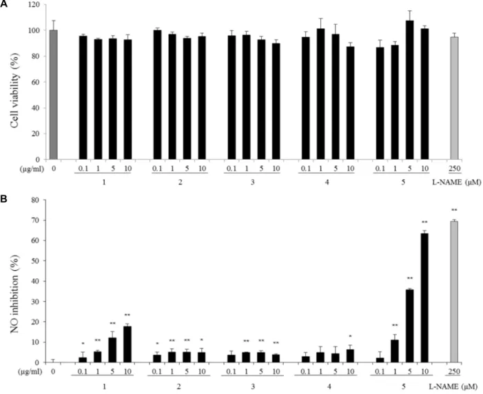
Fig. 2. Effects of compounds 1-5 on cell viability (A) and NO inhibition (B) in LPS-stimulated RAW264.7 cells. NO production was assayed in the media of cells stimulated with LPS (100 ng/ml) for 24 hr. Values are mean ± SEM of three independent experiments. *p<0.05, **p<0.01 vs. LPS alone.
isolated from S. koreana, cytotoxicity and NO production were measured in unstimulated and LPS-stimulated RAW 264.7 cells. RAW 264.7 cells were incubated with LPS (100 ng/ml) in the presence of the compounds for 24 hr. NO pro- duction drastically increased upon LPS treatment from 2.43 nM to 34.87 nM (data not shown), and cells pretreated with all compounds at 10 μg/ml showed significantly increased inhibition of NO production in culture media. Kaempferol- 3-o-[2'',6''-di-o-(trans-p-coumaroyl)]-β-D-glucopyranoside (5) showed the strongest inhibition rates of 2.13±3.13, 10.98±2.83, 35.86±0.49, and 63.35±1.61% at concentrations of 0.1, 1, 5, and 10 μg/ml, respectively (Fig. 2B). Quercetin (1) also reduced NO production in a dose-dependent manner but was less effective than compound 5. On the other hand, quercitrin (2), hyperin (3), and Quercetin-3-O-(6''-O-galloyl)- β-D-galactopyranoside (4) were less effective than quercetin
(1) or kaempferol-3-o-[2'',6''-di-o-(trans-p-coumaroyl)]-β-D- glucopyranoside (5) regarding inhibition of NO production.
In other words, at a concentration of 10 μg/ml, inhibition rates of quercetin (1), quercitrin (2), hyperin (3), querce- tin-3-O-(6''-O-galloyl)-β-D-galactopyranoside (4), and kaem- pferol-3-o-[2'',6''-di-o-(trans-p-coumaroyl)]-β-D-glucopyr- anoside (5) were 17.17±1.17, 5.0±1.92, 3.92±0.36, 6.32±2.15, and 63.35±1.61%, respectively. The NO synthase inhibitor N(G)-nitro-L-arginine methyl ester (L-NAME) was used as a positive control and showed an NO inhibition rate of 69.39±0.93% at 250 μM. Cytotoxicities of the compounds were determined to ensure that the observed reduction of NO production was not due to cell death. Neither the tested compounds nor 0.1% DMSO significantly affected cell via- bility under the tested conditions (Fig. 2A).
Of the compounds tested in our study, kaempferol-3-
A
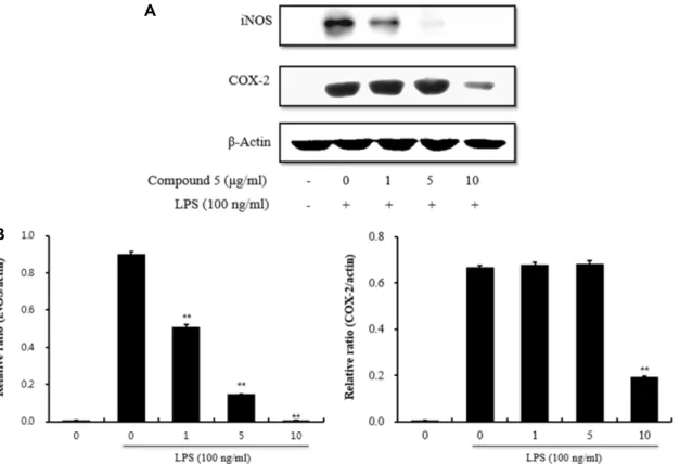
B
Fig. 3. Inhibition of iNOS and COX-2 expression by compound 5. (A) RAW264.7 cells were pretreated with different concentrations of compound 5 for 1 hr and then stimulated with LPS (100 ng/ml) for 24 hr. β-actin expression was used as an internal control for Western blot analysis. (B) All values were normalized based on β-actin expression. The results were expressed as mean ± SD of three independent experiments. ** p<0.01 vs. LPS alone.
o-[2'',6''-di-o-(trans-p-coumaroyl)]-β-D-glucopyranoside (5) exhibited significantly high inhibitory activity on NO pro- duction in activated macrophages compared to the other four quercetin backbone constituents. The results suggest a difference in bioactivity between the quercetin backbone and kaempferol backbone, and these data are similar to those reported previously by others [2, 3].
Effects of kaempferol-3-o-[2'',6''-di-o-(trans-p- coumaroyl)]-β-D-glucopyranoside from S. koreana on LPS-induced iNOS and COX-2 protein expressions
We next performed Western blot analysis to determine whether or not the inhibitory effects of kaempferol-3-o- [2'',6''-di-o-(trans-p-coumaroyl)]-β-D-glucopyranoside from S. Koreana on pro-inflammatory mediators could be attrib- uted to modulation of iNOS and COX-2 expression. In un- stimulated RAW 264.7 cells, iNOS and COX-2 proteins were not detected, whereas LPS treatment remarkably up-regu- lated their protein levels. Further, pre-treatment with kaem- pferol-3-o-[2'',6''-di-o-(trans-p-coumaroyl)]-β-D-glucopyr- anoside inhibited LPS-induced iNOS and COX-2 up-
regulation. However, kaempferol-3-o-[2'',6''-di-o-(trans-p- coumaroyl)]-β-D-glucopyranoside did not affect expression of β-actin, a housekeeping protein (Fig. 3A). Kaempferol- 3-o-[2'',6''-di-o-(trans-p-coumaroyl)]-β-D-glucopyranoside exhibited a strong inhibitory effect against iNOS expression.
Since the level of iNOS protein expression correlated with NO accumulation, this result suggests that kaempferol-3-o- [2'',6''-di-o-(trans-p-coumaroyl)]-β-D-glucopyranoside inhibi- ted NO production by reducing iNOS expression. In addi- tion, the level of COX-2 protein increased upon LPS treat- ment and was antagonized by kaempferol-3-o-[2'',6''-di-o- (trans-p-coumaroyl)]-β-D-glucopyranoside at 10 μg/ml. Fig.
3B shows the densitometric analysis of the immunoblot for iNOS and COX-2 protein expression. Kaempferol-3-o-[2'',6''- di-o-(trans-p-coumaroyl)]-β-D-glucopyranoside (0.1-10 μg/
ml) dose-dependently reduced LPS-induced iNOS ex- pression, and COX-2 protein expression was significantly in- hibited at 10 μg/ml.
Previous studies have demonstrated that methanol extract of leaves from S. koreana exhibit significant anti-inflamma- tory activities via COX-2 and iNOS by blocking NF-κB ex-
pression [10]. Further, syringaresinol [7], a phenolic com- pound isolated from the stems of this plant, was shown to inhibit NO production in activated macrophages. Kaempferol- 3-o-[2'',6''-di-o-(trans-p-coumaroyl)]-β-D-glucopyranoside, identified for the first time in the present study from S. kore- ana, has not been previously reported to have anti-in- flammatory activity, although its presence has been reported in Querces dentate [19] and Eryngium yuccifolium [21].
In conclusion, five phenolic compounds, including two newly identified flavonols from leaves of S. koreana (= S.
pseudocamellia), were isolated and their NO inhibitory activ- ities were measured to identify the major active constituent responsible for the efficacy of the extract against inflamma- tory reactions. Among the separated compounds in the EtOAc fraction, including quercetin, quercitrin, hyperin, quercetin-3-O-(6''-O-galloyl)-β-D-galactopyranoside, and kaem- pferol-3-o-[2'',6''-di-o-(trans-p-coumaroyl)]-β-D-glucopyr- anoside, kaempferol-3-o-[2'',6''-di-o-(trans-p-coumaroyl)]-β- D-glucopyranoside showed significantly higher potent NO inhibitory activity than the other quercetin backbone com- pounds in LPS-treated RAW 264.7 cells. Thus, these data suggest that kaempferol-3-o-[2'',6''-di-o-(trans-p-coumaroyl)]- β-D-glucopyranoside might be the major active compound responsible for the anti-inflammatory effect of S. koreana for the first time.
Acknowledgements
This research was supported by Basic Science Research Program through the National Research Foundation of Korea (NRF), funded by the Ministry of Science, ICT &
Future Planning (No. 2015RICIA2A01055125),and by a grant from the regional innovation center program of the Ministry of Trade, Industry and Energy at the Skin Biotechnology Center of Kyung Hee University, Korea.
References
1. Alderton, W. K., Cooper, C. E. and Knowles, R. G. 2001.
Nitric oxide synthases: structure, function and inhibition.
Biochem. J. 357, 593-615.
2. García-Mediavilla, V., Crespo, I., Collado, P. S., Esteller, A., Sánchez-Campos, S., Tuñón, M. J. and González-Gallego, J.
2007. The anti-inflammatory flavones quercetin and kaemp- ferol cause inhibition of inducible nitric oxide synthase, cu- clooxygenase-2 and reactive C-protein, and down-regu- lation of the nuclear factor kappaB pathway in Chang liver cells. Eur. J. Pharmacol. 557, 221-229.
3. Hämäläinen, M., Nieminen, R., Vuorela, P., Heinonen, M.
and Moilanen, E. 2007. Anti-inflammatory effects of fla- vonoids: genistein, kaempferol, quercetin, and daidzein in- hibit STAT-1 and NF-kB activations, whereas flavones, iso- rhamnetin, naringenin, and pelargonidin inhibit only NF-kB activation along with their inhibitory effect on iNOS ex- pression and NO production in activated macrophages.
Mediators Inflamm. 2007, 45673.
4. Ha, S., Choi, Y., Jeon, Y., Kang, S., Zee, O. and Kwak, J.
2013. Phenolic compounds from the flower of Stewartia pseu- do-camellia and their inhibitory effects on the release of β- hexosaminidase in RBL-2H3 cells. Planta Med. 79, PJ20.
5. Hossen, M. J., Jeon, S. H., Kim, S. C., Kim, J. H., Jeong, D., Sung, N. Y., Yang, S., Baek, K. S., Kim, J. H., Yoon, D.
H., Song, W. O., Yoon, K. D., Cho, S. H., Lee, S., Kim, J.
H. and Cho, J. Y. 2015. In vitro and in vivo anti-inflammatory activity of Phyllanthus acidus methanolic extract. J. Ethnopha- macol. 168, 217-228.
6. Jeong, R. H., Lee, D. Y., Cho, J. G., Lee, S. M., Kang, H.
C., Seo, W. D., Kang, H. W., Kim, J. Y. and Baek, N. I. 2011.
A new flavonolignan from the aerial parts of Oryza sativa L. inhibits nitric oxide production in RAW 264.7 macro- phage cells. J. Appl. Biol. Chem. 54, 865-870.
7. Kim, M. H., Jang, J. H., Oh, M. H., Heo, J. H. and Lee, M.
W. 2014. The comparison of DPPH-scavenging capacity and anti-inflammatory effects of phenolic compounds isolated from the stem of Stewartia koreana Nakai. Nat. Prod. Res. 28, 1409-1412.
8. Kim, S. K., Kim, H. J., Choi, S. E., Park, K. H., Choi, H.
K. and Lee, M. W. 2008. Anti-oxidative and inhibitory activ- ities on nitric oxide (NO) and prostaglandin E2 (COX-2) pro- duction of flavonoids from seeds of Prunus tomentosa Thun- berg. Arch. Pharm. Res. 31, 424-428.
9. Lee, J. M., Kim, H. M., Lee, S., Han, S., Cho, S. H. and Lee, S. 2010. Determination of hyperin in the fruits of Acanthopa- nax species by high performance liquid chromatography.
Nat. Prod. Sci. 16, 39-42.
10. Lee, T. H., Kwak, H. B., Kim, H. H., Lee, Z. H., Chung, D. K., Baek, N. I. and Kim, J. 2007. Methanol extracts of Stewartia koreana inhibit cycloocygenase-2 (COX-2) and in- ducible nitric oxide synthase (iNOS) gene expression by blocking NF-kappaB transactivation in LPS-activated RAW 264.7 cells. Mol. Cells 23, 398-404.
11. Lee, T. H., Lee, G. W., Kim, C. W., Bang, M. H., Beak, N.
I., Kim, S. H., Chung, D. H. and Kim, J. Y. 2010. Stewartia koreana extract stimulates proliferation and migration of hu- man endothelial cells and induces neovasculization in vivo.
Phytother. Res. 24, 20-25.
12. Lee, T. H., Lee, G. W., Park, K. H., Mohamed, M. A., Bang, M. H., Baek, Y. S., Son, Y., Chung, D. K., Baek, N. I. and Kim, J. 2014. The stimulatory effects of Stewartia koreana ex- tract on the proliferation and migration of fibroblasts and the wound healing activity of the extract in mice. Int. J. Mol.
Med. 34, 145-152.
13. Lee, T. H., Lee, S. M., Lee, D. Y., Son, Y., Chung, D. K., Baek, N. I. and Kim, J. 2011. A glycosidic spinasterol from
초록:노각나무 잎에서 분리된 플라보노이드에 의한 대식세포에서 산화질소 생성 억제효과 이승수1․방면호1․박세호2․정대균1․양선아3*
(1경희대학교 피부생명공학센터, 2계명대학교 자연과학연구소, 3계명대학교 식품가공학전공)
노각나무(Stewartia koreana) 잎 에틸아세테이트 분획으로부터 quercetin (1), quercitrin (2), hyperin (3), querce- tin-3-O-(6''-O-galloyl)-β-D-galactopyranoside (4), kaempferol-3-o-[2'',6''-di-o-(trans-p-coumaroyl)]-β-D-glucopyr- anoside (5)의 5종의 플라보노이드를 분리하였으며, 이들 5종 성분의 염증 반응에 대한 활성을 분석하기 위하여 LPS를 처리한 대식세포에서 산화질소(NO) 생성 억제활성을 측정하였다. 이들 5종 성분 중 compound 4, 5는 노각 나무에서 처음으로 분리된 것으로 항염증 활성에 대한 보고도 없다. 분광분석법으로 확인된 노각나무 잎 유래 성분들은 LPS 처리한 대식세포의 NO 생성을 유의적으로 저해하였으며, 특히 kaempferol-3-o-[2'',6''-di-o-(trans- p-coumaroyl)]-β-D-glucopyranoside (5)는 가장 강한 억제효과(17.17%, 5.0%, 3.92%, 6.32% and 63.35% inhibition of compound 1, 2, 3, 4 and 5 at 10 μg/ml)를 나타냈다. 또한, 이러한 NO 생성 억제효과는 inducible nitric oxide synthase(iNOS) 단백질 발현 억제를 통한 것으로 나타났다. 따라서, 본 연구에서 새로 분리된 플라보놀인 kaemp- ferol-3-o-[2'',6''-di-o-(trans-p-coumaroyl)]-β-D-glucopyranoside (5)는 노각나무 잎의 항염증 활성을 나타내는 주 요 물질로 사료된다.
Koreana stewartia promotes procollagen production and in- hibits matrix metalloproteinase-1 expression in UVB-irradi- ated human dermal fibroblasts. Biol. Pharm. Bull. 34, 768-773.
14. Mosmann, T. 1985. Rapid colorimetric assay for cellular growth and survival: Application to proliferation and cyto- tocixity assay. J. Immunol. Methods 65, 55-63.
15. Park, C. K., Kim, H. J., Kwak, H. B., Lee, T. H., Bang, M.
H., Kim, C. M., Lee, Y., Chung, D. K., Baek, N. I., Kim, J., Lee, Z. H. and Kim, H. H. 2007. Inhibitory effects of Stewartia koreana on osteoclast differentiation and bone resorption. Int. Immunopharmacol. 7, 1507-1516.
16. Roh, H. J., Noh, H. J., Na, C. S., Kim, C. S., Kim, K. H., Hong, C. Y. and Lee, K. R. 2015. Phenolic compounds from the leaves of Stewartia pseudocamellia Maxim and their whitening activities. Biomol. Ther. 23, 283-289.
17. Statti, G. A., Conforti, F., Menichini, F., Marrelli, M., Gangale, C., Tundis, R., Loizzo, M. R., Bonesi, M. and Menichini, F.
2011. Protective effect of Hypericum calabricum Sprengel on oxidative damage and its inhibition of nitric oxide in lip-
opolysaccharide-stimulated RAW264.7 macrophages. Biol.
Res. 44, 213-218.
18. Tracey, K. J. and Cerami, A. 1993. Tumor necrosis factor, other cytokines and disease. Annu. Rev. Cell Biol. 9, 317-343.
19. Yamashita, N., Etoh, H., Sakata, K., Yahi, A., Ina, H. and Ina, K. 1989. An acylated kaempferol glucoside isolated from Quercus dentate as a repellent against the blue mussel Mytilus edulis. Agr. Biol. Chem. 53, 1383-1385.
20. Yang, Z. G., Jia, L. N., Shen, Y., Ohmura, A. and Kitanaka, S. 2011. Inhibitory effects of constituents from Euphorbia lu- nulata on differentiation of 3T3-L1 cells and nitric oxide pro- duction in RAW264.7 cells. Molecules 16, 8305-8318.
21. Zhang, Z., Li, S., Ownby, S., Wang, P., Yuan, W., Zhang, W. and Scott Beasley, R. 2008. Phenolic compounds and rare polyhydroxylated triterpenoid saponins from Eryngium yuccifolium. Phytochemistry 69, 2070-2080.
22. Zhou, Y. J., Xu, S. X., Che, Q. M. and Sun, Q. S. 2001.
Flavonoids from the leaves of Quercus dentate. Indian J.
Chem. 40B, 394-398.